Total Suture Technique for Neer Type II & V Distal Clavicle Fractures: A Comprehensive Clinical and Surgical Guide
Key Takeaway
The Total Suture Technique is an innovative surgical method for unstable Neer Type II & V distal clavicle fractures. It involves using strong sutures to reconstruct or augment coracoclavicular ligament stability, anchoring the clavicle to the coracoid process. This technique provides stable fixation, promotes natural healing, and uniquely avoids the complications and second surgery associated with traditional metal implant removal.
Introduction and Epidemiology
Fractures of the clavicle constitute a significant proportion of orthopedic trauma, accounting for approximately 2.6% to 10% of all fractures. Distal clavicle fractures, involving the lateral one-third, represent about 15% of all clavicular fractures, with a potentially higher incidence observed in specific athletic populations. Despite their relative rarity, the complexity of their management and profound implications for shoulder function underscore their clinical importance, particularly for unstable fracture patterns. The modified Neer classification subdivides distal clavicle fractures into five primary types. Among these, Neer Type II and Type V fractures pose unique therapeutic challenges due to their inherent instability and elevated risk of non-union, frequently mandating surgical intervention to restore optimal functional outcomes.
Neer Type I fractures are defined as extra-articular fractures occurring lateral to the coracoclavicular (CC) ligaments. Given the integrity of the CC ligaments, these are generally considered stable fractures and typically achieve favorable healing with non-operative modalities. Neer Type III fractures are intra-articular fractures involving the acromioclavicular (AC) joint, extending lateral to the CC ligaments and into the AC joint articular surface. The intact CC ligaments typically confer relative stability to this type as well. Neer Type IV fractures are specifically epiphyseal fractures observed in skeletally immature patients, where the epiphysis remains tethered to the CC ligaments, also generally considered stable.
Conversely, Neer Type II and Type V fractures are unequivocally categorized as unstable patterns. The fundamental pathophysiology driving their instability involves the disruption or avulsion of the coracoclavicular ligaments, which abrogates or functionally compromises the crucial connection between the proximal clavicular segment and the scapula.
-
Neer Type II Fractures
Neer Type II fractures are further stratified into IIA and IIB subtypes:
- Type IIA Fractures: The fracture line is situated medial to the coracoclavicular ligaments. In this subtype, the CC ligaments typically remain attached to the distal clavicular fragment. The potent superior traction forces exerted by muscle groups such as the sternocleidomastoid and trapezius on the proximal clavicular segment, coupled with the inferior displacement of the distal clavicular segment and its attached shoulder girdle due to upper limb gravity, result in characteristic superior displacement of the proximal fragment, manifesting as significant vertical instability.
- Type IIB Fractures: The fracture line lies between the conoid and trapezoid ligaments. In this configuration, the conoid ligament is commonly detached from the proximal fragment, while the trapezoid ligament may be detached from either the distal or proximal fragment, or both. This fracture pattern similarly exhibits pronounced superior displacement of the proximal clavicular segment, with its inherent instability substantially increasing the risk of non-union.
-
Neer Type V Fractures
Neer Type V fractures are comminuted patterns characterized by a large medial fragment to which the entire coracoclavicular ligament complex (both conoid and trapezoid) remains robustly attached. However, the distal clavicular fragments and the associated shoulder complex displace inferiorly due to gravity, leading to marked superior migration of the proximal clavicular segment, again demonstrating significant vertical instability.
The critical disruption of the coracoclavicular ligaments, combined with the unopposed pull of powerful muscle groups (e.g., sternocleidomastoid, trapezius) on the proximal fragment and the gravitational forces acting on the distal fragment and scapula, creates profound vertical instability. This displacement and instability contribute to non-union rates as high as 30-40% with non-operative management, particularly in active young patients, where functional recovery is often suboptimal. Consequently, for displaced Neer Type II and Type V distal clavicle fractures, surgical stabilization has become the predominant and widely accepted standard of care.
Historically, various internal fixation techniques have been employed, including locking plates, hook plates, tension band wiring, intramedullary screws, K-wires, cortical buttons, or suture-based coracoclavicular fixation. While locking plates and hook plates have achieved satisfactory union rates, they possess inherent drawbacks. Locking plates can be challenging for stabilizing small distal fragments, and hook plates, while providing immediate stability, carry a risk of subacromial impingement, rotator cuff irritation or injury, and frequently necessitate a mandated second-stage surgical removal to mitigate long-term complications. Other techniques, such as K-wire fixation, are largely discouraged as sole fixation methods due to risks of migration and potentially life-threatening complications.
In recent years, advancements in biomechanical materials and minimally invasive techniques have led to the increasing adoption of suture-based fixation, often augmented with cortical buttons. These techniques aim to provide robust coracoclavicular ligament reconstruction or augmentation while minimizing the complications associated with traditional metallic internal fixation. This guide meticulously details a contemporary "Total Suture Technique" for displaced Neer Type II and Type V distal clavicle fractures. This approach endeavors to provide effective stabilization and promote union while obviating the need for secondary implant removal, offering a comprehensive clinical and surgical roadmap for orthopedic surgeons.
Surgical Anatomy and Biomechanics
A profound understanding of the distal clavicle's anatomy, its surrounding soft tissue structures, and their biomechanical interplay is paramount for the successful management of unstable Neer Type II and Type V distal clavicle fractures.
Distal Clavicle Morphology and Articulations
The distal clavicle encompasses the lateral one-third of the bone, characterized by its flatter, broader morphology compared to the cylindrical shaft. It articulates with the acromion of the scapula to form the acromioclavicular (AC) joint and connects to the coracoid process via the coracoclavicular (CC) ligaments.
- Acromioclavicular Joint: This is a diarthrodial, plane synovial joint formed by the articular facet on the distal clavicle and the medial aspect of the acromion. It allows limited gliding and rotational movements, crucial for scapulothoracic motion and shoulder girdle flexibility. The joint capsule is composed of fibrous tissue and lined by a thin synovial membrane. Its primary stability in the horizontal plane is provided by the superior, inferior, anterior, and posterior acromioclavicular ligaments. The superior AC ligament is the strongest and plays a critical role in resisting shear forces. An articular disc, often incomplete or meniscoid, may be present within the joint.
- Coracoid Process: This is a robust, hook-like projection originating from the superior aspect of the scapular neck, anterior and inferior to the glenoid. It serves as a vital anchor point for numerous ligaments and muscles, including the coracoclavicular ligaments, coracoacromial ligament, coracohumeral ligament, pectoralis minor, coracobrachialis, and the short head of the biceps brachii. Its base provides a stable anatomical landmark for reconstruction.
Coracoclavicular Ligament Complex
The coracoclavicular (CC) ligament complex is the primary stabilizer of the distal clavicle, particularly against superior displacement relative to the scapula. It consists of two distinct components: the conoid and trapezoid ligaments.
- Conoid Ligament: This more medial and posterior ligament is triangular or fan-shaped. It originates from the posteromedial aspect of the base of the coracoid process and inserts onto the conoid tubercle on the inferomedial aspect of the distal clavicle. Its fibers run predominantly vertically, providing primary resistance to superior displacement of the clavicle.
- Trapezoid Ligament: Located more laterally and anteriorly, this ligament is quadrilateral in shape. It originates from the superior aspect of the coracoid process, anterior to the conoid attachment, and inserts onto the trapezoid line on the inferolateral aspect of the distal clavicle. Its fibers run in a more oblique direction, contributing to both vertical and horizontal stability, resisting both superior and posterior migration of the clavicle.
Together, these ligaments form an inverted trapezoid, with their insertions separated by approximately 2 cm on the clavicle and their origins on the coracoid being distinct but closely related. The average distance from the trapezoid insertion to the lateral end of the clavicle is 2.5 cm, while the conoid insertion is approximately 4.5 cm from the lateral end.
Associated Musculature and Neurovasculature
- Muscles: The trapezius and sternocleidomastoid muscles exert significant superior pull on the proximal clavicular segment. The deltoid muscle, particularly its anterior and middle fibers, originates from the distal clavicle and acromion, contributing to the muscle envelope around the fracture site. When the CC ligaments are disrupted, the unopposed pull of the trapezius and sternocleidomastoid on the proximal fragment, combined with the downward pull of gravity on the distal fragment and the upper extremity, dictates the characteristic vertical displacement seen in Neer Type II and V fractures.
- Neurovascular Structures: The supraclavicular nerves, branches of the cervical plexus, traverse superficially over the clavicle and are at risk during surgical approaches. Deeper and more medially, the subclavian artery and vein, along with the brachial plexus, lie in close proximity to the clavicle's undersurface. While direct injury is rare in distal clavicle surgery, awareness of these structures is critical during medial dissection or extensive drilling.
Biomechanics of Instability and Suture-Based Reconstruction
In Neer Type II and V fractures, the integrity of the CC ligaments is compromised.
* In Type IIA, the fracture occurs medial to both ligaments, leaving them attached to the distal fragment. The proximal fragment is pulled superiorly.
* In Type IIB, the fracture occurs between the ligaments, often detaching the conoid from the proximal fragment. Both proximal and distal fragments are unstable.
* In Type V, the fracture is comminuted, but the CC ligaments remain attached to a large medial fragment. The distal fragments still displace inferiorly.
The goal of the total suture technique is to functionally restore the coracoclavicular relationship. By passing high-strength sutures around the coracoid process and through the clavicle, the technique biomechanically replicates the functions of the conoid and trapezoid ligaments, resisting superior migration of the clavicle and providing vertical stability. The repair of the deltotrapezial fascia further contributes to horizontal stability and overall shoulder girdle integrity. This suture-based construct allows for controlled micromotion, potentially promoting biological healing, while avoiding the complications associated with metallic implants.
Indications and Contraindications
Precise patient selection is crucial for optimizing outcomes with the total suture technique for distal clavicle fractures. Surgical intervention is primarily reserved for unstable patterns.
Indications for Operative Management
- Displaced Neer Type II Fractures (IIA and IIB): These fractures inherently exhibit vertical instability due to CC ligament disruption and muscle pull. Displacement typically defined as > 4-5 mm of superior migration of the proximal fragment relative to the distal fragment or coracoid.
- Displaced Neer Type V Fractures: Characterized by comminution and CC ligament attachment to a large medial fragment, resulting in superior displacement of the medial fragment and inferior displacement of the distal fragments and shoulder girdle.
- Clinical Sagging Deformity: A palpable and visible step-off or deformity, even if radiographic displacement seems borderline, often warrants surgical correction, especially in younger, active patients.
- High Functional Demand Patients: Athletes, laborers, or individuals requiring robust shoulder function where non-union or malunion would severely compromise activity.
- Acute Fractures: Generally, surgical fixation is more predictable when performed within 2-3 weeks of injury before significant fibrotic changes occur.
- Non-Union of Neer Type II or V Fractures: Persistent pain, instability, or functional deficit from a failed non-operative trial or previous inadequate fixation. Often requires debridement and bone grafting in conjunction with suture fixation.
- Associated Acromioclavicular Joint Instability: If the AC joint itself is disrupted in addition to the distal clavicle fracture, suture techniques can concurrently address AC joint reduction and stabilization.
- Fractures with Inadequate Bone Stock for Plate Fixation: In cases of severe comminution of the distal fragment where conventional plate fixation would be tenuous, suture-based techniques provide an alternative.
Indications for Non-Operative Management
- Minimally Displaced Neer Type II or V Fractures: Defined as vertical displacement < 2-3 mm. These are rare but can be observed. Close radiographic follow-up is essential to monitor for progressive displacement.
- Neer Type I, III, and IV Fractures: These types are generally stable due to intact CC ligaments and respond well to conservative treatment, including sling immobilization and early range of motion.
- Patients with Significant Comorbidities: Medical conditions that significantly increase surgical risk and anesthetic complications, especially in low-demand individuals.
- Elderly, Low-Demand Patients: Individuals with minimal symptoms and low functional expectations, where the risks of surgery outweigh the potential benefits.
Contraindications for Operative Management
- Active Infection: Absolute contraindication. Surgery should be deferred until infection is cleared.
- Severe Local Skin Compromise: Open wounds, severe blistering, or impending skin necrosis over the surgical site. Delay surgery until skin integrity is optimized.
- Uncontrolled Systemic Medical Conditions: Conditions such as uncontrolled diabetes, severe cardiovascular disease, or coagulopathy that significantly elevate surgical risk.
- Extensive Coracoid Comminution: If the coracoid process is severely fractured or osteoporotic to the extent that it cannot reliably hold sutures, suture-based fixation may be compromised.
- Inadequate Soft Tissue Coverage: In cases of severe open fractures or avulsion injuries where sufficient soft tissue cannot be achieved for wound closure.
- Patient Refusal: Patient's informed decision to decline surgical intervention.
Operative vs Non-Operative Indications Summary
| INDICATION CATEGORY | OPERATIVE MANAGEMENT | NON-OPERATIVE MANAGEMENT |
|---|---|---|
| Fracture Type & Displacement | - Displaced Neer Type II (IIA, IIB) fractures (vertical displacement > 4-5 mm). - Displaced Neer Type V fractures. - Clinically significant superior migration/deformity of the clavicle. - Non-union of Neer Type II or V fractures. |
- Minimally displaced Neer Type II or V fractures (vertical displacement < 2-3 mm, rare). - Stable Neer Type I, III, and IV fractures. |
| Patient Factors | - Young, active, high-demand patients (athletes, laborers). - Patients intolerant of functional deficit or cosmetic deformity. - Acute fractures (typically within 2-3 weeks of injury). |
- Elderly, low-demand patients with minimal symptoms and functional expectations. - Patients with significant comorbidities precluding surgery. - Patients who explicitly refuse surgical intervention after informed consent. |
| Associated Injuries | - Concomitant AC joint instability requiring stabilization. - Extensive comminution of the distal clavicle precluding stable plate fixation. |
- No associated injuries requiring surgical intervention. |
| Local Factors | - Adequate bone stock of coracoid and clavicle for suture passage. - Intact skin and soft tissue envelope. |
- Active infection. - Severe local skin compromise (open wounds, severe blistering). - Extensive coracoid comminution preventing reliable suture purchase. - Inadequate soft tissue coverage. |
| Systemic Factors | - Medically stable patients able to tolerate general anesthesia and surgery. | - Uncontrolled systemic medical conditions (e.g., severe coagulopathy, uncontrolled diabetes, acute cardiac events). |
Pre Operative Planning and Patient Positioning
Thorough preoperative planning is critical for successful outcomes in distal clavicle fracture repair, particularly when utilizing a total suture technique. This phase encompasses comprehensive patient evaluation, detailed imaging review, and meticulous surgical setup.
Patient Evaluation
- History: A detailed history should include the mechanism of injury (e.g., direct fall onto the shoulder, motor vehicle accident), dominant hand, pre-existing shoulder pain or pathology, occupational demands, athletic aspirations, and any significant medical comorbidities (e.g., diabetes, osteoporosis, smoking history which negatively impacts bone healing). The timing of injury is crucial, as acute repairs generally yield better results.
- Physical Examination: A thorough examination involves inspection for swelling, ecchymosis, and any open wounds or skin tenting. Palpation will reveal tenderness over the fracture site and often a step-off deformity at the distal clavicle and AC joint. Assessment of skin integrity, especially over the fracture fragments, is paramount. A comprehensive neurovascular assessment of the ipsilateral upper extremity must be performed, including sensation in the supraclavicular nerve distribution. Active and passive range of motion of the shoulder, elbow, and wrist should be documented, acknowledging that pain often severely limits shoulder motion.
- Imaging Studies:
- Plain Radiographs: Standard views include an anteroposterior (AP) view of the shoulder, a 15-20 degree cephalic tilt view (AP clavicle view) to project the clavicle above the scapular spine, and an AC joint specific view (Zanca view – 10-15 degree cephalic tilt, allowing for a better appreciation of the AC joint space). An axillary lateral view is also valuable to assess posterior displacement or comminution, particularly relative to the coracoid. Bilateral weight-bearing views are useful for comparing superior migration with the contralateral side, though often too painful in acute injuries.
- Computed Tomography (CT) Scan: A CT scan with 3D reconstructions is highly recommended for complex or comminuted distal clavicle fractures, especially Neer Type V. It provides invaluable information regarding the degree of comminution, fragment size, intra-articular involvement, and the exact relationship of the clavicle to the coracoid process, aiding in precise preoperative planning for suture tunnel placement and reduction strategy. It helps to identify any subtle fractures of the coracoid process itself.
- Magnetic Resonance Imaging (MRI): While not routinely indicated for acute fractures, an MRI can be useful if there is suspicion of concomitant rotator cuff tears, labral injuries, or significant soft tissue damage, which might influence rehabilitation protocols or require concurrent addressing.
Pre-Operative Discussion and Informed Consent
A detailed discussion with the patient is essential. This includes:
* Explanation of the fracture pattern and the rationale for surgical intervention.
* Description of the total suture technique, emphasizing its advantages (no implant removal) and potential limitations.
* Review of potential risks, including but not limited to infection, non-union/malunion, neurovascular injury (supraclavicular nerve paresthesia), implant failure (suture cut-out or breakage), post-traumatic AC joint arthritis, pain, stiffness, and the possibility of revision surgery.
* Realistic expectations regarding functional recovery and return to activity.
* Discussion of the proposed rehabilitation protocol.
Anesthesia and Patient Positioning
- Anesthesia: General endotracheal anesthesia is typically employed. An interscalene brachial plexus block can be a valuable adjunct for postoperative pain management, reducing opioid requirements and facilitating early rehabilitation.
- Patient Positioning: The beach chair position is generally preferred for distal clavicle fracture fixation.
- Advantages: This position provides excellent exposure to the superior aspect of the shoulder, clavicle, and AC joint. It allows for easy access to the coracoid process. The surgeon can freely manipulate the arm to aid in reduction and assess range of motion, and it facilitates arthroscopic visualization if a concomitant arthroscopic procedure is planned.
- Setup: The patient is placed supine on the operating table, and the torso is gradually elevated to approximately 45-70 degrees. The head is securely positioned in a headrest, ensuring the neck is neutral and avoiding excessive rotation or lateral flexion to prevent brachial plexus stretch. The ipsilateral arm should be draped free to allow for full manipulation and assessment of shoulder motion. Adequate padding of all pressure points (sacrum, heels, opposite elbow, etc.) is crucial.
- Precautions: Careful attention must be paid to blood pressure monitoring in the beach chair position, as it can predispose to cerebral hypoperfusion. Lowering the legs slightly can help mitigate this risk. Ensure the eyes are taped shut and protected.
- Imaging Access: A C-arm fluoroscopy unit should be readily available and positioned to obtain AP and lateral views of the shoulder and clavicle to confirm reduction and suture placement intraoperatively.
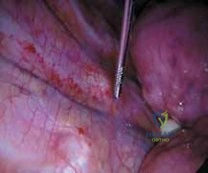
Detailed Surgical Approach and Technique
The total suture technique for Neer Type II and V distal clavicle fractures aims to achieve anatomical reduction and stable coracoclavicular fixation using high-strength suture material, obviating the need for metallic implants across the fracture site or within the AC joint. This approach relies on careful surgical planning, meticulous soft tissue handling, and precise suture placement.
Incision and Superficial Dissection
- Skin Incision: A longitudinal incision is typically made directly over the distal clavicle, centered over the fracture site and extending approximately 5-7 cm. This allows adequate visualization of the distal clavicle, AC joint, and access to the coracoid. Some surgeons prefer a sabre-cut incision.
- Pearls: The incision should be carefully planned to minimize tension and optimize cosmesis. Avoid excessive undermining of skin flaps to preserve vascularity.
- Subcutaneous Dissection: The subcutaneous tissue is dissected, and the platysma muscle (if encountered superiorly) is carefully incised. The supraclavicular nerves, which typically cross the operative field perpendicularly, should be identified and protected. Gentle retraction rather than sharp dissection around these nerves is paramount to minimize the risk of neuropraxia or permanent sensory deficits (numbness over the anterior shoulder/chest).
Deep Dissection and Fracture Exposure
- Deltoid and Trapezius: The deltotrapezial fascia is carefully incised along the fracture line or detached in a sleeve fashion if viable. The fracture hematoma is evacuated. The distal clavicular fragments and the proximal clavicular segment are exposed. The AC joint capsule and surrounding ligaments (if intact or reparable) are identified.
- Coracoid Process Exposure: Access to the coracoid process is critical. This is achieved by carefully splitting the deltoid fibers, typically along the interval between the anterior and middle deltoid, or by detaching the deltoid origin from the clavicle and acromion, preserving a cuff for later repair. The interval between the pectoralis major and deltoid can also be used, or a subpectoralis approach can be considered for a more inferior exposure, depending on surgeon preference and fracture configuration.
- Caution: During dissection towards the coracoid, care must be taken to protect the neurovascular structures located inferiorly and medially (axillary nerve, brachial plexus, subclavian vessels). The coracoid tip itself should be avoided as it hosts several muscle and ligament attachments, and the base is a stronger anchor point.
- Identification: The coracoid process is palpated and visually identified. Its superior surface and base are prepared for suture passage.
Fracture Reduction
- Anatomical Alignment: The primary goal is to achieve an anatomical reduction of the distal clavicle. This involves restoring normal length, rotation, and especially vertical alignment relative to the acromion and coracoid.
- Technique: The proximal fragment, often displaced superiorly, is carefully manipulated inferiorly. The distal fragment(s) and associated shoulder girdle are simultaneously elevated to match the proximal fragment. Reduction clamps (e.g., Verbrugge clamp, point-to-point clamp) can be used to provisionally hold the fracture fragments in place. K-wires can be inserted perpendicular to the fracture line to maintain reduction temporarily, particularly for comminuted fractures, but care must be taken to avoid interfering with suture passage.
- Confirm Reduction: Intraoperative fluoroscopy (AP, Zanca, and axillary views) is used to confirm satisfactory reduction of the clavicle and restoration of the coracoclavicular distance.
Suture Placement and Fixation (Total Suture Technique)
The "Total Suture Technique" typically refers to methods that rely solely on high-strength sutures, possibly augmented by cortical buttons, to stabilize the coracoclavicular interval, without requiring a metal plate directly across the fracture site or an intramedullary device. The choice of suture material (e.g., FiberWire, UltraBraid) should be high-strength non-absorbable.
-
Coracoid Preparation and Suture Passage:
- Direct Coracoid Tunneling: Two small drill holes (e.g., 2.5 mm or 3.0 mm) are typically placed through the base of the coracoid process, carefully aiming to exit dorsally, medial to the tip, avoiding its posterior aspect where the suprascapular nerve lies. The holes should be approximately 1.5-2 cm apart, simulating the footprint of the conoid and trapezoid ligaments. A curved suture passer (e.g., a "corkscrew" suture retriever or an aneurysm needle) is then used to pass the high-strength sutures through these tunnels from inferior to superior.
- Coracoid Sling/Loop (Circumcoracoid technique): Alternatively, for patients with smaller coracoids or concern for drilling-related complications, sutures can be passed around the base of the coracoid, creating a sling. This typically involves using a curved passer or blunt dissection to carefully navigate the sutures inferior to the coracoid. This approach is technically more challenging due to the proximity of neurovascular structures.
- Cortical Button Augmentation (Optional but common): While the technique is "total suture," cortical buttons are often used on the inferior aspect of the coracoid to prevent suture cut-through or fraying, effectively creating a "suture-button" construct. A single large tunnel or two smaller tunnels are drilled through the coracoid, and a cortical button (e.g., Dog Bone, TightRope) is passed from inferior to superior, anchored on the inferior cortex of the coracoid. The sutures then pass through the clavicle. This provides a broad, stable anchor point on the coracoid.
-
Clavicular Suture Passage:
- Transosseous Clavicular Tunnels: For Neer Type IIA and V, two transosseous tunnels (e.g., 2.5 mm) are drilled through the proximal clavicular fragment, aligning with the trajectory of the conoid and trapezoid ligaments. The tunnels should be spaced approximately 1.5-2 cm apart and positioned accurately to recreate the ligamentous attachments. The sutures, having been passed through or around the coracoid, are then passed from inferior to superior through these clavicular tunnels.
- Distal Fragment Capture (Neer Type IIB): In Neer Type IIB fractures, where the fracture line lies between the ligaments, it may be necessary to capture both the most medial fragment (to which the conoid was attached) and the distal fragment (to which the trapezoid was attached) with separate suture limbs or to create tunnels within the viable distal fragment itself if large enough. Often, the suture construct aims to fix the proximal clavicular fragment directly to the coracoid, with additional sutures potentially used to stabilize the most distal fragments to the main proximal fragment or the reconstructed AC joint.
- Augmented AC Ligament Repair: If there is associated AC joint instability or injury to the superior AC ligaments, additional sutures can be placed in a figure-of-eight fashion through the acromion and distal clavicle or using transosseous tunnels to directly repair the AC joint capsule. This provides horizontal stability.
-
Suture Tensioning and Knot Tying:
- With the fracture provisionally reduced and held, the sutures are incrementally tensioned and tied down on the superior aspect of the clavicle. This must be performed meticulously to achieve stable reduction without over-compression.
- Key steps: While an assistant holds the shoulder in an anatomically reduced position (reducing the vertical displacement), the surgeon progressively tensions and ties the sutures. This recreates the anatomical relationship between the clavicle and coracoid, restoring the coracoclavicular distance to approximately 11-13 mm (as measured on the contralateral side or by anatomical estimation).
- Multiple passes and knot security are essential. Some techniques advocate for a "double cinch" or "locking" knot configuration to enhance stability.
- After knot tying, the stability of the construct is assessed by gently ranging the shoulder.
Soft Tissue Repair and Closure
- Deltotrapezial Fascia Repair: The deltoid and trapezius attachments to the clavicle and AC joint capsule are meticulously repaired. This is a critical step, as these soft tissues contribute significantly to the dynamic and static stability of the shoulder girdle and help prevent late displacement or loosening of the suture construct. Use strong, non-absorbable sutures for this repair.
- Layered Closure: The subcutaneous tissues are closed in layers using absorbable sutures. The skin is then closed with staples or fine absorbable sutures, ensuring good apposition of wound edges.
- Dressing: A sterile dressing is applied, and the arm is placed in a sling or shoulder immobilizer.
Complications and Management
Despite advancements in surgical techniques, complications can still arise following the total suture technique for distal clavicle fractures. Meticulous surgical execution and appropriate postoperative care are crucial in minimizing these risks.
Common Complications and Management Strategies
| Complication | Incidence (Approximate) | Description | Salvage Strategies and Management |
|---|---|---|---|
| Non-Union / Malunion | 5-15% (for unstable types) | Failure of fracture fragments to heal after 6 months (non-union) or healing in an unacceptable position (malunion) leading to persistent pain, weakness, and functional limitation. Higher risk with poor reduction, smoking, NSAID use, or severe comminution. | Non-union: - Symptomatic, aseptic non-union: Revision surgery with open reduction and internal fixation (e.g., locking plate), often with autogenous bone grafting (e.g., iliac crest or distal radius) or allograft, and re-establishment of CC stability with a new suture-button construct or other CC reconstruction. - Asymptomatic non-union: Non-operative management with observation and physical therapy. Malunion: - Symptomatic malunion: Corrective osteotomy and revision fixation, potentially with bone grafting. - Asymptomatic malunion: Non-operative management focusing on rehabilitation to maximize function. |
| Infection | 1-5% | Superficial (skin/subcutaneous) or deep (to fracture site/suture material). Manifests as erythema, warmth, pain, purulent drainage. Risk factors include diabetes, smoking, prolonged surgery. | Superficial: Oral antibiotics, local wound care. Deep: Surgical debridement, thorough irrigation, intravenous antibiotics based on culture results. In some cases, removal of suture material (if infected) may be necessary, followed by re-fixation or staged reconstruction once infection is cleared. Repeat debridements may be required. |
| Suture Cut-out / Failure | 2-10% | Suture material cuts through bone (especially clavicle), breaks, or knots unravels, leading to loss of reduction and instability. May occur due to excessive tensioning, inadequate bone quality, or premature aggressive rehabilitation. | Revision surgery with re-reduction and re-fixation. May involve using a different suture configuration, incorporating a cortical button if not used initially, or switching to a plate-based construct if suture options are exhausted or bone quality is severely compromised. Bone grafting might be considered if there is significant bone loss at suture tunnels. Postoperative rehabilitation protocol may need to be modified for slower progression. |
| Neurovascular Injury | <1% (serious), 5-15% (supraclavicular) | Supraclavicular Nerve Injury: Numbness, paresthesia over anterior shoulder/chest wall. Usually temporary neuropraxia from retraction. Axillary Nerve/Brachial Plexus/Subclavian Vessels: Rare but serious injury during deep dissection towards coracoid or drilling. |
Supraclavicular Nerve: Mostly resolves spontaneously. Conservative management with reassurance. Chronic symptoms rarely require neurolysis. Axillary Nerve/Brachial Plexus/Subclavian Vessels: Immediate intraoperative recognition and repair by an experienced surgeon. Postoperative management depends on the extent of injury (observation, nerve grafting, vascular repair). These are devastating complications requiring urgent management. |
| Post-Traumatic AC Joint Arthritis | 10-20% | Degenerative changes within the AC joint due to articular cartilage damage at the time of injury or altered biomechanics post-fixation. Presents as localized pain, especially with overhead activities. | Conservative: NSAIDs, physical therapy, corticosteroid injections. Surgical: Distal clavicle excision (Mumford procedure), either open or arthroscopic, for refractory symptomatic cases. |
| Shoulder Stiffness / Pain | Variable | Restricted range of motion or persistent pain not related to other complications. Can be due to inadequate rehabilitation, adhesions, or unrecognized soft tissue injury. | Aggressive physical therapy, pain management (NSAIDs, analgesics). Consider corticosteroid injections for capsulitis or bursitis. Manipulation under anesthesia or arthroscopic capsular release may be necessary in severe, refractory cases. Thorough investigation for other causes of pain (e.g., rotator cuff pathology). |
| Coracoid Fracture | <1% | Fracture of the coracoid process during drilling or suture tensioning, compromising the anchor point for fixation. | If recognized intraoperatively and stable, can proceed with suture fixation around the remaining intact portion of the coracoid or using a different anchor point. If unstable, may require alternative fixation (e.g., mini-plate for coracoid) or abandonment of the suture-only technique in favor of plate fixation to the clavicle. Prevention is key through careful drilling. |
| Prominent Suture Knots | 2-5% | Palpable or painful knots on the superior surface of the clavicle, causing localized irritation. | If significantly symptomatic and not resolving with time, minor surgical removal of the prominent knot(s) after adequate healing of the fracture. |
General Principles of Complication Management
- Prevention: Meticulous surgical technique, precise anatomical reduction, careful soft tissue handling, appropriate suture tensioning, and adherence to postoperative rehabilitation protocols are the cornerstones of complication prevention.
- Early Recognition: Vigilant monitoring for signs and symptoms of complications during the postoperative period.
- Timely Intervention: Prompt and appropriate management based on the specific complication, ranging from conservative measures to revision surgery.
- Patient Education: Comprehensive education regarding potential complications and the importance of compliance with rehabilitation is essential.
Post Operative Rehabilitation Protocols
A structured and progressive postoperative rehabilitation protocol is integral to achieving optimal functional outcomes and minimizing complications after the total suture technique for Neer Type II and V distal clavicle fractures. The protocol emphasizes protection of the repair while gradually restoring range of motion, strength, and function. Individualization based on patient healing, bone quality, fracture comminution, and surgeon preference is paramount.
Phase I: Immobilization and Controlled Motion (Weeks 0-6)
The primary goal of this phase is to protect the suture repair and allow initial fracture healing while preventing stiffness in uninvolved joints.
- Immobilization:
- Arm Sling/Shoulder Immobilizer: The operated arm is typically immobilized in an arm sling or shoulder immobilizer for 4 to 6 weeks. The duration depends on the surgeon's assessment of initial stability and the complexity of the fracture. For very comminuted fractures or patients with poor bone quality, immobilization may be extended.
- Avoidance: Strict avoidance of active shoulder flexion, abduction, external rotation, and any weight-bearing or lifting with the affected arm. Patients should be educated on log-rolling for transfers and maintaining proper posture.
- Exercises:
- Elbow, Wrist, Hand AROM: Active range of motion exercises for the elbow, wrist, and hand should begin immediately to prevent stiffness and promote circulation.
- Gentle Pendulum Exercises: Starting within the first week, gentle pendulum exercises (Codman's exercises) can be initiated, keeping the trunk stable and allowing the arm to swing passively. These are performed multiple times daily.
- Scapular Retractions: Gentle isometric scapular retractions can be started to promote scapular stability without stressing the fracture site.
- Cervical Range of Motion: Active range of motion for the cervical spine should be maintained to prevent neck stiffness.
- Weight Bearing: No weight bearing or lifting with the affected arm.
- Sleeping: Patients may find comfort sleeping in a semi-reclined position (e.g., in a recliner) or with pillows supporting the arm.
Phase II: Early Motion and Progressive Strengthening (Weeks 6-12)
This phase focuses on gradually restoring active shoulder range of motion and initiating light strengthening exercises, provided radiographic signs of early union are present.
- Transition from Immobilization: The sling is typically discontinued around 6 weeks, or as guided by radiographic healing and clinical comfort.
- Active Range of Motion (AROM):
- Begin progressive active-assisted and active range of motion exercises for the shoulder in all planes (flexion, abduction, internal and external rotation).
- Goals by end of Phase II: Achieve near full passive range of motion and progressive improvement in active range of motion, aiming for at least 75-80% of contralateral shoulder.
- Avoid stressing the repair with end-range forceful movements.
- Light Strengthening:
- Isometric Exercises: Initiate gentle isometric strengthening for the rotator cuff (internal/external rotation, abduction) and deltoid, with the arm at the side and elbow bent.
- Scapular Stabilization: Progress scapular stabilization exercises, including rows and presses against light resistance (e.g., theraband).
- Core Strengthening: Maintain general fitness and core strength.
- Weight Bearing: Still no heavy lifting or carrying objects. Avoid pushing, pulling, or sudden jerking movements.
- Activities of Daily Living (ADLs): Encourage use of the arm for light ADLs within a pain-free range.
Phase III: Advanced Strengthening and Functional Return (Weeks 12-24)
This phase concentrates on restoring full strength, endurance, and preparing the patient for a gradual return to higher-level activities.
- Strengthening:
- Progressive Resistance Exercises: Advance rotator cuff and deltoid strengthening with resistance bands, light weights, and eventually machine weights. Focus on concentric and eccentric control.
- Functional Exercises: Incorporate exercises that mimic daily activities and work-specific or sport-specific movements.
- Proprioception and Neuromuscular Control: Balance and proprioceptive exercises for the shoulder girdle.
- Range of Motion: Full pain-free range of motion should be the goal. Continue stretching exercises as needed.
- Weight Bearing: Gradual increase in weight bearing and lifting capacities.
- Return to Activity:
- Light Activities: Gradual return to light recreational activities.
- Work-Specific Tasks: For laborers, progressive return to work-specific tasks, starting with lighter duties.
- Criteria for Progression: Pain-free, full range of motion, and adequate strength (often assessed using dynamometry or functional tests).
Phase IV: Return to Sport / Full Activity (Months 4-6+)
The final phase focuses on sport-specific training, high-impact activities, and ensuring full readiness for unrestricted return to pre-injury activity levels.
- Sport-Specific Training:
- Gradual reintroduction to sport-specific drills, throwing programs (if applicable), and overhead activities.
- Emphasis on technique, endurance, and injury prevention strategies.
- Full Activity: Return to full contact sports or heavy labor generally not before 4-6 months, and only after meeting objective criteria:
- Pain-free full range of motion.
- Strength symmetry (at least 90% compared to the contralateral side).
- No tenderness at the fracture site.
- Radiographic evidence of complete union.
- Confidence and absence of apprehension during dynamic activities.
-
Long-Term Maintenance: Encourage ongoing shoulder strengthening and flexibility exercises to maintain function and prevent recurrence.
-
Important Considerations:
- Pain as a Guide: Rehabilitation should always be pain-guided. Any sharp or increasing pain should lead to a temporary reduction in activity level.
- Radiographic Healing: Regular radiographic follow-up is essential to monitor fracture healing and guide progression through rehabilitation phases.
- Patient Compliance: Patient education and compliance with the home exercise program are critical for successful outcomes.
- Physical Therapist Collaboration: Close collaboration with an experienced physical therapist specializing in shoulder rehabilitation is vital.
Summary of Key Literature and Guidelines
The surgical management of unstable distal clavicle fractures, particularly Neer Type II and V, has evolved significantly, with a growing body of literature supporting various techniques. The "Total Suture Technique," often utilizing high-strength sutures and sometimes augmented by cortical buttons, has emerged as a viable and increasingly popular option, aiming to restore biomechanical stability while mitigating common complications associated with traditional plating.
Biomechanical Studies and Rationale
Biomechanical studies have consistently demonstrated that coracoclavicular (CC) ligament reconstruction or augmentation with high-strength sutures can restore stability comparable to that of the native CC ligaments and, in some constructs, to that achieved with hook plates or locking plates. For instance, studies by Wellmann et al. (2009) and Kim et al. (2011) showed that transosseous CC suture fixation effectively resists superior displacement and restores load-sharing capacity, particularly when augmented with cortical buttons. The rationale for suture-based techniques lies in their ability to dynamically stabilize the clavicle to the coracoid, allowing for micromotion at the fracture site which may promote biological healing, while avoiding the potential pitfalls of rigid plate fixation such as stress shielding or the need for secondary removal. The total suture technique, by eliminating permanent metallic implants across the fracture or AC joint, directly addresses the issue of hardware-related complications such as subacromial impingement and hardware prominence.
Clinical Outcomes and Comparative Studies
Clinical outcomes with suture-based CC fixation have been largely favorable, reporting good-to-excellent functional scores (e.g., Constant, ASES scores), high union rates, and low complication profiles.
* Union Rates: Union rates for suture-based techniques generally range from 90% to 100%, comparable to those reported for locking plates and hook plates. Factors influencing union include patient compliance, smoking status, and meticulous surgical technique.
* Complications: The incidence of specific complications such as infection, non-union, and re-operation rates are generally low, in line with other surgical modalities. A distinct advantage of the total suture technique is the near elimination of hook plate-specific complications (subacromial impingement, rotator cuff irritation) and the need for routine secondary hardware removal. However, suture-specific complications such as suture cut-out or breakage can occur, particularly if tunnels are too close to fracture lines or if rehabilitation is too aggressive.
* Functional Outcomes: Patients typically achieve good to excellent functional recovery, with satisfactory return to sport and work activities. Studies comparing suture-button techniques with hook plates have shown similar functional outcomes, but with a significantly lower rate of re-operation for implant removal in the suture-button groups. This translates into substantial cost savings and reduced patient morbidity.
Current Guidelines and Recommendations
While no single set of universally adopted guidelines dictates the "gold standard" for unstable distal clavicle fractures, the trend in major orthopedic centers leans towards stable anatomical reduction and fixation.
* Surgical Preference: For displaced Neer Type II and V fractures, surgical intervention is strongly recommended due to high rates of symptomatic non-union with non-operative management.
* Technique Evolution: The move away from hook plates as a primary treatment for distal clavicle fractures, in favor of locking plates or suture-based CC fixation, is increasingly evident in the literature and clinical practice. Many surgeons now consider suture-button techniques as a first-line option, particularly when the distal fragment is adequate for clavicular tunnel placement or for Neer Type IIA fractures.
* Evidence for Total Suture: The growing body of evidence supports the total suture technique for its ability to provide stable fixation, facilitate biological healing, and minimize hardware-related complications, thereby improving patient satisfaction and reducing the need for secondary procedures. It is particularly advantageous in scenarios where avoiding implant removal is a priority, such as in high-performance athletes or patients with significant travel limitations.
* Combined Approaches: In cases of severe comminution or very small distal fragments, the total suture technique can be combined with other methods, such as a small fragment plate for direct fracture reduction and compression, in a load-sharing manner. However, the true "total suture" technique strictly avoids bridging plates across the fracture.
Limitations and Future Directions
Despite its advantages, the total suture technique is not without its limitations. It can be technically demanding, requiring precise anatomical exposure and meticulous suture passage and tensioning. The risk of suture cut-out, though low, remains a consideration, especially in osteoporotic bone.
Future research directions include:
* Long-term Comparative Studies: Larger, multi-center prospective studies comparing the total suture technique with other established methods (e.g., locking plates) with longer follow-up to definitively establish long-term outcomes, re-operation rates, and incidence of post-traumatic arthritis.
* Optimal Suture Configuration: Further biomechanical and clinical studies to determine the optimal number, configuration, and material of sutures for maximal stability and biological healing.
* Role of Biological Augmentation: Investigation into the use of biological adjuncts (e.g., PRP, bone marrow aspirate concentrates) to enhance fracture healing, particularly in high-risk patients.
* Minimally Invasive Approaches: Development and refinement of fully arthroscopic or minimally invasive techniques for suture-based CC fixation to further reduce soft tissue dissection and improve cosmesis.
In conclusion, the total suture technique represents a robust and effective surgical option for displaced Neer Type II and V distal clavicle fractures. Its ability to provide stable fixation, foster biological union, and largely eliminate the need for secondary implant removal positions it as a highly valuable tool in the orthopedic surgeon's armamentarium for managing these challenging injuries. Continued refinement and evidence-based practice will further solidify its role in contemporary trauma care.
