Elbow Cases: Unmasking Posterolateral Instability Pain
Key Takeaway
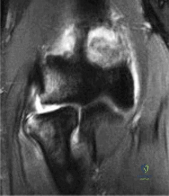
Here are the crucial details you must know about Elbow Cases: Unmasking Posterolateral Instability Pain. Posterolateral rotatory instability (PLRI) is the most likely diagnosis for elbow cases posterolateral pain following dislocation, a positive lateral pivot shift, and chronic lateral elbow pain with popping. Patients often struggle with activities like push-ups. This instability results from failure of lateral ligamentous stabilizers, typically involving the lateral ulnar collateral ligament (LUCL).
A 42-year-old carpenter presents with persistent lateral elbow pain and a sensation of "giving way" for 10 months following a FOOSH injury. He has failed conservative management. Based on the clinical history provided, what is your primary working diagnosis, and which specific physical examination maneuver is essential to confirm it?
Candidate: The most likely diagnosis is Posterolateral Rotatory Instability (PLRI) due to a chronic LUCL deficiency. The gold standard clinical test is the Lateral Pivot Shift test, which demonstrates subluxation of the radial head in extension and reduction upon flexion.
Candidates often confuse this with lateral epicondylitis. A poor answer focuses solely on the pain and forgets to emphasize the *mechanical* nature of the instability or fails to mention the provocative maneuvers that demonstrate the pathognomonic "clunk" of the radial head.
A high-scoring answer identifies PLRI, highlighting that the primary restraint (LUCL) is incompetent. The candidate should describe the Lateral Pivot Shift Test/External Rotation Recurvatum test as the definitive clinical maneuver, explaining that it reproduces the patient's apprehension and the mechanical subluxation of the radial head that reduces spontaneously during flexion.
The imaging obtained includes the following study. How does this diagnostic tool confirm your clinical suspicion, and what specific anatomical structures are being assessed?
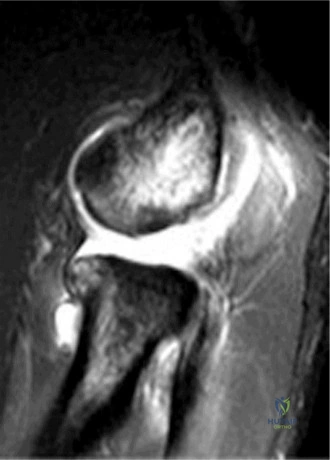
Candidate: This is a dynamic stress fluoroscopy study. It confirms PLRI by demonstrating the objective posterolateral subluxation of the radial head relative to the capitellum under valgus and supination stress, which is the functional hallmark of an incompetent Lateral Ulnar Collateral Ligament (LUCL).
Failing to emphasize that this is a *dynamic* study. Candidates often describe it as a static X-ray or fail to mention why stress is necessary to reveal the laxity that remains hidden in a neutral resting state.
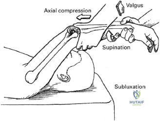
The candidate explicitly links the stress maneuver (valgus, axial compression, and supination) to the visualization of the radial head's subluxation, establishing this as the gold-standard diagnostic tool for quantifying dynamic instability in the outpatient setting.
You have decided to proceed with surgical reconstruction. Describe your approach to the graft fixation and the importance of the isometric point in your technique.
Candidate: I would use a palmaris longus autograft. Placement is critical: the humeral tunnel must be at the isometric point, which is slightly anterior and superior to the center of capitellar rotation. The ulnar tunnels are drilled through the supinator crest. Tensioning must be done in 30-45 degrees of flexion with the forearm pronated.
Ignoring the "isometry" concept. If the graft is not placed at the isometric point, it will tighten in some positions and become lax in others, leading to either restricted ROM or recurrent instability—the "teeter-totter" effect.
The candidate demonstrates an expert understanding of anatomy and biomechanics: the isometric point prevents over-tensioning, while the forearm pronation during fixation ensures the radial head is reduced into the capitellar notch, neutralizing the rotatory force.
This patient is a carpenter. What are the key elements of your postoperative rehabilitation, and what specific activities must they avoid in the first 6 weeks?
Candidate: The brace must be locked to prevent full extension and, critically, avoid supination. He must avoid resisted supination, valgus stress, and lifting. Supination is the movement that most destabilizes the posterolateral rotatory axis.
A general "take it easy" approach is insufficient. Failing to highlight the dangers of active supination in the early phase—which literally pulls the radial head out of the joint—is a critical failure that would likely lead to graft stretch-out.
The candidate provides a structured, phased rehabilitation protocol. They specifically forbid resisted supination and terminal extension with valgus/supination load, noting that the graft needs to protect the joint while it undergoes biological healing and maturation.
