Advanced Avascular Necrosis (AVN) of the Femoral Head: A Detailed Orthopedic Case Study
Key Takeaway
Advanced avascular necrosis (AVN) of the femoral head, often Ficat-Arlet Stage IV, presents with intractable hip pain, antalgic gait, and severely restricted range of motion, especially internal rotation. Diagnosis involves plain radiographs showing subchondral collapse and joint space narrowing, confirmed by MRI revealing classic double-line signs, extensive edema, and cartilage loss. Patient history frequently includes chronic corticosteroid use.
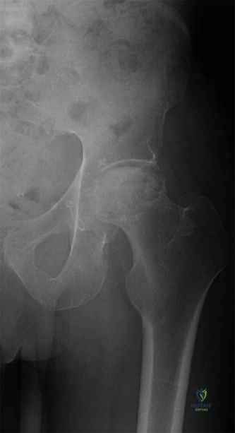
You are presented with this 45-year-old male. His radiographs and MRI are provided below. Based on the clinical history and these images, what is your diagnosis and how do you classify the severity of his condition?
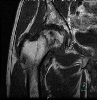
Candidate: The patient has avascular necrosis (AVN) of the femoral head, likely secondary to chronic corticosteroid use. The MRI shows a "double-line sign" and subchondral collapse. It is Ficat-Arlet Stage IV disease.
Simply stating "AVN" and the stage. Candidates often forget to mention the functional impact or fail to link the etiology (steroids/alcohol) systematically. They also frequently miss the importance of the secondary acetabular changes in advanced stage IV disease.
The patient presents with symptomatic, advanced-stage Avascular Necrosis (AVN) of the right femoral head. Radiographically and on MRI, this is Ficat-Arlet Stage IV, evidenced by the pathognomonic "crescent sign," loss of femoral head sphericity (collapse), and secondary acetabular degenerative changes. The etiology is likely multifactorial, secondary to chronic high-dose corticosteroid use and prior alcohol consumption, which have led to failed microvascular repair following his index core decompression.
The patient has already failed a core decompression 3 years ago. Given his age of 45 and his current stage of disease, discuss your surgical strategy and the specific rationale for your implant choices.
Candidate: Since he has failed joint preservation, I would recommend a total hip arthroplasty (THA). I would use a cementless femoral stem and a ceramic-on-ceramic bearing to maximize longevity for a young patient.
Failing to address the specific bone quality issues seen in AVN, such as acetabular bone stock or the risk of future revision. Candidates often jump to "ceramic-on-ceramic" without discussing the trade-offs (e.g., squeaking, brittle fracture risk) or the importance of pre-operative templating for potential metal augments.
Because the patient has entered the salvage phase (Stage IV), the goal is now definitive reconstruction. A primary cementless Total Hip Arthroplasty (THA) is the gold standard. 1. Fixation: I would utilize a modern tapered cementless stem to restore native offset and biomechanics. 2. Bearing: A highly cross-linked polyethylene or ceramic-on-ceramic surface is preferred to mitigate wear in a young, active patient. 3. Planning: Crucially, I would template for potential acetabular bone loss or rim deficiency identified on CT, ensuring I have options for augmentative fixation or screw placement if the primary press-fit is insufficient due to the secondary osteoarthritic changes.
You have performed the procedure and are now closing the patient. Discuss the specific steps taken to mitigate the risk of post-operative dislocation, given the posterior approach used.
Candidate: I would make sure to repair the posterior capsule and the short external rotators back to the greater trochanter. I would also tell the patient to follow hip precautions for 6 weeks.
Mentioning "precautions" but ignoring the surgical technique. The examiner wants to hear about anatomical reattachment (suturing through bone tunnels or drill holes) and verifying tension/offset intra-operatively.
Dislocation risk in the posterior approach is primarily managed through surgical technique rather than just patient compliance: 1. Anatomic Repair: Meticulous reattachment of the posterior capsule and short external rotators (piriformis, obturators) is mandatory, ideally through transosseous drill holes in the posterior greater trochanter. 2. Soft Tissue Tensioning: I must confirm intra-operative stability through a dynamic "shuck test" to ensure appropriate neck length and offset are achieved, preventing impingement. 3. Component Positioning: Ensuring the cup is placed in the "safe zone" of abduction and anteversion to maximize the jump distance. 4. Patient Education: While mechanical stability is primary, formal patient education on avoiding combined flexion, adduction, and internal rotation is standard practice for the first 6–8 weeks.
