Radial Bow: Comprehensive Review of Surgical Anatomy, Biomechanics & Restoration for Optimal Forearm Function

17 Apr 2026 16 min read 102 Views
Illustration of radial bow radial - Dr. Mohammed Hutaif

Key Takeaway

The radial bow is the inherent curvature of the radial diaphysis, crucial for forearm pronation and supination by maintaining interosseous space and optimal DRUJ kinematics. Its anatomical restoration is paramount in managing forearm fractures, as disruption or malunion severely compromises rotational function, leading to significant disability and impaired upper limb function.
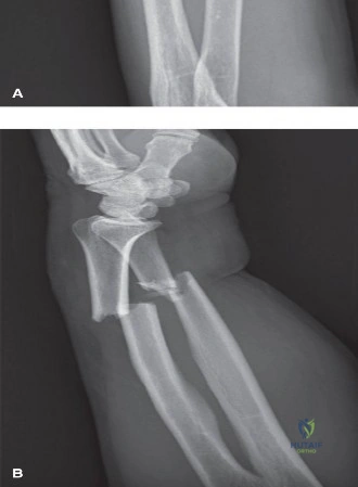
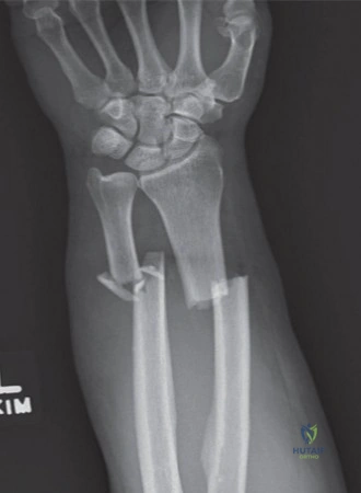
Introduction and Epidemiology

The forearm, a functionally critical segment of the upper extremity, is comprised of the radius and ulna, articulated proximally by the proximal radioulnar joint (PRUJ) and distally by the distal radioulnar joint (DRUJ). Its unique anatomy, characterized by the inherent curvature of the radial diaphysis – the radial bow – is fundamental for the complex kinematics of pronation and supination. This elegant anatomical feature allows the radius to rotate around a relatively fixed ulnar axis, creating a stable yet highly mobile platform for hand function. Disruption of this radial bow, whether through acute trauma leading to fracture, malunion, or iatrogenic miscontouring during osteosynthesis, inevitably compromises forearm rotation and can severely impair overall upper limb function.

Forearm fractures, particularly diaphyseal fractures involving both the radius and ulna, are common injuries, accounting for approximately 10-15% of all adult fractures. They represent a significant challenge due to the high biomechanical demands placed on the forearm and the critical need to restore precise anatomical relationships. In adults, these fractures typically result from high-energy trauma, often leading to significant displacement and instability. The incidence exhibits a bimodal distribution, affecting young, active individuals and the elderly, with distinct injury mechanisms and associated comorbidities. In younger cohorts, sports injuries and motor vehicle accidents are prevalent etiologies, while falls are more common in older populations.

The primary objective in managing diaphyseal forearm fractures is the restoration of forearm length, axial alignment, and most critically, the intrinsic radial bow. Failure to achieve anatomical reduction, particularly the restoration of the radial length and curvature, leads to malunion, which can manifest as persistent pain, restriction of pronation/supination, and ultimately, functional disability. This comprehensive review aims to delineate the intricate surgical anatomy and biomechanics of the radial bow, outline contemporary operative strategies, and discuss potential complications and their management, underscoring the paramount importance of anatomical restoration for optimal functional outcomes. Furthermore, we will explore the evolving role of advanced imaging and three dimensional preoperative templating in addressing complex diaphyseal deformities.

Surgical Anatomy and Biomechanics

The radial bow is a characteristic curvature of the radial shaft, typically exhibiting both a dorsal and a lateral concavity. This double-curved morphology is essential for maintaining the interosseous space and facilitating the intricate rotational mechanics of the forearm. The radius is convex dorsally in its proximal third, relatively straight in its middle third, and then convex ventrally in its distal third. Laterally, the radius has a gentle curve, being convex laterally in its mid-diaphysis. These curvatures ensure that the radius maintains sufficient distance from the ulna throughout the arc of pronation and supination, preventing impingement of the interosseous membrane and the surrounding musculature.

Image

Key Anatomical Features

The shaft of the radius exhibits a distinct posterior and lateral bow. The average dorsal bow is approximately 10-15 degrees, with the apex typically located around the junction of the proximal and middle thirds. The lateral bow is less pronounced but equally critical. In contrast, the ulna is relatively straight, acting as the stable axis around which the radius rotates. The olecranon and coronoid processes articulate with the humerus, forming the hinge-like humeroulnar joint, which contributes to elbow stability.

A fibrous sheet connecting the radius and ulna, the Interosseous Membrane (IOM) plays a vital role in longitudinal stability, load sharing between the two bones, and facilitating muscle attachment. Its fibers are oriented obliquely, largely responsible for transmitting axial forces from the radius to the ulna. Preservation or anatomical reconstruction of the IOM is critical for maintaining longitudinal stability, particularly in the setting of Essex-Lopresti injuries. The central band of the IOM is the most robust portion, originating from the proximal radius and inserting on the distal ulna, effectively transferring compressive loads from the wrist to the elbow.

Biomechanics of Forearm Rotation

The axis of forearm rotation is an imaginary line passing from the center of the radial head proximally to the fovea of the ulnar head distally. During pronation and supination, the radial diaphysis swings around the relatively stationary ulna. The magnitude and location of the radial bow are the primary determinants of the interosseous space volume.

Classic biomechanical studies by Schemitsch and Richards defined the normal parameters of the radial bow. The maximum radial bow averages 15.3 millimeters (range, 10 to 22 mm), and the location of this maximum bow is situated at approximately 60% of the total radial length, measured from the bicipital tuberosity to the radiocarpal joint. Alterations in these parameters have direct kinematic consequences. A loss of the normal radial bow decreases the interosseous space, leading to bony impingement between the radius and ulna during rotation.

Matthews et al. demonstrated that angular deformities of less than 10 degrees in any plane do not significantly restrict forearm rotation. However, deformities exceeding 20 degrees, particularly those involving a loss of the radial bow, result in a severe and clinically significant loss of pronation and supination. Furthermore, failure to restore the anatomical location of the maximum radial bow shifts the axis of rotation, increasing tension on the interosseous membrane and the radioulnar ligaments, which can lead to chronic DRUJ instability and early post-traumatic arthrosis.

Indications and Contraindications

The management of diaphyseal forearm fractures requires a nuanced understanding of fracture morphology, soft tissue integrity, and patient-specific functional demands. Because the forearm functions essentially as a complex joint, diaphyseal fractures of the radius and ulna are treated with the same principles applied to intra-articular fractures: exact anatomical reduction and absolute stability.

Operative Decision Making

Surgical intervention is the gold standard for the vast majority of adult diaphyseal forearm fractures. Non-operative management is reserved for a highly select, narrow subset of injuries or for patients with prohibitive surgical risks.

Parameter Operative Indications Non Operative Indications
Fracture Pattern Displaced fractures of the radius and/or ulna Truly non-displaced, isolated ulnar shaft fractures (Nightstick)
Angulation > 10 degrees of angulation in any plane < 10 degrees of angulation
Displacement > 50% translation of the diaphyseal shaft < 50% translation (isolated ulna only)
Radial Bow Any measurable loss or disruption of the radial bow Intact radial bow parameters
Associated Injuries Galeazzi, Monteggia, Essex-Lopresti fracture-dislocations Isolated injury with stable PRUJ and DRUJ
Soft Tissue Open fractures, impending compartment syndrome Intact soft tissue envelope, minimal swelling
Patient Factors Active adults, high functional demands Non-ambulatory, severe medical comorbidities precluding anesthesia

Contraindications to immediate internal fixation include active deep infection at the surgical site, severe soft tissue compromise precluding safe surgical incisions (e.g., extensive blistering, crush injury with tenuous viability), and medically unstable polytrauma patients requiring damage control orthopedics. In such instances, temporary spanning external fixation may be employed until the soft tissue envelope recovers and definitive internal fixation can be safely executed.

Pre Operative Planning and Patient Positioning

Thorough preoperative planning is indispensable for achieving anatomical restoration of the radial bow. The surgeon must meticulously evaluate the fracture pattern, select the appropriate surgical approach, and anticipate the required implants.

Radiographic Evaluation

Standard orthogonal imaging, including true anteroposterior (AP) and lateral radiographs of the forearm, elbow, and wrist, is mandatory. The integrity of the PRUJ and DRUJ must be critically assessed to rule out associated Monteggia or Galeazzi fracture-dislocations.

Contralateral templating is a cornerstone of preoperative planning. Radiographs of the uninjured forearm provide an individualized template of the patient's native radial bow magnitude and location. In complex comminuted fractures or established malunions, computed tomography (CT) with three-dimensional reconstructions is highly recommended. Advanced planning software can mirror the contralateral intact radius, allowing for precise calculation of the required osteotomy angles or the degree of plate contouring necessary to restore the native anatomy.

Positioning and Setup

The patient is positioned supine on the operating table. The injured upper extremity is placed on a radiolucent hand table to facilitate unhindered fluoroscopic imaging. A non-sterile tourniquet is typically applied high on the brachium. The arm is prepped and draped freely to allow full intraoperative assessment of pronation and supination following fracture fixation.

Fluoroscopy should be positioned parallel to the longitudinal axis of the table, entering from the head or the axilla, ensuring that orthogonal views of the entire forearm can be obtained without compromising the sterile field. Preoperative antibiotics are administered within one hour prior to incision, and the tourniquet is inflated only after exsanguination with an Esmarch bandage, unless the presence of an infection or malignancy dictates elevation alone.

Detailed Surgical Approach and Technique

The surgical approach to the radius must be tailored to the location of the fracture and the surgeon's familiarity with the regional neurovascular anatomy. The two primary approaches utilized are the volar (Henry) approach and the dorsal (Thompson) approach.

Image

Volar Henry Approach

The volar approach is highly versatile, providing excellent exposure of the entire volar aspect of the radius from the bicipital tuberosity to the radiocarpal joint. It exploits the internervous plane between the brachioradialis (innervated by the radial nerve) and the flexor carpi radialis (innervated by the median nerve).

  1. Incision: A longitudinal incision is made along the volar aspect of the forearm, tracing a line from the biceps tendon proximally to the radial styloid distally.
  2. Superficial Dissection: The deep fascia is incised in line with the skin incision. The interval between the brachioradialis and the FCR is identified and developed. The radial artery and its accompanying venae comitantes are identified beneath the brachioradialis. The artery must be carefully mobilized and retracted ulnarly with the FCR.
  3. Deep Dissection (Proximal Radius): To expose the proximal third of the radius, the recurrent radial artery (the "leash of Henry") must be identified, ligated, and divided. This allows the brachioradialis to be retracted laterally, exposing the supinator muscle. The supinator is sharply detached from its ulnar origin and reflected laterally. This maneuver protects the posterior interosseous nerve (PIN), which courses within the substance of the supinator.
  4. Deep Dissection (Middle and Distal Radius): In the middle third, the pronator teres insertion is identified and can be partially elevated if necessary for plate placement. Distally, the flexor pollicis longus (FPL) and the pronator quadratus are elevated from the volar surface of the radius to expose the fracture site.

Dorsal Thompson Approach

The dorsal approach is typically reserved for fractures of the proximal and middle thirds of the radius, particularly those with dorsal comminution or when a dorsal plate application is mechanically advantageous. It utilizes the internervous plane between the extensor carpi radialis brevis (ECRB, radial nerve) and the extensor digitorum communis (EDC, posterior interosseous nerve).

  1. Incision: A straight incision is made from the lateral epicondyle to the dorsal radial tubercle (Lister's tubercle).
  2. Superficial Dissection: The fascia is incised, and the interval between the ECRB and EDC is developed.
  3. Deep Dissection: The supinator muscle is exposed. The critical step in this approach is the identification and protection of the PIN. The nerve emerges from the supinator approximately 1 cm proximal to the distal edge of the muscle. The supinator must be carefully split along the course of the nerve, or elevated off the radius from ulnar to radial, ensuring the nerve remains protected within the muscle belly during retraction.

Plate Contouring and Fixation

The standard of care for diaphyseal forearm fractures is open reduction and internal fixation (ORIF) utilizing 3.5 mm dynamic compression plates (DCP), limited contact dynamic compression plates (LC-DCP), or locking compression plates (LCP).

Restoration of the radial bow is achieved through meticulous plate contouring. Pre-contoured anatomical plates are commercially available and offer a reliable template for restoring the native curvature. However, when utilizing standard straight plates, the surgeon must manually contour the plate using bending presses. The plate must be bent to match the apex dorsal and apex lateral curves of the native radius.

Applying a straight plate to the curved radial diaphysis will inevitably flatten the radial bow, narrowing the interosseous space and restricting rotation. Conversely, over-contouring the plate can lead to excessive bowing, which may impinge on the ulna or alter the kinematics of the DRUJ. The contoured plate is typically applied to the volar surface of the radius via the Henry approach, as the volar surface is relatively flat distally and provides a broad surface for fixation.

Fixation principles dictate the achievement of absolute stability for simple fracture patterns (AO/OTA Type A and B) utilizing lag screws and neutralization plates, or compression plating techniques. For complex, comminuted fractures (AO/OTA Type C), bridge plating techniques are employed to preserve the soft tissue envelope and vascularity of the fracture fragments, focusing on the restoration of length, alignment, and rotation rather than anatomical reduction of every butterfly fragment. A minimum of six cortices (three bicortical screws) of fixation is required proximal and distal to the fracture, though eight cortices are biomechanically preferable in the diaphyseal bone.

Intraoperative assessment of the radial bow is critical. Following provisional fixation, the surgeon must take the forearm through a full, passive arc of pronation and supination. Any crepitus, resistance, or restriction in motion compared to the uninjured side suggests an anatomical mismatch, soft tissue interposition, or hardware impingement, necessitating immediate plate revision or re-contouring.

Complications and Management

Despite meticulous surgical technique, complications following surgical management of radial diaphysis fractures can occur. Anticipation, early recognition, and appropriate salvage strategies are essential for preserving upper extremity function.

Adverse Events and Salvage

Complication Incidence Etiology and Risk Factors Management and Salvage Strategy
Radioulnar Synostosis 2% - 8% High-energy trauma, closed head injury, single-incision approach for both bones, severe soft tissue stripping, delayed surgery. Excision of heterotopic bone once mature (typically >6 months post-injury). Prophylaxis with Indomethacin or low-dose radiation therapy post-excision.
Nonunion 2% - 10% Inadequate fixation stability, poor soft tissue envelope, infection, smoking, severe comminution. Revision ORIF with rigid plating, autologous bone grafting (iliac crest), and decortication. Rule out indolent infection.
Malunion (Loss of Bow) Variable Failure to contour plate, loss of reduction, unrecognized comminution leading to shortening or angular deformity. Corrective closing or opening wedge osteotomy. Preoperative 3D templating and patient-specific instrumentation (PSI) are highly recommended.
Infection 1% - 3% Open fractures, prolonged surgical time, medical comorbidities (diabetes, immunosuppression). Acute: Irrigation and debridement, hardware retention if stable, targeted IV antibiotics. Chronic: Hardware removal, radical debridement, antibiotic spacers, staged reconstruction.
Nerve Injury (PIN/Median) 1% - 5% Iatrogenic retraction injury, direct laceration during supinator elevation (PIN), or retractor placement (Median). Most are neuropraxias that resolve spontaneously within 3-6 months. Exploration indicated if nerve was directly visualized and lacerated, or if no recovery is noted by 4-6 months (EMG/NCS evaluation).
Hardware Failure Rare Premature weight-bearing, nonunion leading to fatigue failure of the plate, insufficient working length. Revision ORIF with longer, stronger implants (e.g., 3.5mm broad plates or dual plating), bone grafting to address underlying nonunion.

Loss of forearm rotation is the most frequent functional complaint following diaphyseal fractures. When evaluating a patient with restricted rotation, the surgeon must differentiate between soft tissue contracture, heterotopic ossification, and bony malunion. CT imaging is paramount in this diagnostic algorithm. If a malunion with a loss of the radial bow is identified as the primary culprit, a corrective osteotomy is indicated. The osteotomy must be meticulously planned to restore the maximum radial bow to its anatomical location (60% of radial length) and magnitude (~15mm).

Post Operative Rehabilitation Protocols

The rehabilitation of the forearm following diaphyseal fracture fixation is a delicate balance between protecting the osteosynthesis and preventing debilitating joint stiffness. The protocol must be individualized based on the stability of the fixation, the quality of the host bone, and the patient's compliance.

Phased Recovery

Phase I: Early Mobilization (Weeks 0-2)
Assuming rigid internal fixation has been achieved, early active and active-assisted range of motion (ROM) is initiated within the first few days postoperatively. The arm is typically placed in a bulky soft dressing or a removable volar splint for comfort. The primary focus is on digital ROM to prevent tendon adhesions and reduce edema. Gentle, pain-free pronation and supination exercises are commenced, along with elbow flexion and extension. Weight-bearing and lifting are strictly prohibited.

Phase II: Intermediate Phase (Weeks 2-6)
Sutures are removed at approximately two weeks. The removable splint is discontinued. Active ROM exercises are progressed. Passive stretching may be cautiously introduced if motion is lagging, but aggressive manipulation must be avoided to prevent stress on the healing fracture and mitigate the risk of heterotopic ossification. Radiographs are obtained at 2 and 6 weeks to monitor alignment and assess for early signs of callus formation.

Phase III: Strengthening Phase (Weeks 6-12)
Once clinical and radiographic evidence of fracture consolidation is present (typically bridging callus on at least three of four cortices), progressive strengthening exercises are initiated. Isometric exercises for the forearm musculature are followed by isotonic exercises using light resistance. The patient may begin to incorporate the injured extremity into light activities of daily living.

Phase IV: Return to Function (Months 3-6)
Full, unrestricted weight-bearing and return to heavy manual labor or contact sports are typically delayed until advanced radiographic healing is confirmed, often between 3 to 6 months post-injury. Patients must be counseled that maximal recovery of grip strength and rotational endurance may take up to a year. Persistent deficits in pronation and supination, despite aggressive therapy, warrant a thorough reassessment for malunion or hardware impingement.

Summary of Key Literature and Guidelines

The contemporary management of diaphyseal forearm fractures and the critical importance of the radial bow are deeply rooted in foundational biomechanical research and evolving clinical guidelines.

Landmark Biomechanical Studies

The seminal work by Schemitsch and Richards (1992) remains the cornerstone of our understanding of radial bow anatomy. Their cadaveric and radiographic analysis established the normative values for the magnitude (15.3 mm) and location (60% of radial length) of the maximum radial bow. Crucially, they correlated the restoration of these specific parameters with functional outcomes, demonstrating that failure to restore the normal magnitude and location of the bow directly correlates with a proportional loss of forearm rotation.

Matthews et al. (1982) provided critical insights into the tolerance of the forearm axis to angular deformity. Their biomechanical models demonstrated that while the forearm can tolerate minor angular deformities (<10 degrees) without significant kinematic penalty, deformities exceeding 20 degrees, particularly those that narrow the interosseous space, result in severe mechanical block to pronation and supination. This study underscores the absolute necessity of precise plate contouring.

Contemporary Clinical Guidelines

Current AO/OTA guidelines emphasize the necessity of absolute stability for simple diaphyseal fractures, advocating for the use of 3.5 mm LC-DCP or LCP constructs. The literature consistently supports the superiority of operative intervention over conservative management for displaced adult forearm fractures, citing significantly lower rates of nonunion and superior functional outcomes.

Recent advancements in the literature focus on the utility of three-dimensional templating and patient-specific instrumentation (PSI) for the correction of complex diaphyseal malunions. Studies have shown that PSI significantly improves the accuracy of radial bow restoration compared to conventional freehand osteotomy techniques, translating to superior recovery of pronation and supination in the salvage setting. Furthermore, the routine use of dual incisions (separate volar Henry and dorsal ulnar approaches) is universally recommended over single-incision approaches to minimize the devastating complication of radioulnar synostosis.


Dr. Mohammed Hutaif
Medically Verified Content
Prof. Dr. Mohammed Hutaif
Consultant Orthopedic & Spine Surgeon
Article Contents