Comprehensive Introduction and Patho-Epidemiology
Hip arthroscopy has undergone an exponential evolution over the past two decades, transitioning from a niche diagnostic modality to a highly sophisticated, joint-preserving therapeutic intervention. Unlike the knee or shoulder, the hip joint presents a uniquely hostile environment for arthroscopic navigation. It is a highly constrained, deep ball-and-socket articulation, enveloped by a massive muscular envelope and fortified by the thickest capsuloligamentous complex in the human body. Consequently, the procedure is characterized by a notoriously steep learning curve. The foundational pillar of a successful hip arthroscopy is the safe, precise, and reproducible establishment of surgical portals. Mastery of portal placement is not merely a preliminary step; it is the definitive prerequisite that dictates the trajectory of instrumentation, the quality of visualization, and the ultimate success of the operation.
The patho-epidemiology driving the surge in hip arthroscopy volumes is predominantly centered around Femoroacetabular Impingement (FAI) syndrome and associated acetabular labral tears. FAI is a dynamic, pathomechanical process wherein morphological abnormalities of the proximal femur (cam morphology) or the acetabulum (pincer morphology) lead to abnormal contact forces during terminal ranges of motion. Epidemiological studies indicate that asymptomatic cam morphology is present in up to 30% of the general population, with a significantly higher prevalence in high-level athletes participating in sports requiring repetitive hip flexion and internal rotation. Over time, these repetitive shear and compressive forces precipitate labral detachment, chondral delamination, and early-onset osteoarthritis. The advent of advanced arthroscopic techniques has allowed orthopedic surgeons to address these pathomorphologies directly, reshaping the osseous anatomy and restoring the labral seal without the morbidity associated with open surgical dislocation.
However, the transition to minimally invasive techniques is not without profound risks. The deep anatomical location of the hip necessitates the use of long, rigid trocars and specialized instrumentation that must traverse complex neurovascular planes. Improper portal placement can lead to catastrophic iatrogenic injuries, including the irreversible scuffing of the femoral head articular cartilage, iatrogenic laceration of the acetabular labrum, or permanent neuropraxia of major neurovascular structures. The arthroscopic "Safe Zone" is a narrow, unforgiving anatomical window. Therefore, a profound understanding of three-dimensional pelvic anatomy, coupled with meticulous preoperative planning and precise fluoroscopic execution, is mandatory for the modern joint-preserving orthopedic surgeon.
This comprehensive chapter meticulously details the anatomical landmarks, biomechanical positioning, and step-by-step surgical techniques required to safely establish standard and accessory hip arthroscopy portals. It synthesizes current evidence-based practices, landmark literature, and advanced clinical guidelines to provide an exhaustive reference for orthopedic residents, fellows, and practicing surgeons striving for excellence in hip arthroscopy.
Detailed Surgical Anatomy and Biomechanics
The safe establishment of hip arthroscopy portals requires an intimate, three-dimensional understanding of the extra-articular neurovascular anatomy, the muscular envelope, and the intra-articular capsuloligamentous structures. The concept of the arthroscopic "Safe Zone" was initially popularized by J.W. Thomas Byrd, who elegantly mapped the safe trajectories for portal placement relative to the surrounding neurovascular bundles. This safe zone is generally located laterally and anteriorly, bounded by the lateral femoral cutaneous nerve (LFCN) anteriorly and the sciatic nerve posteriorly.
The Lateral Femoral Cutaneous Nerve (LFCN) represents the most frequently injured structure during hip arthroscopy. Arising from the lumbar plexus (L2-L3), the LFCN typically exits the pelvis medial to the Anterior Superior Iliac Spine (ASIS), passing under the inguinal ligament to provide sensory innervation to the anterolateral thigh. However, its anatomical course is highly variable. The nerve often arborizes into multiple branches (gluteal, femoral, and obturator branches) shortly after exiting the pelvis. The classic anterior portal places the terminal branches of the LFCN at extreme risk, as the nerve lies an average of 3 to 4 cm medial to the standard anterior portal trajectory. Variations in branching patterns mean that the LFCN or its branches can cross directly over the intended path of the anterior or mid-anterior portals, necessitating strict adherence to superficial skin incisions and blunt dissection techniques down to the fascial layer.
Posteriorly, the sciatic nerve dictates the posterior boundary of the safe zone. Exiting the greater sciatic foramen below the piriformis muscle, the sciatic nerve courses distally in the posterior thigh. The standard posterolateral portal is established approximately 1 cm posterior and 1 cm superior to the tip of the greater trochanter. In a neutral anatomical position, the distance from the posterolateral portal to the sciatic nerve is approximately 2.5 to 3.0 cm. However, biomechanical positioning plays a critical role in mitigating this risk. By placing the operative leg in 15 degrees of internal rotation during setup, the greater trochanter is translated anteriorly. This maneuver effectively increases the distance between the posterolateral portal trajectory and the sciatic nerve by an additional 1 to 1.5 cm, providing a vital margin of safety.
Proximally, the superior gluteal nerve provides motor innervation to the gluteus medius, gluteus minimus, and tensor fasciae latae. It courses approximately 4 to 5 cm proximal to the tip of the greater trochanter. The anterolateral portal, which serves as the primary viewing portal, is established 1 cm superior and 1 cm anterior to the greater trochanter. When established correctly, the trajectory of the anterolateral portal remains well distal to the superior gluteal nerve, rendering it the safest of all standard portals. Furthermore, the robust capsular anatomy—comprising the iliofemoral ligament (the strongest ligament in the human body), the pubofemoral ligament, and the ischiofemoral ligament—must be understood. The capsule is thickest anteriorly and superiorly, requiring significant force to penetrate. The zona orbicularis, a circumferential collar of capsular fibers around the femoral neck, acts as a critical stabilizer and must be respected during capsulotomy to prevent postoperative microinstability.
Exhaustive Indications and Contraindications
The indications for hip arthroscopy have expanded significantly, driven by advancements in instrumentation, optics, and a deeper understanding of hip pathomechanics. However, stringent patient selection remains the most critical determinant of postoperative success. The primary indication for hip arthroscopy is symptomatic Femoroacetabular Impingement (FAI) syndrome that has failed a comprehensive trial of conservative management, including targeted physical therapy, activity modification, and intra-articular diagnostic/therapeutic injections. FAI presents in three morphological variants: cam (femoral-sided), pincer (acetabular-sided), and mixed (a combination of both), with mixed being the most common clinical presentation.
Beyond FAI, hip arthroscopy is highly indicated for the management of isolated acetabular labral tears, often traumatic or degenerative in nature. The labrum serves a critical biomechanical role, deepening the acetabulum by 21%, increasing the articular surface area by 22%, and maintaining a pressurized intra-articular fluid seal that protects the underlying articular cartilage from consolidation and shear stress. Arthroscopic labral repair or reconstruction is aimed at restoring this vital fluid seal. Additional indications include the removal of intra-articular loose bodies, synovial biopsy or synovectomy for conditions such as synovial chondromatosis or pigmented villonodular synovitis (PVNS), debridement of the ligamentum teres, and the management of septic arthritis in the native hip.
Conversely, absolute contraindications to hip arthroscopy are primarily centered around advanced joint degeneration and severe structural instability. Patients with Tonnis Grade 2 or 3 osteoarthritis, characterized by less than 2 mm of joint space on a weight-bearing anteroposterior pelvic radiograph, have uniformly poor outcomes following hip arthroscopy and are better served by total hip arthroplasty. Severe acetabular dysplasia (Lateral Center Edge Angle < 20 degrees) is an absolute contraindication for isolated arthroscopy, as addressing the labral tear without correcting the underlying structural instability will inevitably lead to rapid failure and accelerated joint subluxation. In such cases, a periacetabular osteotomy (PAO) is the gold standard.
| Category | Indications for Hip Arthroscopy | Contraindications for Hip Arthroscopy |
|---|---|---|
| Absolute | Symptomatic FAI (Cam, Pincer, Mixed) failing conservative care | Advanced Osteoarthritis (Tonnis Grade 2 or 3; joint space < 2mm) |
| Absolute | Acute or chronic symptomatic acetabular labral tears | Severe Acetabular Dysplasia (LCEA < 20°) without concomitant PAO |
| Absolute | Intra-articular loose bodies / Synovial Chondromatosis | Active local or systemic infection (unless performing I&D for septic hip) |
| Absolute | Ligamentum Teres avulsion or symptomatic hypertrophy | Advanced Avascular Necrosis (AVN) with femoral head collapse |
| Relative | Borderline Dysplasia (LCEA 20°-25°) with microinstability | Obesity (BMI > 35) complicating portal placement and traction |
| Relative | Extra-articular impingement (Subspine, Ischiofemoral) | Severe joint stiffness or arthrofibrosis preventing adequate distraction |
| Relative | Gluteus medius/minimus tears (Peritrochanteric space access) | Prior open hip surgery with extensive heterotopic ossification |
Pre-Operative Planning, Templating, and Patient Positioning
Meticulous preoperative planning and flawless patient positioning are non-negotiable prerequisites for safe hip arthroscopy. The procedure demands a highly choreographed setup to achieve the requisite joint distraction while mitigating the immense risks associated with prolonged traction. Preoperative templating begins with high-quality imaging, including an AP pelvis, Dunn lateral, and false-profile views, supplemented by fine-cut MRI or MR arthrography. The surgeon must meticulously evaluate the alpha angle, lateral center edge angle, Tonnis angle, and the presence of retroversion (cross-over sign, ischial spine sign). This templating dictates the specific areas of the femoral neck and acetabular rim that will require osteochondroplasty, thereby influencing the choice of accessory portals.
While hip arthroscopy can be performed in the lateral decubitus position, the supine position is the most widely utilized globally. The supine position offers superior ease of patient setup, excellent and reproducible fluoroscopic visualization, and a more straightforward anatomical orientation for the surgeon. The patient is placed supine on a specialized hip distraction table. The establishment of the "distraction vector" is the most critical biomechanical element of the setup. To safely access the central compartment of the hip—the space between the femoral head and the acetabular fossa—the femoral head must be distracted away from the acetabulum. A minimum of 10 mm of joint distraction is strictly required to safely introduce rigid trocars and instruments without causing catastrophic iatrogenic chondral injury.

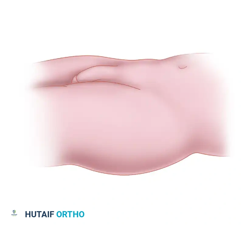
The perineal post is the fulcrum against which traction is applied. A well-padded, oversized perineal post (typically greater than 10 cm in diameter) is positioned slightly lateralized against the medial thigh of the operative leg. It is a critical surgical error to allow the post to rest directly in the perineum or against the genitalia, as this drastically increases the risk of pudendal nerve neuropraxia and soft tissue necrosis. The operative leg is positioned in slight flexion (10° to 15°), neutral abduction, and approximately 15° of internal rotation. The internal rotation is biomechanically critical; it horizontalizes the femoral neck, thereby maximizing the safe space for anterior portal placement, and simultaneously translates the greater trochanter anteriorly, protecting the sciatic nerve from the posterolateral portal trajectory.
Traction application must be methodical. Gross traction is applied first to tension the limb, followed by fine traction under live fluoroscopy. The goal is to break the vacuum seal of the hip joint, which is often visualized as a radiolucent "crescent sign" or "vacuum phenomenon" in the joint space on the fluoroscopic monitor. Surgical warning: Traction time must be strictly monitored by the circulating nurse and the surgeon. To minimize the risk of perineal necrosis, sciatic neuropraxia, and pudendal neuropraxia, continuous traction should ideally not exceed 90 to 120 minutes. If the central compartment work is not completed within this timeframe, traction must be released for 10 to 15 minutes to allow for tissue perfusion before reapplication.
Step-by-Step Surgical Approach and Portal Placement Technique
The establishment of portals must be an exquisitely methodical process, relying on a combination of tactile feedback, precise fluoroscopic confirmation, and meticulous angulation. The standard supine setup traditionally utilizes three primary portals: the anterolateral (AL), the anterior (AP) or mid-anterior (MAP), and occasionally the posterolateral (PL) portal. The anterolateral portal is the universal "workhorse" of hip arthroscopy, established first under fluoroscopic guidance, serving as the primary viewing portal for the central compartment.
Step 1: Establishing the Anterolateral (AL) Portal
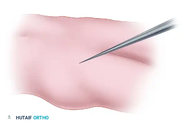
Before skin preparation, bony landmarks—specifically the Anterior Superior Iliac Spine (ASIS) and the borders of the Greater Trochanter (GT)—are meticulously palpated and marked. Following verification of adequate joint distraction (minimum 10 mm) via fluoroscopy, a superficial 5 mm stab incision is made at the marked AL portal site, located approximately 1 cm superior and 1 cm anterior to the anterior edge of the greater trochanter. A 17-gauge, 6-inch spinal needle is introduced. The trajectory is critical: the needle is directed approximately 45° cephalad and 30° posterior, aiming precisely for the lateral aspect of the femoral head-neck junction. Under live fluoroscopy, the needle is advanced until it rests on the thick capsule. The surgeon then applies controlled force to pop through the capsule, feeling a distinct tactile "give." If the joint vacuum is intact, a drop of saline placed in the hub of the needle will be instantly sucked into the joint—the classic "drop sign." The joint is then distended with 10 to 20 cc of normal saline or local anesthetic, pushing the capsule away from the labrum. A flexible Nitinol guidewire is passed through the needle, the needle is removed, and a cannulated dilator and arthroscopic sheath are passed over the wire with a twisting motion to breach the capsule. The 70° arthroscope is then introduced.
Step 2: Establishing the Mid-Anterior Portal (MAP)
The classic anterior portal has largely been superseded by the Mid-Anterior Portal (MAP) due to a superior safety profile regarding the LFCN and an optimized trajectory for anchor placement along the anterior acetabular rim. The MAP is located approximately 2 to 3 cm distal and slightly lateral to the classic anterior portal. This second portal is established under direct intra-articular visualization combined with fluoroscopy. With the arthroscope in the AL portal, the surgeon visualizes the "anterior triangle," bounded by the anterior labrum, the femoral head, and the anterior capsule. A spinal needle is introduced at the MAP site. The trajectory is directed 45° cephalad and 30° toward the midline.
The surgeon watches the needle pierce the capsule on the monitor, ensuring it enters the joint safely away from the delicate labrum and the femoral head articular cartilage. Once the trajectory is perfected and triangulation is achieved, a Nitinol wire is passed, followed by a cannulated working sheath. Superficial skin incisions and blunt dissection with a hemostat down to the fascia are mandatory here to protect the arborizing branches of the LFCN.
Step 3: The Interportal Capsulotomy
To maneuver instruments freely between the AL and MAP portals and to access the peripheral compartment later in the case, the thick capsule between them must be incised. An arthroscopic scalpel (e.g., Beaver blade or specialized disposable blade) is introduced through the anterior working portal. Under direct vision from the AL portal, the surgeon incises the capsule connecting the two portals. This cut runs parallel to the acetabular rim, approximately 5 to 8 mm away from the chondrolabral junction to leave a sufficient cuff of capsular tissue for subsequent closure. This interportal capsulotomy allows for the introduction of larger instruments, shavers, and burrs without capsular restriction, and is a critical step in exposing the cam lesion during peripheral compartment work.
Complications, Incidence Rates, and Salvage Management
Despite meticulous technique, hip arthroscopy carries a distinct complication profile, primarily driven by the requisite traction, the deep anatomical dissection, and the high-pressure fluid environment. The overall complication rate in hip arthroscopy ranges from 1.5% to 5%, with the vast majority being transient neuropraxias. Understanding the pathophysiology of these complications is essential for rapid identification and salvage management.
Neurovascular injuries are the most frequently encountered complications. Lateral Femoral Cutaneous Nerve (LFCN) neuropraxia is the most common, reported in up to 30% of cases in some historical cohorts, though modern techniques utilizing the mid-anterior portal have reduced this incidence significantly. It manifests as numbness, tingling, or dysesthesia over the anterolateral thigh. It is typically caused by direct trauma during portal placement, fluid extravasation compressing the nerve, or traction stretch. The vast majority of LFCN neuropraxias are transient and resolve spontaneously within 3 to 6 months. Pudendal nerve neuropraxia, manifesting as perineal numbness or sexual dysfunction, is exclusively a traction-related complication caused by compression against the perineal post. Sciatic nerve injury is exceptionally rare but devastating, typically resulting from prolonged traction or aberrant placement of the posterolateral portal without adequate internal rotation of the limb.
Iatrogenic cartilage and labral damage represent severe technical failures. Forcing a rigid trocar into a tightly constrained joint without adequate distraction or without a guidewire will inevitably scuff the femoral head or puncture the labrum. These chondral injuries can lead to accelerated localized osteoarthritis. Avoidance requires strict adherence to the 10 mm distraction rule and always distending the joint with fluid prior to passing dilators.
Fluid extravasation is a unique and potentially life-threatening complication. The hip joint requires high pump pressures (often 50-70 mmHg) to maintain visualization and tamponade bleeding from the muscular envelope and cancellous bone. During prolonged surgeries, particularly those involving extensive capsulotomies or iliopsoas fractional lengthening, massive volumes of fluid can extravasate into the thigh, causing massive swelling and hypothermia. Rarely, fluid can track superiorly along the iliopsoas fascia into the retroperitoneal space, leading to intra-abdominal fluid accumulation and subsequent Abdominal Compartment Syndrome. Surgeons must proactively monitor core body temperature and periodically palpate the patient's abdomen during cases exceeding two hours.
| Complication | Estimated Incidence | Avoidance Strategy | Salvage Management / Prognosis |
|---|---|---|---|
| LFCN Neuropraxia | 5% - 30% | Use MAP instead of classic AP; superficial skin incisions; blunt dissection. | Observation. Usually transient, resolving in 3-6 months. Gabapentin for severe dysesthesia. |
| Pudendal Neuropraxia | 1% - 5% | Well-padded, lateralized perineal post; limit traction to < 120 mins. | Observation. Typically resolves in 2-6 weeks. Urology consult if persistent dysfunction occurs. |
| Sciatic Nerve Injury | < 1% | 15° internal rotation during setup; limit traction; careful PL portal placement. | Immediate release of traction. EMG at 6 weeks if no recovery. Variable long-term prognosis. |
| Iatrogenic Chondral Scuffing | 1% - 3% | Ensure >10mm distraction; use "drop sign"; never plunge trocars blindly. | Debridement of loose flaps. If severe, microfracture may be required. May accelerate OA. |
| Fluid Extravasation / Abdominal Compartment Syndrome | < 0.5% | Monitor pump pressures (<70 mmHg); palpate abdomen; minimize surgical time. | Stop fluid flow immediately. Diuretics. In severe ACS, emergent exploratory laparotomy is required. |
| Portal Site Infection | < 0.5% | Meticulous sterile technique; preoperative prophylactic antibiotics. | Superficial: Oral antibiotics. Deep/Intra-articular: Emergent arthroscopic I&D and IV antibiotics. |
Phased Post-Operative Rehabilitation Protocols
The surgical intervention in hip arthroscopy represents only half of the therapeutic equation; the subsequent rehabilitation protocol is equally critical in determining the ultimate clinical outcome. Postoperative rehabilitation must be highly structured, phased, and tailored to the specific intra-articular procedures performed. The overarching goals are to protect the healing tissues (labral repair, capsular closure, microfracture), prevent intra-articular adhesions, restore normal arthrokinematics, and progressively return the patient to high-level function.
Phase 1: Protection and Early Mobility (Weeks 0-4)
The primary objective in the immediate postoperative phase is the protection of the surgical repairs while preventing capsular scarring and synechiae formation at the portal entry sites. Weight-bearing status is heavily dictated by the procedure. Following a standard labral repair and osteochondroplasty, patients are typically restricted to 20 lbs flat-foot weight-bearing using crutches for 2 to 4 weeks. If a microfracture was performed for chondral defects, strict touch-down weight-bearing is extended to 6 to 8 weeks. Early passive range of motion (PROM) is initiated on postoperative day one. Continuous Passive Motion (CPM) machines or stationary cycling with zero resistance are frequently utilized. A critical component of Phase 1 is daily circumduction exercises; the therapist passively moves the hip in large circular motions to prevent the capsule from scarring down to the underlying femoral neck, a common complication that severely limits postoperative rotation.
Phase 2: Gait Normalization and Early Strengthening (Weeks 4-8)
As the biological healing of the labrum and capsule progresses, the focus shifts to weaning off crutches and normalizing the gait pattern. Patients transition to full weight-bearing as tolerated. Aquatic therapy is highly beneficial during this phase, utilizing the buoyancy of water to reduce joint reactive forces while allowing for functional movement. Strengthening exercises focus on the core, gluteus medius, and deep external rotators. Isometrics progress to isotonic exercises. However, active hip flexion (e.g., straight leg raises) is often restricted or heavily modified during this phase to prevent anterior hip pain and avoid excessive stress on the healing anterior capsule and hip flexor tendons.
Phase 3: Advanced Strengthening and Neuromuscular Control (Weeks 8-12)
By the third month, the capsuloligamentous complex has achieved significant tensile strength. Rehabilitation intensifies to include closed kinetic chain exercises, proprioceptive training, and advanced core stabilization. Exercises such as single-leg squats, lunges, and dynamic balance board training are incorporated. The goal is to correct the compensatory movement patterns that the patient likely developed prior to surgery due to chronic FAI.
Phase 4: Return to Sport and High-Level Function (Weeks 12+)
The final phase involves sport-specific or occupation-specific training. Progression to this phase requires a pain-free full range of motion, symmetrical muscle strength (at least 90% of the contralateral limb on isokinetic testing), and no reactive effusion following activity. Plyometrics, cutting, and pivoting drills are gradually introduced. The timeline for a full return to competitive sports is typically between 4 to 6 months, though this can vary significantly based on the severity of the initial pathology and the specific demands of the sport.
Summary of Landmark Literature and Clinical Guidelines
The rapid evolution of hip arthroscopy is heavily underpinned by a robust body of landmark literature and evolving clinical guidelines. A profound understanding of these foundational texts is essential for any academic orthopedic surgeon.
J.W. Thomas Byrd's pioneering work in the late 1990s and early 2000s established the anatomical basis for safe portal placement. His seminal papers describing the lateral approach and the mapping of the "safe zones" relative to the lateral femoral cutaneous nerve and sciatic nerve remain the gold standard for arthroscopic orientation. Byrd demonstrated that the anterolateral portal, established under fluoroscopy, provides the safest reproducible access to the constrained joint.
The understanding of FAI pathomechanics was revolutionized by Reinhold Ganz and his colleagues in Bern, Switzerland. While Ganz originally described the open surgical dislocation, his conceptual framework of cam and pincer impingement provided the rationale for modern arthroscopic osteochondroplasty. Marc Philippon subsequently translated these concepts into the arthroscopic realm, publishing extensively on the techniques of arthroscopic labral repair and the critical importance of restoring the labral fluid seal. Philippon's outcome studies demonstrated that arthroscopic labral repair yielded significantly superior long-term patient-reported outcomes compared to historical labral debridement.
More recently, the biomechanical importance of the hip capsule has been a major focus of orthopedic literature. Studies by Martin and colleagues, as well as biomechanical models from various academic centers, have demonstrated that failure to repair the interportal or T-capsulotomy can lead to iatrogenic microinstability, particularly in patients with borderline dysplasia or ligamentous laxity. This has led to a paradigm shift in clinical guidelines, with routine capsular closure now recommended by the majority of high-volume hip arthroscopists.
From an evidence-based medicine perspective, the UK FASHIoN (Femoroacetabular Impingement Trial) published in The Lancet provided Level 1 evidence supporting the efficacy of hip arthroscopy. The randomized controlled trial demonstrated that arthroscopic surgery for FAI resulted in significantly improved hip-related quality of life compared to personalized physiotherapy at 12 months post-randomization. Furthermore, the Warwick Agreement, an international consensus statement on FAI syndrome, standardized the diagnostic criteria and treatment algorithms, emphasizing that surgery is highly indicated for symptomatic patients with distinct morphological FAI who have failed conservative care. These landmark studies and consensus guidelines collectively form the academic foundation upon which modern hip arthroscopy portals and techniques are executed.
