Osteotomy for Femoral Malunion: Comprehensive Surgical Principles
Key Takeaway
Femoral malunion alters lower extremity biomechanics, leading to joint degeneration and functional impairment. Corrective osteotomy aims to restore mechanical alignment, length, and rotation. This comprehensive guide details the surgical principles of femoral osteotomy, including preoperative planning, surgical approaches, and fixation strategies using interlocking intramedullary nails or compression plating. Special considerations for pediatric remodeling and adult biomechanical restoration are also thoroughly examined to optimize postoperative outcomes.
Comprehensive Introduction and Patho-Epidemiology
Femoral malunion represents a highly complex and biomechanically debilitating sequela stemming from either the historical reliance on conservatively managed femoral shaft fractures or the failure of contemporary operative stabilization techniques. In the modern era of orthopedic traumatology, the incidence of severe femoral malunion has decreased with the ubiquitous adoption of interlocking intramedullary nailing; however, it remains a formidable challenge when encountered. Malunions frequently arise in the context of polytrauma where "damage control orthopedics" necessitates rapid, provisional external fixation, or in resource-limited settings where skeletal traction remains a primary treatment modality. Furthermore, comminuted fractures, unrecognized segmental bone loss, and patient non-compliance with weight-bearing restrictions contribute significantly to the epidemiological burden of this pathology.
The pathomechanics of a malunited femur are rarely isolated to a single plane; rather, they present as multiplanar deformities encompassing angular deviations (varus/valgus, procurvatum/recurvatum), rotational malalignment (internal or external torsion), and translational shifts, almost universally accompanied by a clinically significant leg-length discrepancy (LLD). This complex three-dimensional distortion fundamentally alters the mechanical axis of the lower extremity. The mechanical axis, defined by a line drawn from the center of the femoral head to the center of the ankle joint, normally bisects the knee joint just medial to the tibial spines. In the presence of a diaphyseal malunion, this weight-bearing axis is abruptly shifted, precipitating asymmetric loading across the articular cartilage of the knee and hip joints.
The long-term pathophysiological consequences of an uncorrected femoral malunion are profound and well-documented. A severe varus malunion, for example, displaces the mechanical axis medially, exponentially increasing the dynamic contact pressures within the medial compartment of the knee. Over time, this abnormal stress concentration overwhelms the viscoelastic dampening properties of the meniscus and articular cartilage, predisposing the patient to early-onset, accelerated osteoarthritis. Similarly, rotational malunions—particularly excessive internal or external torsion exceeding 15 to 20 degrees—profoundly alter patellofemoral tracking mechanics. This rotational incongruence leads to lateral patellar facet overload, chronic anterior knee pain, patellar subluxation, and significantly increased energy expenditure during the gait cycle due to compensatory pelvic and spinal kinematics.
Addressing these multiplanar deformities requires a deep understanding of both spatial geometry and bone biology. The primary objective of a corrective osteotomy is not merely aesthetic realignment, but the precise restoration of the lower extremity's mechanical axis and rotational profile to optimize joint kinematics and prevent premature arthropathy. The orthopedic surgeon must approach this pathology with the rigor of a reconstructive architect, recognizing that the malunited callus is often highly sclerotic, poorly vascularized, and mechanically unyielding. Consequently, the surgical intervention demands exhaustive preoperative planning, meticulous soft-tissue handling, and the application of rigid, biomechanically sound internal fixation to ensure a successful clinical outcome.
Detailed Surgical Anatomy and Biomechanics
A profound mastery of femoral surgical anatomy and the muscular deforming forces acting upon the diaphysis is an absolute prerequisite for executing a successful corrective osteotomy. The femur is the longest and strongest bone in the human body, characterized by an anterior physiological bow (radius of curvature typically ranging from 114 to 120 cm) that must be respected and restored during operative reconstruction. The vascular supply to the femoral diaphysis is dual-sourced, relying on the nutrient artery system (primarily derived from the perforating branches of the profunda femoris artery) which supplies the inner two-thirds of the cortex via the endosteal network, and the periosteal vessels which supply the outer one-third. In a long-standing malunion, the medullary canal is frequently obliterated by dense endosteal bone, rendering the localized cortex heavily dependent on the periosteal blood supply. Therefore, meticulous subperiosteal dissection that preserves the osteogenic cambium layer is critical to prevent iatrogenic avascular necrosis and subsequent nonunion of the osteotomy.
The characteristic deformity patterns observed in femoral malunions are dictated by the powerful, predictable muscular forces acting on the proximal and distal fragments. In proximal third diaphyseal malunions, the proximal fragment is typically driven into flexion, abduction, and external rotation. This is driven by the unopposed pull of the iliopsoas (inserting on the lesser trochanter), the gluteus medius and minimus (inserting on the greater trochanter), and the short external rotators. Conversely, the distal fragment is drawn proximally and into adduction by the massive adductor musculature (inserting along the linea aspera). In distal third malunions, the distal fragment is characteristically pulled into recurvatum (extension) by the strong origin of the medial and lateral heads of the gastrocnemius muscle on the posterior femoral condyles. Understanding these vector forces is essential, as the surgeon must actively overcome these muscular contractures during the intraoperative reduction phase, often requiring targeted soft-tissue releases such as fractional lengthening of the fascia lata or release of the linea aspera.
Biomechanically, the evaluation of a femoral malunion relies heavily on the principles of deformity analysis pioneered by Paley and others. The mechanical axis deviation (MAD) must be quantified to understand the magnitude of joint overload. Furthermore, the orientation of the knee joint line relative to the mechanical axis is assessed using the mechanical lateral distal femoral angle (mLDFA, normal range 85-90 degrees) and the mechanical medial proximal femoral angle (mMPFA, normal range 80-89 degrees). When a diaphyseal deformity alters these angles, the surgeon must identify the Center of Rotation of Angulation (CORA). The CORA represents the intersection of the proximal and distal anatomical axes.
The relationship between the planned osteotomy site and the CORA dictates the biomechanical success of the correction. If the osteotomy is performed exactly at the CORA (which usually corresponds to the apex of the malunion), angular correction alone will perfectly realign the mechanical axis without introducing secondary translation. However, if surgical constraints (such as poor skin envelope or the need for optimal hardware purchase) dictate that the osteotomy must be performed at a level proximal or distal to the CORA, the surgeon must incorporate a calculated compensatory translation into the surgical correction. Failure to recognize this biomechanical imperative will result in a limb that appears straight but suffers from a persistent, parallel shift of the mechanical axis, ultimately failing to relieve the abnormal joint contact pressures that necessitated the surgery.
Exhaustive Indications and Contraindications
The decision to proceed with a corrective osteotomy for a femoral malunion requires a nuanced risk-benefit analysis, balancing the patient's current functional deficits against the inherent morbidities of a major reconstructive procedure. The primary indication for surgical intervention is the presence of a symptomatic deformity that disrupts the mechanical axis, compromises joint kinematics, or causes an intolerable leg-length discrepancy. Specifically, angular deformities exceeding 10 to 15 degrees in the coronal plane (varus/valgus) or 15 to 20 degrees in the sagittal plane (procurvatum/recurvatum) are generally accepted thresholds for intervention, as deformities of this magnitude predictably lead to symptomatic joint overload and early-onset osteoarthritis. Rotational malunions are notoriously poorly tolerated if they exceed 15 degrees of internal rotation or 20 degrees of external rotation, leading to severe patellofemoral tracking dysfunction, anterior knee pain, and an awkward, energy-inefficient foot-progression angle during ambulation.
Pediatric considerations introduce a dynamic variable into the indications for osteotomy: the biological remodeling potential of the immature skeleton. Guided by the Hueter-Volkmann law, the physes of a growing child possess a remarkable capacity to correct angular deformities over time. In children younger than 13 years of age, a femoral malunion of up to 25 degrees in the sagittal or coronal plane may remodel sufficiently to restore normal joint alignment, provided the deformity is close to the rapidly growing distal femoral physis. However, rotational deformities and significant leg-length discrepancies do not remodel reliably, regardless of the patient's age. Therefore, while a "watch and wait" approach is often indicated for angular deformities in young children, persistent symptomatic rotational malalignment or severe multiplanar deformities that immediately impair gait mechanics warrant surgical correction.
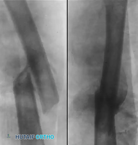
Figure A: Demonstrates a severely malunited fracture of the femur with significant overlapping of fragments and angular deformity in an 11-year-old boy. At this degree of shortening and angulation, spontaneous remodeling alone is insufficient to restore normal biomechanics.
Contraindications to corrective osteotomy must be rigorously respected to prevent catastrophic surgical failures. Absolute contraindications include the presence of active, untreated deep infection or osteomyelitis at the planned surgical site. Severe, unmanageable medical comorbidities that preclude safe administration of anesthesia or systemic recovery also represent absolute barriers to elective reconstruction. Relative contraindications include advanced, end-stage osteoarthritis of the ipsilateral hip or knee; in such cases, correcting the diaphyseal deformity may not relieve the patient's pain, and the surgeon should instead consider deformity-correcting total joint arthroplasty. Furthermore, severe osteoporosis, compromised soft-tissue envelopes (e.g., extensive scarring from previous trauma or burns), and active smoking are significant relative contraindications that exponentially increase the risk of hardware failure, wound dehiscence, and nonunion.
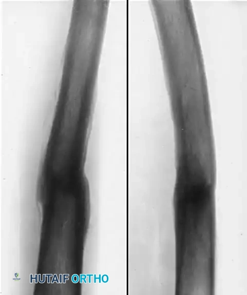
Figure B: Radiographic appearance five months after open reduction and corrective osteotomy. The procedure involved the insertion of a Kirschner wire through the distal femur and the application of a spica cast incorporating the wire to maintain length and alignment. Through this intervention, the length of the limb and the functional mechanics of the knee were successfully regained, allowing subsequent growth to finalize the remodeling process.
| Deformity Parameter | Threshold for Osteotomy Indication | Primary Clinical Symptoms & Sequelae |
|---|---|---|
| Coronal Plane (Varus/Valgus) | > 10 - 15 degrees | Medial/Lateral compartment overload, early OA, thrust during gait. |
| Sagittal Plane (Procurvatum/Recurvatum) | > 15 - 20 degrees | Altered knee flexion/extension arcs, compensatory lumbar lordosis. |
| Axial Plane (Rotational Torsion) | > 15° Internal / > 20° External | Patellofemoral instability, anterior knee pain, out-toeing/in-toeing. |
| Leg-Length Discrepancy (LLD) | > 2.0 - 2.5 cm | Pelvic obliquity, secondary scoliosis, increased energy expenditure. |
Pre-Operative Planning, Templating, and Patient Positioning
Meticulous, exhaustive preoperative planning is the absolute cornerstone of a successful corrective osteotomy; intraoperative improvisation in the face of a complex malunion is a recipe for disaster. The evaluation begins with a rigorous clinical assessment. The surgeon must evaluate the patient's dynamic gait, noting any varus/valgus thrust, Trendelenburg sign, or abnormal foot progression angle. The rotational profile is quantified by measuring hip internal and external rotation in both the prone (extension) and seated (flexion) positions, comparing the affected limb to the contralateral normal side. True leg-length discrepancy is measured using calibrated blocks under the short limb until the pelvis is leveled, differentiating it from apparent LLD caused by pelvic obliquity or contractures.
Radiographic analysis demands high-quality, standardized imaging. Full-length, weight-bearing anteroposterior (AP) and lateral radiographs of both lower extremities (from hips to ankles) are mandatory. These films allow for the precise measurement of the mechanical axis deviation (MAD), the joint line convergence angle (JLCA), the mLDFA, and the mMPFA. Advanced imaging is non-negotiable for multiplanar deformities. A computed tomography (CT) scan with standardized rotational cuts through the femoral neck, femoral condyles, and tibial plafond is the gold standard for quantifying femoral version and confirming the exact degree of rotational malalignment. The Jeanmart method or similar standardized CT protocols should be utilized to calculate the angle between the axis of the femoral neck and the posterior condylar axis.
Digital templating using modern orthopedic software is utilized to simulate the osteotomy. The surgeon must identify the CORA and determine whether a single-cut osteotomy (opening wedge, closing wedge, or transverse) or a more complex multi-apical correction is required. The level of the osteotomy is selected based on the CORA, the quality of the surrounding bone, and the requirements of the planned fixation construct. If an intramedullary nail is planned, the surgeon must template the diameter and length of the nail, paying close attention to the radius of curvature of the implant versus the patient's native femoral bow. The exact location of the interlocking screws must be templated to ensure adequate purchase in both the proximal and distal fragments after the deformity is corrected.
Patient positioning and operating room setup are critical logistical steps that directly impact the execution of the surgery. For diaphyseal femoral osteotomies planned for antegrade intramedullary nailing, the patient is typically positioned supine on a radiolucent flat table (e.g., Jackson table) or a specialized fracture table. The flat table is often preferred for complex multiplanar corrections as it allows for easier manipulation of the limb and more accurate intraoperative assessment of rotational alignment and leg length. A bump is placed under the ipsilateral hip to elevate the greater trochanter, and the torso is adducted to provide unimpeded access to the piriformis fossa or greater trochanteric entry point. The fluoroscopy unit (C-arm) must be positioned to allow unhindered orthogonal views of the entire femur, from the hip joint to the knee, without compromising the sterile field.
Step-by-Step Surgical Approach and Fixation Technique
The surgical approach to the femoral diaphysis must be carefully selected to provide adequate exposure of the malunion site while minimizing iatrogenic soft-tissue trauma. The lateral or anterolateral approach is most commonly utilized. The lateral approach involves an incision along the line connecting the greater trochanter to the lateral epicondyle. After incising the fascia lata, the vastus lateralis is elevated anteriorly off the lateral intermuscular septum. The surgeon must meticulously identify, ligate, or cauterize the perforating branches of the profunda femoris artery as they cross the septum; failure to do so can result in massive postoperative hematoma. The periosteum is then incised longitudinally for a distance of 6 to 8 cm over the apex of the deformity. Meticulous subperiosteal elevation is performed to expose the sclerotic callus, ensuring the preservation of the osteogenic cambium layer attached to the elevated periosteal flaps.
Executing the osteotomy cut requires technical precision and consideration of the bone's density. In long-standing malunions, the callus is often exceedingly dense and sclerotic. While a motorized oscillating or reciprocating saw can be used, it generates significant heat. If a saw is utilized, it must be accompanied by continuous, copious chilled saline irrigation to prevent thermal necrosis of the osteotomy margins, which would severely compromise bone healing. Alternatively, and often preferably in highly sclerotic bone, the drill-and-osteotome technique is employed. The surgeon drills multiple transverse holes through the bone at the planned osteotomy site using a 2.5 mm or 3.2 mm drill bit. A sharp, broad osteotome is then driven through the plane of the drill holes to complete the cut. This technique ensures a perfectly planar cut, reduces physical exertion, and virtually eliminates the risk of thermal necrosis.
Once the bone is divided, the deformity is corrected using manual force and specialized reduction clamps (e.g., Jungbluth clamps or heavy pointed reduction forceps). This step frequently requires the systematic release of contracted soft tissues. The linea aspera may need to be released posteriorly, and the fascia lata may require fractional lengthening laterally to permit restoration of anatomic length and alignment. A critical step follows: recanalizing the medullary canal. In chronic malunions, the endosteal canal is often completely obliterated by dense bone. The surgeon must use rigid reamers, sharp curettes, or a cannulated drill over a heavy guide wire to meticulously open the medullary space in both the proximal and distal fragments, ensuring that the anatomical axis of both segments is restored to accept an intramedullary device.
Rigid internal fixation is mandatory, as the corrected osteotomy is inherently unstable against the massive deforming forces of the thigh musculature. Interlocking intramedullary (IM) nailing is the gold standard for diaphyseal osteotomies. A ball-tipped guide wire is passed from the proximal entry point, across the osteotomy, and centered into the distal epiphysis. The canal is sequentially reamed to 1.0 mm to 1.5 mm larger than the planned nail diameter. The nail is inserted and secured with multiple proximal and distal interlocking screws. The IM nail acts as a load-sharing device positioned precisely along the mechanical axis, biomechanically favoring early weight-bearing and robust secondary bone healing via callus formation.
Alternatively, for periarticular malunions or situations where the medullary canal cannot be safely recanalized, heavy-duty broad dynamic compression plating (DCP) or locking compression plating (LCP) is utilized. The plate is applied to the tension band side of the femur (typically the lateral cortex) and secured with a minimum of eight to ten cortices of fixation above and below the osteotomy. Regardless of the fixation method, the biological environment of the sclerotic malunion is compromised. Therefore, the liberal application of autologous cancellous bone graft (harvested from the iliac crest or utilizing the Reamer-Irrigator-Aspirator (RIA) system) circumferentially around the osteotomy site is strongly recommended to provide the osteogenesis, osteoinduction, and osteoconduction necessary for reliable union.
Complications, Incidence Rates, and Salvage Management
Surgeons undertaking corrective osteotomies for femoral malunion must be acutely aware of the high complication profile associated with these complex reconstructive procedures. The biological and mechanical environment of a malunited femur is inherently hostile to rapid healing. The most prevalent and concerning complication is nonunion or delayed union. The sclerotic nature of the osteotomy site, combined with the disruption of the endosteal blood supply during recanalization, creates a precarious environment for osteogenesis. If rigid fixation is not achieved, or if the biological preparation (decortication and bone grafting) is inadequate, the osteotomy will fail to unite. Management of a nonunion typically requires a salvage operation involving hardware removal, aggressive debridement of fibrous tissue, recanalization, exchange nailing (using a larger diameter nail), and the application of copious autologous bone graft or orthobiologics (e.g., BMP-2).
Hardware failure is a direct consequence of either premature weight-bearing in the face of a delayed union or fundamentally inadequate initial fixation. Intramedullary nails can experience fatigue fracture at the level of the osteotomy, and interlocking screws can break or back out. Plates applied to the lateral femur are subjected to massive cyclical bending moments; if the medial cortex is not rigidly apposed (creating a structural void), the plate will inevitably undergo fatigue failure. Salvage management for hardware failure requires immediate revision surgery. Broken nails must be meticulously extracted, which may require specialized extraction tools or opening the femur at the site of the broken implant. The nonunion site is then stabilized with a more robust construct, often combining an intramedullary nail with a supplementary augmentation plate (nail-plate combination) to maximize biomechanical stability.
Neurovascular injury is a catastrophic complication that demands meticulous surgical technique to prevent. The profunda femoris artery and its perforating branches are highly vulnerable during the lateral approach and during the placement of diaphyseal screws. Iatrogenic injury can lead to massive hemorrhage or the formation of a pseudoaneurysm. The sciatic nerve is at risk during aggressive posterior soft-tissue retraction or if a drill bit plunges excessively past the posterior cortex. Furthermore, acute correction of a severe, long-standing leg-length discrepancy (lengthening greater than 3-4 cm acutely) can cause traction neuropraxia of the sciatic or peroneal nerve. If significant lengthening is required, gradual distraction osteogenesis using a circular external fixator or a motorized intramedullary lengthening nail is indicated over acute correction.
Infection, while less common in elective osteotomies than in acute trauma, remains a devastating complication. Deep surgical site infection (SSI) necessitates aggressive, rapid intervention. The principles of management include immediate surgical debridement, irrigation, and the acquisition of deep tissue cultures. If the internal fixation remains rigidly stable, it may be retained while the patient undergoes a prolonged course of targeted intravenous antibiotic therapy. However, if the hardware is loose or if the infection is refractory, the implants must be removed, the medullary canal aggressively reamed and irrigated, and a temporary antibiotic-impregnated cement spacer (or coated nail) inserted until the infection is eradicated, followed by definitive reconstruction.
| Complication | Estimated Incidence | Primary Etiology / Risk Factor | Salvage Management Strategy |
|---|---|---|---|
| Nonunion / Delayed Union | 5% - 12% | Sclerotic bone, inadequate grafting, micro-motion. | Exchange nailing, augmentation plating, autologous bone grafting (RIA/Iliac Crest). |
| Hardware Failure | 3% - 8% | Premature weight-bearing, medial cortical void, undersized implant. | Hardware extraction, revision to stiffer construct (nail-plate combo), structural grafting. |
| Neurovascular Injury | < 2% | Plunging drill bits, aggressive retraction, acute over-lengthening. | Vascular repair, nerve exploration. Prevent via gradual distraction for large LLDs. |
| Deep Infection | 1% - 3% | Prolonged operative time, poor soft tissue envelope, hematoma. | Aggressive I&D, retention of stable hardware + IV antibiotics; removal if loose/refractory. |
Phased Post-Operative Rehabilitation Protocols
The postoperative rehabilitation protocol following a femoral osteotomy is not a monolithic pathway; rather, it is a highly individualized, phased progression dictated entirely by the biomechanical stability of the fixation achieved intraoperatively and the biological quality of the host bone. The primary goals of rehabilitation are the prevention of thromboembolic events, the restoration of full, pain-free range of motion (ROM) in the adjacent hip and knee joints, the prevention of profound muscle atrophy, and the safe, progressive return to functional weight-bearing. Clear, continuous communication between the orthopedic surgeon and the physical therapy team is essential to prevent premature loading that could jeopardize the reconstruction.
Phase 1: Immediate Postoperative Period (Weeks 0-2)
During the initial inflammatory phase of healing, the focus is on pain control, edema management, and the prevention of arthrofibrosis. Regardless of the fixation method, aggressive deep vein thrombosis (DVT) prophylaxis (pharmacological and mechanical) is initiated immediately. Active and active-assisted ROM exercises for the hip and knee are commenced on postoperative day one. Continuous Passive Motion (CPM) machines may be utilized if knee stiffness is a concern. If rigid interlocking intramedullary nailing was achieved (a load-sharing construct), patients are typically permitted immediate toe-touch or partial weight-bearing (10 to 15 kg) using crutches or a walker. Conversely, if plate fixation was utilized (a load-bearing construct susceptible to cyclical fatigue), weight-bearing is strictly restricted to toe-touch only to protect the hardware.
Phase 2: Early Healing and Muscle Activation (Weeks 2-6)
As the patient transitions into the reparative phase of bone healing, the emphasis shifts toward muscle activation and early strengthening. Isometric exercises for the quadriceps, hamstrings, and gluteal musculature are intensified. Closed-kinetic-chain exercises may be carefully introduced for patients with IM nails, provided they remain within their prescribed weight-bearing limits. Radiographic evaluation (AP and lateral views) is performed at the 6-week mark to assess for early signs of bridging callus formation. If early callus is visualized and the patient is clinically pain-free at the osteotomy site, weight-bearing may be cautiously advanced in the IM nail cohort. Plated osteotomies generally remain protected with strict weight-bearing limitations until more robust radiographic healing is confirmed.
Phase 3: Progressive Loading and Gait Normalization (Weeks 6-12)
This phase is characterized by the progression toward full weight-bearing, contingent upon radiographic evidence of progressive union. At the 12-week postoperative visit, a clinical and radiographic assessment is critical. If bridging callus is present across at least three of four cortices on orthogonal radiographs, patients are cleared to progressively wean off assistive devices and transition to full, unassisted weight-bearing. Physical therapy focuses heavily on gait retraining, eliminating any residual Trendelenburg lurch or compensatory mechanisms developed during the period of malunion. Advanced strengthening, proprioceptive training, and stationary cycling are incorporated to restore baseline lower extremity function.
Phase 4: Advanced Function and Return to Activity (Weeks 12+)
The final phase of rehabilitation focuses on returning the patient to their pre-injury level of occupational and recreational activity. Radiographs are obtained at 6 months to confirm solid, mature bony union and the maintenance of the corrected mechanical alignment. Once solid union is achieved, patients may initiate plyometric exercises, running, and sport-specific drills. Return to heavy manual labor or high-impact contact sports is generally permitted only after 6 to 9 months, ensuring that the bone has sufficiently remodeled to withstand extreme physiological loads without risk of re-fracture or hardware failure.
Summary of Landmark Literature and Clinical Guidelines
The surgical principles governing the correction of femoral malunions are deeply rooted in decades of rigorous biomechanical research and clinical outcome studies. The foundational literature on deformity correction is heavily indebted to the work of Dr. Dror Paley, whose comprehensive codification of the Center of Rotation of Angulation (CORA) principles revolutionized the way orthopedic surgeons conceptualize and execute osteotomies. Paley's geometric mapping ensures that surgeons can predictably restore the mechanical axis without introducing iatrogenic translation, a concept that remains the bedrock of modern preoperative templating guidelines.
The superiority of interlocking intramedullary nailing for diaphyseal osteotomies is supported by extensive clinical literature. Landmark studies by Winquist and Hansen in the 1980s established the biomechanical advantages of IM nailing for acute fractures, principles which were subsequently adapted for malunion surgery by Kempf and others. These studies demonstrated that the load-sharing nature of the IM nail, positioned centrally along the mechanical axis, significantly reduces the bending moments across the osteotomy site compared to eccentric plate fixation. Consequently, IM nailing is associated with higher union rates, lower hardware failure rates, and the ability to permit earlier postoperative weight-bearing, leading to its designation as the gold standard in contemporary clinical guidelines.
Recent literature has focused on optimizing the biological environment of the osteotomy. Studies evaluating the use of the Reamer-Irrigator-Aspirator (RIA) system have shown that harvesting large volumes of highly osteogenic autograft from the femoral canal significantly enhances union rates in sclerotic malunions, matching or exceeding the efficacy of traditional iliac crest bone grafting while minimizing donor-site morbidity. Furthermore, current clinical guidelines emphasize the critical importance of correcting rotational malalignment. Research by Sanders and others has highlighted that uncorrected femoral torsion greater than 15 degrees correlates strongly with poor clinical outcomes and persistent patellofemoral pain, reinforcing the mandate for routine preoperative CT version
