Comprehensive Master Guide · Medically Reviewed

AAOS & ABOS Upper Extremity MCQs (Set 4): Shoulder, Elbow, Wrist, Hand & Nerve Review | 2025-2026 Boards

Master your 2025-2026 AAOS and ABOS boards with our interactive Upper Extremity MCQs. Review shoulder, elbow, wrist, hand, and nerve topics today!

21 Detailed Chapters
59 min read
Updated: Apr 2026
Dr. Mohammed Hutaif
Medically Reviewed by
Prof. Dr. Mohammed Hutaif
Verified Content Expert Reviewed

Quick Medical Answer

This high-yield question set (Set 4) for the AAOS/ABOS exams focuses on critical upper extremity topics. It covers the diagnosis and management of shoulder and elbow pathology, common wrist and hand fractures (e.g., distal radius, scaphoid), and key peripheral nerve entrapment syndromes, essential for board preparation.

Upper Extremity 2005 MCQs - Part 4

AAOS & ABOS Upper Extremity MCQs (Set 4): Shoulder, Elbow, Wrist, Hand & Nerve Review | 2025-2026 Boards

Comprehensive 100-Question Exam


00:00

Start Quiz

Question 1

A 52-year-old man has shoulder pain and stiffness after undergoing a "mini-lateral" rotator cuff repair 6 months ago. Examination reveals that he is afebrile with normal vital signs. There is slight erythema but no drainage from the incision. Range of motion is limited in all planes, and there is weakness with resisted external rotation and abduction. Radiographs show a well-positioned metal implant within the greater tuberosity. Laboratory studies reveal a WBC count of 8,400/mm3 (normal 3,500 to 10,500/mm3) and an erythrocyte sedimentation rate of 63 mm/h (normal up to 20 mm/h). What is the next most appropriate step in management?


Explanation

Deep sepsis of the shoulder following rotator cuff repair is an uncommon problem. Patients with infections of this type typically report persistent pain and are not systemically ill. They may have signs of local wound problems such as erythema, drainage, and dehiscence. Laboratory studies can be helpful in making an accurate diagnosis. Most patients will not show a significant elevation of the WBC count; however, an elevated erythrocyte sedimentation rate is nearly always present and should alert the clinician to the presence of infection. Aspiration of both subacromial and glenohumeral joint spaces is necessary to confirm the diagnosis. The most effective treatment for deep shoulder sepsis following rotator cuff repair involves extensive surgical debridement, removing all suspicious soft tissue as well as implants. Administration of appropriate antibiotic therapy is needed for complete control of the infection. Mirzayan R, Itamura JM, Vangsness CT, et al: Management of chronic deep infection following rotator cuff repair. J Bone Joint Surg Am 2000;82:1115-1121. Settecerri JJ, Pitnu MA, Rock MG, et al: Infection after rotator cuff repair. J Shoulder Elbow Surg 1994;8:105.

Question 2

A 21-year-old pitcher reports shoulder pain with hard throwing. He notes that the pain occurs in the early acceleration phase of his throw. Given his history, what structures are at greatest risk for injury?


Explanation

Internal impingement in the thrower's shoulder occurs in the abducted, externally rotated position as described by Walch and associates. The injury is thought to occur from repetitive contact between the posterosuperior portion of the labrum and glenoid against the articular side of the rotator cuff and greater tuberosity. Paley KJ, Jobe FW, Pink MM, et al: Arthroscopic findings in the overhand throwing athlete: Evidence for posterior internal impingement of the rotator cuff. Arthroscopy 2000;16:35-40. Jazrawi LM, McCluskey GM III, Andrews JR: Superior labral anterior and posterior lesions and internal impingement in the overhead athlete. Instr Course Lect 2003;52:43-63.

Question 3

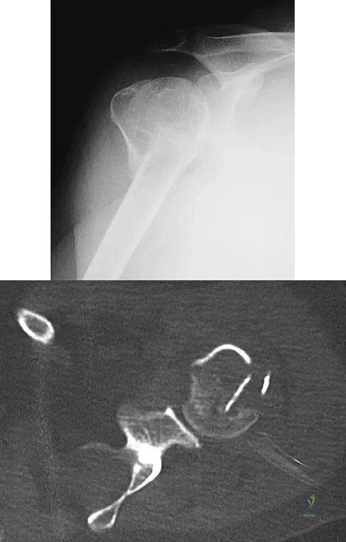
A 30-year-old man landed on his shoulder in a fall off his mountain bike. An AP radiograph and CT scan are shown in Figures 34a and 34b. Management should consist of


Explanation

The radiograph shows a valgus impacted four-part fracture. The humeral head is deeply depressed into the metaphysis but is still articulating with the glenoid as seen on the CT scan. Unlike a "classic" four-part fracture in which the head is dislocated out of the glenoid and devoid of any soft-tissue attachments (high risk of osteonecrosis), this valgus impacted head will have a medial soft-tissue hinge with a lower risk of osteonecrosis. It is most amenable to open reduction and internal fixation with minimal soft-tissue stripping techniques. Bone grafting may be necessary on occasion. Nonsurgical management for displaced proximal humeral fractures generally results in a poor outcome. This patient does not have a humeral head defect. A hemiarthroplasty is not indicated. Jakob RP, Miniaci A, Anson PS, et al: Four-part valgus impacted fractures of the proximal humerus. J Bone Joint Surg Br 1991;73:295-298.

Question 4

A 22-year-old professional baseball pitcher has had pain in the axillary region of his dominant shoulder for the past several weeks. While throwing a pitch during a game, he notes a sharp pulling sensation with a "pop" in his shoulder. Examination the following day reveals tenderness along the posterior axillary fold and pain and weakness with resisted extension of the shoulder. What is the most likely cause of his symptoms?


Explanation

Injury to the latissimus dorsi tendon recently has been reported as a cause of pain in the thrower's shoulder. The etiology of this injury is felt to be eccentric overload during the follow-through of the throwing motion. Recommended management for this unusual injury consists of a short period of rest, followed by physical therapy to restore shoulder motion and strength. Throwing is allowed when the athlete demonstrates full, pain-free motion and good strength and balance of the rotator cuff and scapular rotator muscles. Currently there are no defined indications for surgical repair. Schickendantz MS, Ho CP, Keppler L, et al: MR imaging of the thrower's shoulder: Internal impingement, latissimus dorsi/subscapularis strains and related injuries. Magn Reson Imaging Clin N Am 1999;7:39-49.

Question 5

When comparing the addition of a trough at the greater tuberosity to direct repair of cortical bone, simulated rotator cuff repair in animal models has shown what type of change in the strength of the repair?


Explanation

There was no difference observed in the healing of tendon to bone when comparing healing to cortical bone and to a cancellous trough.

Question 6

Figures 35a and 35b show the radiographs of a 20-year-old man who is unable to rotate his dominant forearm. Examination reveals that the arm is fixed in supination. To regain motion, management should consist of


Explanation

The patient has a proximal synostosis; therefore, resection of the synostosis is considered the best option to regain motion. While forearm osteotomy can place the hand in a more functional position, rotation will not be restored. Proximal radial excision can provide forearm rotation; however, this procedure is reserved for patients who have a proximal radioulnar synostosis that is too extensive to allow a safe resection, involves the articular surface, and is associated with an anatomic deformity. Motion will not be restored with dynamic splinting. Kamineni S, Maritz NG, Morrey BF: Proximal radial resection for posttraumatic radioulnar synostosis: A new technique to improve forearm rotation. J Bone Joint Surg Am 2002;84:745-751.

Question 7

A 20-year-old-man sustained a scapular fracture after attempting to grab a beam as he fell through a ceiling at a job site 3 months ago. A clinical photograph is shown in Figure 36. He now reports pain in the anterior shoulder and difficulty with overhead activities. What nerve roots make up the involved peripheral nerve?

Upper Extremity Board Review 2005: High-Yield MCQs (Set 4) - Figure 5


Explanation

The patient sustained an injury to the long thoracic nerve, which supplies the serratus anterior. Branches of C5 and C6 enter the scalenus medius, unite in the muscle, and emerge as a single trunk and pass down the axilla. On the surface of the serratus anterior, the long thoracic nerve is joined by the branch from C7 and descends in front of the serratus anterior, providing segmental innervation to the serratus anterior.

Question 8

A 20-year-old collegiate baseball pitcher has persistent deep shoulder pain. Examination reveals normal strength, 130 degrees of external rotation in abduction, 10 degrees of internal rotation in abduction, mild dynamic scapular winging, and equivocal findings on provocative tests for labral tears. Management should consist of


Explanation

Although management of shoulder pain in the throwing athlete is controversial, there are some general principles. Initial management generally includes rest from throwing, restoring normal joint function, specifically motion and strength as well as eliminating pain. In this patient, examination reveals excessive external rotation and decreased internal rotation. This pattern is common in pitchers; however, the total arc of motion should remain close to 180 degrees in abduction. In this patient, the total arc is 140 degrees. Treatment should first focus on restoring a 180-degree arc with posterior scapular stretching, as well as pain control and muscle rehabilitation. Injections and surgery are generally reserved for patients who fail to respond to rest and rehabilitation.

Question 9

Which of the following best describes the most common anatomic variation seen in the glenoid labrum and the middle glenohumeral ligament in the anterosuperior quadrant of the shoulder??


Explanation

Wide variations in the anatomy of the anterosuperior portion of the labrum and the middle glenohumeral ligament have been reported and are more common than previously thought. The labrum attached to the glenoid rim and a flat/broad middle glenohumeral ligament is the most common "normal" variation. A cord-like middle glenohumeral ligament is often associated with the presence of a sublabral hole. An anterosuperior labrum confluent with a cord-like middle glenohumeral ligament and no labral attachment to bone is the configuration of the Buford complex. The prevalence of each variation from one recent study is as follows: #1: 86.6%; #2: 3.3%; #3: 8.6%; and #4: 1.5%. Rao AG, Kim TK, Chronopoulos E, et al: Anatomical variants in the anterosuperior aspect of the glenoid labrum. J Bone Joint Surg Am 2003;85:653-659. Ilahi OA, Labbe MR, Cosculluela P: Variants of the anterosuperior glenoid labrum and associated pathology. Arthroscopy 2002;18:882-886.

Question 10

A 21-year-old hockey player who has recurrent shoulder subluxations undergoes an anterior capsulorrhaphy under general anesthesia, and an interscalene block is used to relieve postoperative pain. At the 1-week follow-up examination, he reports loss of sensation over the lateral region of the shoulder and is unable to actively contract the deltoid muscle. The remainder of the examination is normal. What is the best course of action at this time?


Explanation

The patient has an axillary nerve injury, which is relatively uncommon after surgery for instability. This type of injury generally is the result of a stretch injury rather than transection or a hematoma. Therefore, observation is indicated in the early postoperative period. After approximately 6 weeks, electromyography can be used to confirm and document the point of injury. Interscalene blocks can cause prolonged nerve injury but usually are not limited to the axillary nerve.

Question 11

A 10-year-old boy has had a prominent scapula for the past year. He reports crepitus and aching over the area, but only when he is active. A radiograph and CT scans are shown in Figures 37a through 37c. What is the most likely diagnosis?


Explanation

The findings are typical for an osteochondroma. It is found as an outgrowth of bone and cartilage from those bones that arise from enchondral ossification. It may be flat, verrucous, or with a long stalk and cauliflower-like cap. Osteochondromas can become symptomatic secondary to irritation of the adjacent musculature. They cease to proliferate when epiphyseal growth ceases.

Question 12

In patients who have undergone nonsurgical management for idiopathic adhesive capsulitis, long-term follow-up studies have shown which of the following results?


Explanation

Results have been satisfactory in many patients; however, at long-term follow-up, examination of the affected shoulder often shows some decrease in range of motion compared with the contralateral side. Although range of motion often improves over time, it does not return to normal in 60% of patients. Pain improves but is often increased compared with the contralateral side. Griggs SM, Ahn A, Green A: Idiopathic adhesive capsulitis: A prospective functional outcome study of nonoperative treatment. J Bone Joint Surg Am 2000;82:1398-1407.

Question 13

Which of the following statements best describes the relationship between tissue response to thermal capsulorrhaphy and the type of device used?


Explanation

Although radiofrequency devices and lasers differ fundamentally in the way they generate heat within a tissue, both classes of devices are capable of producing temperatures within the critical temperature range (65 to 75 degrees C) for collagen denaturation and subsequent tissue shrinkage. When it comes to cell viability and tissue response, heat is heat. Once critical temperatures are reached, cells will die at 45 degrees C, collagen will become denatured at 60 degrees C, and tissue ablation will occur at 100 degrees C no matter what the source of thermal energy. Therefore, claims of a better or different type of heat have little bearing on the biologic response of the tissue. Histologic, ultrastructural, and biomaterial alterations induced by laser and radiofrequency energy have been shown to be similar. Arnoczky SP, Aksan A: Thermal modification of connective tissues: Basic science considerations and clinical implications. J Am Acad Orthop Surg 2000;8:305-313. Hayashi K, Markel MD: Thermal modification of joint capsule and ligamentous tissues: The use of thermal energy in sports medicine. Operative Techniques Sports Med 1998;6:120-125.

Question 14

A 35-year-old man has profound deltoid weakness after sustaining a traumatic anterior shoulder dislocation 6 weeks ago. Electromyographic (EMG) studies confirm an axillary nerve injury. Follow-up examination at 3 months reveals no recovery of function. What is the best course of action?


Explanation

Documenting the status of recovery at this time is appropriate; therefore, repeat EMG studies should be conducted to check for early signs of reinnervation. Timing of nerve exploration in this setting is debated, with authors suggesting exploration if there is no sign of recovery at 6 to 9 months. Perlmutter GS: Axillary nerve injury. Clin Orthop 1999;368:28-36. Artico M, Salvati M, D'Andrea V, et al: Isolated lesions of the axillary nerves: Surgical treatment and outcome in twelve cases. Neurosurgery 1991;29:697-700. Vissar CP, Coene LN, Brand R, et al: The incidence of nerve injury in anterior dislocation of the shoulder and its influence on functional recovery: A prospective clinical and EMG study. J Bone Joint Surg Br 1999;81:679-685.

Question 15

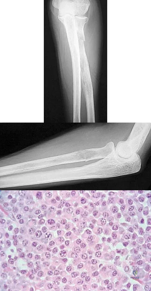
A 65-year-old woman has had chronic aching discomfort involving her elbow for the past 6 months. Radiographs and a biopsy specimen are shown in Figures 38a through 38c. What is the most likely diagnosis?


Explanation

The histologic features of multiple myeloma are distinctive for this lesion. The plasma cells are round or oval and have an eccentric nucleus and prominent nucleolus. These characteristics and a clear area next to the eccentric nucleus representing the prominent Golgi center are pathognomonic for plasma cells. Lymphoma is in the differential diagnosis; the most frequent types that occur in bone are large cell or mixed small and large cell types. The histologic appearance of the specimen is not consistent with the other choices.

Question 16

Which of the following clinical findings is commonly associated with symptomatic partial-thickness rotator cuff tears?


Explanation

In symptomatic partial-thickness rotator cuff tears, a painful arc with active range of motion is common, impingement signs are usually positive, and the lift-off test is normal. Active and passive range of motion measurements are often equal, although active range of motion can be painful. External rotation lag signs are often seen with larger full-thickness tears. Hertel R, Ballmer FT, Lambert SM, Gerber C: Lag signs in the diagnosis of rotator cuff rupture. J Shoulder Elbow Surg 1996;5:307-313. McConville OR, Iannotti JP: Partial thickness tears of the rotator cuff: Evaluation and management. J Am Acad Orthop Surg 1999;7:32-43. Gerber C, Krushell RJ: Isolated rupture of the tendon of the subscapularis muscle: Clinical features in 16 cases. J Bone Joint Surg Br 1991;73:389-394.

Question 17

A 65-year-old woman landed on her nondominant left shoulder in a fall. An AP radiograph is shown in Figure 39. Management should consist of

Upper Extremity Board Review 2005: High-Yield MCQs (Set 4) - Figure 12


Explanation

The radiograph reveals a four-part fracture-dislocation of the proximal humerus. Humeral hemiarthroplasty and tuberosity repair is the treatment of choice because the risk of osteonecrosis is high after attempted repair of this injury. Glenoid resurfacing is reserved for acute fractures in which there is significant preexisting glenoid arthrosis, such as in patients with rheumatoid arthritis. Neer CS II: Displaced proximal humeral fractures: II. Treatment of three- and four-part displacement. J Bone Joint Surg Am 1970;52:1090-1103.

Question 18

To avoid damage to the ascending branch of the anterior humeral circumflex artery during open reduction and internal fixation of a proximal humeral fracture, the blade plate should be placed in what position?


Explanation

The pectoralis major tendon inserts lateral to the biceps tendon, which runs in the bicipital groove. The primary vascular supply of the articular surface of the humeral head is derived from the anterior circumflex humeral artery, which continues into the arcuate artery once it enters the bone. The entry point is on the anterolateral aspect of the humerus just medial to the greater tuberosity within the bicipital groove. To avoid compromising circulation, the blade plate should be placed lateral to the bicipital groove and pectoralis major tendon insertion. Loebenberg M, Plate AM, Zuckerman J: Osteonecrosis of the humeral head. Instr Course Lect 1999;48:349-357.

Question 19

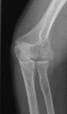
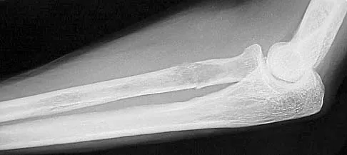
An otherwise healthy 13-year-old boy sustains the fracture shown in Figure 40 while throwing a fastball. Management should consist of

Upper Extremity Board Review 2005: High-Yield MCQs (Set 4) - Figure 13


Explanation

Nonsurgical management such as a functional brace, hanging arm cast, or sugar tong splint is the treatment of choice for a fracture of the humeral shaft that is the result of throwing. The fracture surface typically is wide and the degree of displacement is not large; therefore, surgery is not indicated in most patients. Ogawa K, Yoshida A: Throwing fracture of the humeral shaft: An analysis of 90 patients. Am J Sports Med 1998;26:242-246.

Question 20

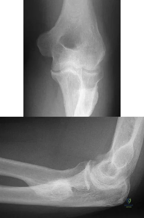
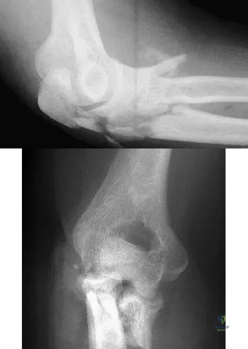
A 24-year-old man sustains an injury to his right elbow after falling 10 feet. Radiographs are shown in Figures 41a and 41b. Treatment should consist of


Explanation

Transolecranon fracture-dislocations are most effectively managed with open reduction and internal fixation, followed by early aggressive range of motion. Concomitant injury to the collateral ligament is rare, and stability is achieved by anatomic reconstruction of the olecranon fracture with rigid fixation. The need for collateral ligament repair or a hinged external fixator is uncommon in this fracture pattern.

Question 21

After closed reduction of the dislocation shown in Figure 42, it is essential to avoid placing the upper extremity in what position for the first 4 to 6 weeks?

Upper Extremity Board Review 2005: High-Yield MCQs (Set 4) - Figure 16


Explanation

Acute posterior dislocations occur rarely, accounting for less than 5% of acute dislocations. They are most often the result of falls on an outstretched hand. Reduction can be accomplished with flexion of the arm to 90 degrees and adduction to disimpact the humeral head from the glenoid rim. The arm is then externally rotated until the head has cleared the glenoid rim. Following brace immobilization in neutral to 5 to 10 degrees of external rotation and slight abduction, it is critical to avoid internal rotation for 4 to 6 weeks. Burkhead WZ Jr, Rockwood CA Jr: Treatment of instability of the shoulder with an exercise program. J Bone Joint Surg Am 1986;68:724-731.

Question 22

A baseball player has had diffuse scapular soreness for the past 8 weeks. He reports that it began insidiously over several days and gradually has become worse. He denies any history of trauma. Examination reveals drooping of the shoulder, with lateral winging of the scapula at rest. He is otherwise neurologically intact. What is the best course of action?


Explanation

Lateral scapular winging is characteristic of trapezius palsy, whereas medial scapular winging is characteristic of long thoracic nerve palsy. During sports activity, injury to the spinal accessory nerve is rare but may occur with blunt or stretching trauma. Patients often report an asymmetric neckline, drooping shoulder, winging of the scapula, and weakness of forward elevation. Evaluation should include a complete electrodiagnostic examination. Wiater JM, Bigliani LU: Spinal accessory nerve injury. Clin Orthop 1999;368:5-16. Wiater JM, Flatow EL: Long thoracic nerve injury. Clin Orthop 1999;368:17-27. Mariani PP, Santoriello P, Maresca G: Spontaneous accessory nerve palsy. J Shoulder Elbow Surg 1998;7:545-546. Porter P, Fernandez GN: Stretch-induced spinal accessory nerve palsy: A case report. J Shoulder Elbow Surg 2001;10:92-94.

Question 23

Which of the following best describes the mechanical response of the inferior glenohumeral ligament to repetitive subfailure strains?


Explanation

Repetitive subfailure strains have been shown to affect the mechanical behavior of the inferior glenohumeral ligament, producing dramatic declines in the peak load response and length increases that are largely unrecoverable. In another study, anteroinferior subluxation was found to result in nonrecoverable strain in the anteroinferior capsule, varying from 3% to 7% through a range of joint subluxation. Pollock RG, Wang VM, Bucchieri JS, et al: Effects of repetitive subfailure strains on the mechanical behavior of the inferior glenohumeral ligament. J Shoulder Elbow Surg 2000;9:427-435.

Question 24

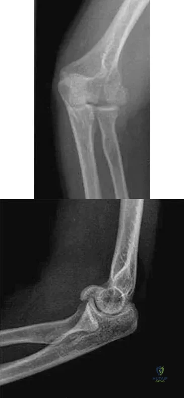
A 38-year-old woman who tripped and fell on her outstretched arm reports pain with movement. Examination reveals swelling. AP and lateral radiographs are shown in Figures 43a and 43b. Management should consist of


Explanation

The patient has a type I (Hahn-Steinthal) capitellar fracture that is best seen on the lateral radiograph. If a fracture fragment is seen proximal to the radial head, a capitellar fracture is the most likely injury because radial head fractures do not migrate proximally. The fragment is large enough for fixation. Excision is the preferred treatment for small shear osteochondral type II (Kocher-Lorenz) capitellar fractures. Closed reduction usually is not successful because of rotation of the displaced fragment. Mehdian H, McKee M: Management of proximal and distal humerus fractures. Orthop Clin North Am 2000;31:115-127.

Question 25

A 15-year-old girl reports popping and clicking at the sternoclavicular joint and an intermittent asymmetrical prominence of the medial head of the clavicle. She denies any history of trauma or other symptoms. Management should consist of


Explanation

Atraumatic subluxation or dislocation of the sternoclavicular joint typically occurs in individuals with generalized ligamentous laxity. It is generally not painful, has no long-term sequelae, and needs no treatment. In fact, it is more likely to be painful following surgery than if managed nonsurgically. Rockwood CA Jr, Odor JM: Spontaneous atraumatic anterior subluxation of the sternoclavicular joint. J Bone Joint Surg Am 1989;71:1280-1288.

Question 26

A 45-year-old manual laborer presents with chronic right wrist pain. Radiographs demonstrate a scaphoid nonunion with radioscaphoid arthritic changes, but the capitolunate joint is preserved. Which of the following is the most appropriate surgical treatment to relieve pain while best preserving his grip strength?


Explanation

This patient has Scaphoid Nonunion Advanced Collapse (SNAC) stage II. Four-corner fusion is preferred over proximal row carpectomy (PRC) in young, heavy laborers as it better preserves grip strength and relies on the spared radiolunate articulation.

Question 27

A 35-year-old male falls from a ladder and sustains a 'terrible triad' injury of the elbow. Operative fixation is planned. What is the most widely accepted sequence of surgical reconstruction to restore elbow stability?


Explanation

The standard protocol for terrible triad reconstruction proceeds from deep to superficial. This involves coronoid fixation first, followed by radial head repair or arthroplasty, and finally lateral ulnar collateral ligament (LUCL) repair.

Question 28

A 65-year-old woman is scheduled to undergo a reverse total shoulder arthroplasty for cuff tear arthropathy. To minimize the risk of scapular notching, how should the glenosphere baseplate optimally be positioned?


Explanation

Scapular notching is a frequent complication in reverse total shoulder arthroplasty. Placing the baseplate with an inferior tilt and inferior translation lowers the center of rotation and prevents mechanical impingement of the humeral component against the scapular neck.

Question 29

During surgical fasciectomy for Dupuytren's contracture, the neurovascular bundle is at risk of iatrogenic injury. The spiral cord is known to displace the neurovascular bundle in which of the following directions?


Explanation

The spiral cord draws the neurovascular bundle centrally, superficially, and proximally. This pathoanatomy makes the bundle highly vulnerable to injury during dissection near the metacarpophalangeal and proximal interphalangeal joints.

Question 30

A 28-year-old male sustained a closed midshaft humerus fracture and was placed in a coaptation splint in the emergency department. Two weeks later in clinic, he presents with a complete inability to extend his wrist or fingers, a deficit that was strictly documented as absent during his initial ED exam. What is the most appropriate next step in management?


Explanation

A secondary radial nerve palsy that develops after a closed reduction, splinting, or bracing of a humeral shaft fracture is a classic indication for surgical exploration. This is to ensure the nerve has not been entrapped within the fracture site.

Question 31

A 22-year-old collision athlete presents with recurrent anterior shoulder instability. A 3D CT scan reveals 30% anterior glenoid bone loss. What is the most appropriate surgical intervention to minimize the risk of recurrent instability?


Explanation

Glenoid bone loss greater than 20-25% is considered a critical defect and a contraindication to isolated soft-tissue Bankart repair. A coracoid transfer (Latarjet procedure) is the standard of care to restore the osseous arc and provide a sling effect.

Question 32

A surgeon is performing a single-incision anterior approach to repair an acute distal biceps tendon rupture. Which of the following nerves is at the greatest risk of iatrogenic injury during this specific approach?


Explanation

The lateral antebrachial cutaneous nerve (LABC) is the most commonly injured nerve during a single-incision anterior approach to the distal biceps. In contrast, the posterior interosseous nerve (PIN) is at higher risk during a two-incision approach if retractors are placed improperly.

Question 33

A 45-year-old female presents with severe basilar thumb pain and a positive grind test. In primary osteoarthritis of the thumb carpometacarpal (CMC) joint, which muscle acts as the primary deforming force leading to proximal, radial, and dorsal subluxation of the first metacarpal base?


Explanation

The abductor pollicis longus (APL) inserts on the dorsal-radial base of the first metacarpal. As the anterior oblique ligament (beak ligament) attenuates in CMC arthritis, the APL pulls the metacarpal shaft proximally, radially, and dorsally.

Question 34

A 55-year-old man presents with advanced scapholunate advanced collapse (SLAC) wrist. During surgical planning, the surgeon relies on the fact that a specific carpal articulation is characteristically spared from degenerative changes due to its concentric spherical anatomy. Which joint is this?


Explanation

In a SLAC wrist, the radiolunate joint is characteristically spared from arthritis because of its congruent, spherical articulation that does not experience abnormal shear forces. This sparing is the biomechanical basis for performing a four-corner fusion.

Question 35

A 30-year-old carpenter sustains a sharp volar laceration to his right index finger, resulting in a zone II flexor tendon injury. Following a 4-strand repair, a dynamic early active motion protocol is initiated. What is the primary biological and mechanical benefit of this protocol compared to prolonged static splinting?


Explanation

Early active motion protocols promote intrinsic tendon healing over extrinsic healing, thereby decreasing peritendinous adhesions. This significantly reduces the work of flexion and improves final digit range of motion without unacceptably increasing the rupture rate.

Question 36

A 70-year-old man presents with chronic, massive, irreparable rotator cuff tears of the supraspinatus and infraspinatus. Physical examination reveals pseudoparalysis of the shoulder with active forward elevation limited to 40 degrees. MRI demonstrates Goutallier stage 4 fatty infiltration of the torn tendons, but the teres minor and subscapularis remain intact. Which of the following is the most appropriate surgical management?


Explanation

Reverse total shoulder arthroplasty is the treatment of choice for elderly patients with massive, irreparable rotator cuff tears and pseudoparalysis. It restores the mechanical advantage of the deltoid, reliably improving active forward elevation.

Question 37

A 24-year-old gymnast falls from a height onto an extended, ulnar-deviated wrist. A lateral radiograph demonstrates the 'spilled teacup' sign with volar displacement of the lunate. She complains of severe pain and tingling in her thumb, index, and middle fingers. What is the most critical initial step in management?


Explanation

The patient has sustained a lunate dislocation complicated by acute median nerve compression. The most critical initial step is an emergent closed reduction to restore carpal alignment, decompress the median nerve, and relieve tension on the carpal blood supply before definitive surgical stabilization.

Question 38

A 60-year-old mechanic presents with intrinsic muscle wasting of the hand, a positive Froment sign, and numbness in the small and ulnar half of the ring fingers. He is diagnosed with severe cubital tunnel syndrome. Which structure typically forms the anatomical roof of the cubital tunnel?


Explanation

The roof of the cubital tunnel is formed by the cubital tunnel retinaculum, also known as Osborne's ligament or the arcuate ligament. It bridges the medial epicondyle to the olecranon.

Question 39

A 40-year-old man presents with a volar shear fracture of the distal radius (Barton's fracture). The surgeon elects to perform open reduction and internal fixation utilizing the standard volar Henry approach. The internervous plane for this approach is found between which two muscles?


Explanation

The volar Henry approach utilizes the internervous plane between the brachioradialis (innervated by the radial nerve) and the flexor carpi radialis (innervated by the median nerve).

Question 40

A 21-year-old collegiate baseball pitcher reports sudden medial elbow pain and an audible pop during the acceleration phase of throwing. Valgus stress testing reveals gross laxity. In surgical reconstruction of the ulnar collateral ligament (UCL), which specific anatomic bundle is the primary restraint to valgus stress from 30 to 120 degrees of elbow flexion?


Explanation

The anterior bundle of the ulnar collateral ligament (UCL) is the primary restraint to valgus stress of the elbow between 30 and 120 degrees of flexion. It is the primary structure reconstructed in 'Tommy John' surgery.

Question 41

A 35-year-old cyclist sustains a comminuted midshaft clavicle fracture. Open reduction and internal fixation with superior plating is performed. Postoperatively, the patient notes a well-demarcated area of numbness over the anterior chest wall just inferior to the incision. Which nerve was most likely injured during the surgical exposure?


Explanation

The supraclavicular nerve branches (medial, intermediate, and lateral) course superficially over the clavicle. They are frequently injured or divided during the standard surgical approach for clavicle plating, leading to anterior chest wall numbness.

Question 42

A 25-year-old male sustains a Type III acromioclavicular (AC) joint separation and opts for nonoperative management after thorough counseling. According to long-term outcome studies, what is the most common persistent patient complaint following nonoperative management of this injury?


Explanation

While functional outcomes and return to sport are generally excellent following nonoperative management of Type III AC separations, the most common long-term complaint is the persistent visible bump or cosmetic deformity over the superior shoulder.

Question 43

A 19-year-old motorcyclist sustains a traction injury to his right shoulder. On examination, his arm hangs at his side with the shoulder adducted, internally rotated, and the forearm pronated (waiter's tip posture). Hand and wrist flexion remain intact. This presentation is most consistent with an injury to which anatomic region of the brachial plexus?


Explanation

This patient exhibits an Erb-Duchenne palsy, which results from an injury to the upper trunk of the brachial plexus (C5-C6 roots). This causes loss of shoulder abduction and external rotation, as well as elbow flexion, leading to the classic 'waiter's tip' posture.

Question 44

A 45-year-old male is struck on the forearm with a heavy pipe. Radiographs reveal an isolated fracture of the proximal third of the ulnar shaft combined with an anterior dislocation of the radial head. What is the appropriate eponymous designation for this specific injury pattern?


Explanation

A Monteggia fracture-dislocation is defined as a fracture of the ulnar shaft associated with a dislocation of the radial head. This is distinct from a Galeazzi fracture, which is a distal radius shaft fracture with a distal radioulnar joint (DRUJ) dislocation.

Question 45

A 35-year-old woman sustains a "terrible triad" injury of the elbow after a fall from a ladder. She is scheduled for operative fixation. To optimally restore elbow stability, what is the generally accepted sequence of surgical reconstruction?


Explanation

Standard surgical management of a terrible triad injury involves a deep-to-superficial (inside-out) approach. The coronoid is fixed first, followed by the radial head (fixation or replacement), and finally the lateral collateral ligament (LCL) is repaired.

Question 46

A 28-year-old carpenter lacerates his index finger flexor tendons in Zone II. During surgical repair, strict adherence to the flexor pulley system anatomy is required. Preservation or reconstruction of which of the following pulleys is most critical to prevent significant tendon bowstringing?


Explanation

The A2 and A4 pulleys are the most mechanically important pulleys in the digital flexor sheath. Loss of the A2 pulley, which arises from the proximal phalanx, leads to significant bowstringing and loss of active flexion excursion.

Question 47

A 45-year-old man presents with chronic wrist pain and is diagnosed with scaphoid nonunion advanced collapse (SNAC). Radiographs reveal arthritis sparing the radiolunate joint but involving the midcarpal joint. According to the SNAC staging system, Stage II disease is characterized by degenerative changes specifically involving which of the following joints?


Explanation

SNAC arthritis progresses predictably: Stage I involves the radial styloid and distal scaphoid. Stage II involves the scaphocapitate joint, while Stage III progresses to involve the capitolunate joint.

Question 48

A 70-year-old woman is 3 years out from a reverse total shoulder arthroplasty. Radiographs demonstrate significant inferior scapular notching. Which of the following technical errors during the index procedure is most strongly associated with this complication?


Explanation

Scapular notching in reverse total shoulder arthroplasty is caused by mechanical impingement of the humeral component against the inferior scapular neck. It is strongly associated with superior placement of the glenosphere and lack of inferior tilt.

Question 49

During a carpal tunnel release, the surgeon encounters an aberrant transligamentous recurrent motor branch of the median nerve (Lanz variation) and inadvertently injures it. Weakness in which of the following muscles will most likely be observed?


Explanation

The recurrent motor branch of the median nerve innervates the thenar musculature: the abductor pollicis brevis, the superficial head of the flexor pollicis brevis, and the opponens pollicis. Adductor pollicis is innervated by the ulnar nerve.

Question 50

A 22-year-old man has a symptomatic scaphoid nonunion. MRI demonstrates avascular necrosis (AVN) of the proximal pole, and CT shows structural collapse (humpback deformity). Which of the following is the most appropriate surgical option to achieve union?


Explanation

For a scaphoid nonunion with proximal pole AVN and structural collapse, a free vascularized bone graft (such as from the medial femoral condyle) provides both the necessary blood supply and structural support. The 1,2 ICSRA graft is less effective in the setting of severe structural collapse and proximal pole AVN.

Question 51

A 34-year-old female hairdresser presents with paresthesias in her medial forearm and hand, worsening when she works with her arms elevated. She is diagnosed with neurogenic thoracic outlet syndrome. Compression of the brachial plexus most commonly occurs at which of the following anatomical locations?


Explanation

Neurogenic thoracic outlet syndrome most frequently results from compression of the brachial plexus roots or trunks within the interscalene triangle. This space is bordered by the anterior scalene, middle scalene, and the first rib.

Question 52

A 32-year-old laborer is diagnosed with Kienbock's disease. Radiographs reveal sclerosis and early collapse of the lunate, but normal carpal alignment (Stage IIIA). His ulnar variance is negative 2 mm. Which of the following is the most appropriate operative treatment?


Explanation

In Stage IIIA Kienbock's disease with negative ulnar variance, joint-leveling procedures such as a radial shortening osteotomy are indicated. This decreases the compressive load transmitted across the radiolunate joint.

Question 53

A 40-year-old bodybuilder sustains an acute distal biceps tendon rupture. He elects to undergo surgical repair. A two-incision (modified Boyd-Anderson) approach is chosen over a single anterior incision. The two-incision approach carries a higher historical risk of which of the following complications?


Explanation

The two-incision approach for distal biceps repair exposes the interosseous membrane and carries a higher risk of radioulnar synostosis compared to a single-incision approach. Conversely, the single-incision approach has a higher rate of LABCN neurapraxia.

Question 54

An 18-year-old football player sustains a high-energy impact to his medial shoulder. He presents to the emergency department in extreme pain, with dysphagia, a sensation of choking, and a clinically absent medial clavicle prominence. What is the most appropriate next step in management?


Explanation

This patient has a posterior sternoclavicular dislocation, a true orthopedic emergency due to potential compression of the trachea, esophagus, and great vessels. Reduction should be attempted in the OR under general anesthesia with a cardiothoracic surgeon available in case of a great vessel laceration.

Question 55

A 65-year-old woman sustains a 4-part proximal humerus fracture. Understanding the vascular anatomy is critical for predicting avascular necrosis. According to recent quantitative anatomical studies, which vessel provides the dominant blood supply to the articular segment of the humeral head?


Explanation

Recent studies (e.g., Hettrich et al.) have demonstrated that the posterior humeral circumflex artery provides the vast majority (approximately 64%) of the blood supply to the humeral head. This updated the classic teaching that the anterior humeral circumflex artery was the dominant supplier.

Question 56

A 24-year-old baseball pitcher complains of deep shoulder pain during the late cocking phase of throwing. MRI arthrogram reveals a Type II SLAP tear. Which of the following physical examination findings is most specific for identifying a SLAP lesion in this patient?


Explanation

The O'Brien (active compression) test is commonly used to evaluate for SLAP lesions. Pain elicited with the arm internally rotated (thumb down) that is relieved when the arm is externally rotated (thumb up) suggests superior labral pathology.

Question 57

A 45-year-old man presents with a claw-hand deformity and profound intrinsic muscle weakness. When asked to pinch a piece of paper between his thumb and index finger, his thumb interphalangeal joint strongly flexes (Froment's sign). This sign represents a compensatory mechanism for weakness in which muscle, and what is its primary innervation?


Explanation

Froment's sign occurs when the flexor pollicis longus (anterior interosseous nerve) compensates for a weakened adductor pollicis (ulnar nerve) during a key pinch. It classically indicates ulnar neuropathy.

Question 58

A 25-year-old man sustains a closed, displaced distal-third spiral fracture of the humeral shaft (Holstein-Lewis fracture). His neurovascular examination in the emergency department is entirely normal. Following closed reduction and placement of a coaptation splint, he develops a complete, dense wrist drop. What is the most appropriate next step in management?


Explanation

A secondary (post-reduction) radial nerve palsy in the setting of a humeral shaft fracture is an absolute indication for immediate surgical exploration. The nerve may be entrapped within the fracture site following the reduction maneuver.

Question 59

A 60-year-old man undergoes subtotal palmar fasciectomy for Dupuytren's contracture. The surgeon carefully identifies the pathologically thickened fascial bands causing a severe proximal interphalangeal (PIP) joint flexion contracture. Which of the following cords is most directly responsible for PIP joint contracture and often displaces the neurovascular bundle centrally?


Explanation

The spiral cord is formed by the pretendinous band, spiral band, lateral digital sheet, and Grayson's ligament. It causes PIP joint contractures and notoriously displaces the digital neurovascular bundle centrally and superficially, placing it at high risk during surgery.

Question 60

A 30-year-old man requires open reduction and internal fixation of a transverse olecranon fracture using tension band wiring. To successfully convert distracting forces at the posterior cortex into compressive forces at the articular surface, the figure-of-eight wire must be placed on which aspect of the olecranon?


Explanation

The biomechanical principle of tension band wiring relies on placing the fixation (wire) on the tension side (dorsal/posterior aspect of the olecranon). Upon active flexion by the triceps, the distracting forces are converted into compressive forces at the articular surface.

Question 61

A 28-year-old gymnast presents with chronic dorsal wrist pain and a positive Watson scaphoid shift test. Suspecting a scapholunate ligament tear, the surgeon discusses potential repair. Which anatomical region of the scapholunate interosseous ligament (SLIL) is the thickest and most critical for preventing pathologic diastasis?


Explanation

The scapholunate interosseous ligament is C-shaped and consists of three components. The dorsal portion is the thickest, strongest, and most critical for maintaining the mechanical stability of the scapholunate articulation.

Question 62

A 29-year-old competitive weightlifter feels a sudden "pop" in his anterior chest while performing a heavy bench press. Examination reveals loss of the anterior axillary fold and weakness with internal rotation. MRI confirms a pectoralis major rupture. What is the most common anatomical location of this tear in this patient population?


Explanation

In weightlifters, pectoralis major ruptures almost exclusively occur as avulsions of the tendon from its insertion on the proximal humerus. These injuries typically require surgical repair to restore strength and cosmesis.

Question 63

A 36-year-old volleyball player complains of vague posterior shoulder pain and profound weakness in external rotation. MRI reveals a large paralabral cyst located in the spinoglenoid notch. Which of the following muscle denervation patterns is most likely present?


Explanation

The suprascapular nerve innervates the supraspinatus before passing through the spinoglenoid notch to innervate the infraspinatus. Therefore, compression at the spinoglenoid notch results in isolated infraspinatus weakness and atrophy.

Question 64

A 21-year-old skier falls while holding a ski pole, sustaining an acute thumb metacarpophalangeal (MCP) joint ulnar collateral ligament (UCL) tear. Ultrasound reveals a Stener lesion. Which anatomical structure interposes between the torn ends of the UCL, preventing conservative healing?


Explanation

A Stener lesion occurs when the torn distal end of the UCL displaces superficial to the adductor pollicis aponeurosis. This interposition prevents the ligament ends from healing naturally, necessitating surgical repair.

Question 65

A 72-year-old woman presents with severe shoulder pain, poor active elevation, and "pseudoparalysis" of the right arm. Radiographs demonstrate severe glenohumeral osteoarthritis with high-riding of the humeral head. MRI confirms a massive, retracted, and fatty-infiltrated tear of the supraspinatus and infraspinatus. What is the most appropriate definitive surgical management?


Explanation

Reverse total shoulder arthroplasty relies on the deltoid muscle to power shoulder elevation, bypassing the deficient rotator cuff. It is the treatment of choice for elderly patients with cuff tear arthropathy and pseudoparalysis.

Question 66

A 35-year-old man presents with a puncture wound on the volar aspect of his index finger. Examination reveals a finger held in slight flexion, fusiform swelling, tenderness along the flexor tendon sheath, and severe pain with passive extension. What is the most appropriate next step in management?


Explanation

The patient exhibits Kanavel's four cardinal signs, highly indicative of purulent flexor tenosynovitis. This is an orthopedic emergency requiring immediate surgical incision, drainage, and broad-spectrum intravenous antibiotics to prevent tendon necrosis.

Question 67

A 40-year-old woman complains of a "snapping" sensation and apprehension in her elbow when pushing herself up from a chair. She has a history of an elbow dislocation treated nonoperatively 2 years ago. On examination, a pivot-shift test reproduces her symptoms. Which structure is most likely deficient?


Explanation

Posterolateral rotatory instability (PLRI) occurs due to insufficiency of the lateral ulnar collateral ligament (LUCL). Patients typically describe clicking, snapping, or apprehension when the elbow is subjected to axial load, supination, and valgus stress.

Question 68

A 25-year-old man falls onto his outstretched hand and presents with severe volar wrist pain and median nerve paresthesias.

A lateral radiograph of the wrist demonstrates the "spilled teacup" sign. What is the most likely diagnosis?


Explanation

The "spilled teacup" sign on a lateral radiograph indicates a lunate dislocation, which almost exclusively displaces volarly into the carpal tunnel. This often causes acute median neuropathy due to direct compression.

Question 69

A 28-year-old weightlifter presents with a dull ache in his right shoulder and noticeable weakness. On examination, performing a wall push-up causes his right scapula to exhibit significant medial winging. Injury to which of the following nerves is the primary cause of this finding?


Explanation

Medial winging of the scapula is caused by serratus anterior muscle weakness, which is innervated by the long thoracic nerve. Lateral winging, in contrast, is typically associated with trapezius weakness due to a spinal accessory nerve injury.

Question 70

When performing a surgical repair of a distal biceps tendon rupture, the choice between a single-incision anterior approach and a two-incision approach affects the complication profile. The single-incision approach carries a significantly higher risk of which of the following complications compared to the two-incision technique?


Explanation

The single-incision anterior approach places the lateral antebrachial cutaneous nerve (LABCN) at a higher risk of neurapraxia due to traction. The two-incision approach historically carries a higher risk of heterotopic ossification and radioulnar synostosis.

Question 71

A 45-year-old man presents with chronic lateral elbow pain. He reports deep, aching pain in the proximal forearm that worsens with repetitive pronation and supination. Examination reveals maximal tenderness approximately 4 cm distal to the lateral epicondyle and pain elicited by resisted middle finger extension. What is the most likely diagnosis?


Explanation

Radial tunnel syndrome is characterized by deep aching pain in the lateral proximal forearm, with point tenderness distal to the lateral epicondyle. Pain with resisted middle finger extension (provoking the extensor carpi radialis brevis) is a classic clinical test differentiating it from lateral epicondylitis.

Question 72

A 22-year-old woman undergoes primary repair of a zone 2 flexor digitorum profundus (FDP) laceration. Postoperatively, she is started on an early active motion protocol. What is the primary benefit of this protocol compared to prolonged immobilization?


Explanation

Early active motion protocols apply controlled stress to the healing tendon, which promotes intrinsic healing and tendon glide. This significantly decreases the formation of restrictive peritendinous adhesions without unacceptably increasing the rupture rate.

Question 73

A 55-year-old man sustains a "terrible triad" injury of the elbow after a fall. Surgical management is planned. What is the standard recommended sequence of surgical repair for this specific injury pattern?


Explanation

The standard surgical approach for a terrible triad injury involves a deep-to-superficial repair sequence. This typically begins with coronoid fixation, followed by radial head repair or replacement, and concludes with lateral ulnar collateral ligament (LUCL) repair.

Question 74

A 20-year-old rugby player felt a pop in his ring finger while grabbing an opponent's jersey. He is unable to actively flex the distal interphalangeal (DIP) joint. Radiographs show a small bony avulsion fragment located at the level of the proximal interphalangeal (PIP) joint. According to the Leddy-Packer classification, what type of injury is this?


Explanation

In Leddy-Packer Type II "Jersey finger" injuries, the avulsed FDP tendon retracts to the level of the PIP joint, held up by the intact vinculum longum. Type I retracts to the palm, and Type III features a large bony fragment trapped at the A4 pulley.

Question 75

A 55-year-old manual laborer presents with persistent shoulder pain due to a symptomatic Type II SLAP tear that has failed conservative management. Given his age and occupation, which surgical procedure is associated with the most reliable clinical outcome and lowest rate of stiffness?


Explanation

In patients older than 40-45 years, biceps tenodesis provides more reliable pain relief and functional outcomes for symptomatic SLAP tears compared to SLAP repair. SLAP repairs in older patients are associated with higher rates of postoperative stiffness and failure.

Question 76

A 30-year-old man presents with chronic central dorsal wrist pain. Radiographs demonstrate ulnar minus variance and sclerosis of the lunate without architectural collapse (Lichtman Stage II). Which of the following is the most appropriate joint-leveling procedure for this patient?


Explanation

In Kienböck disease with ulnar negative variance and no carpal collapse (Lichtman Stage I, II, or IIIa), joint leveling reduces compressive loads on the lunate. Radial shortening osteotomy is preferred over ulnar lengthening due to lower nonunion rates.

Question 77

A 35-year-old woman complains of volar forearm pain and numbness in the thumb, index, and long fingers. Which of the following physical examination findings best differentiates Pronator Syndrome from Carpal Tunnel Syndrome?


Explanation

The palmar cutaneous branch of the median nerve arises proximal to the carpal tunnel and supplies sensation to the thenar eminence. Consequently, thenar sensation is typically decreased in Pronator Syndrome but spared in Carpal Tunnel Syndrome.

Question 78

A 60-year-old woman with severe basilar thumb arthritis undergoes a ligament reconstruction and tendon interposition (LRTI) procedure. What is the most common radiographic complication associated with this surgery?


Explanation

Subsidence (proximal migration) of the first metacarpal is the most common radiographic complication following an LRTI procedure. Despite this radiographic finding, clinical outcomes and pain relief generally remain satisfactory.

Question 79

A 19-year-old collegiate pitcher complains of medial elbow pain during the late cocking and early acceleration phases of throwing. A moving valgus stress test is positive. Which specific bundle of the ulnar collateral ligament (UCL) is the primary restraint to valgus stress and is most likely injured?


Explanation

The anterior bundle of the ulnar collateral ligament is the primary static restraint to valgus stress at the elbow from 20 to 120 degrees of flexion. It is the structure most commonly injured in throwing athletes.

Question 80

A 24-year-old man presents with a scaphoid waist fracture nonunion 9 months after injury. MRI reveals avascular necrosis of the proximal pole. What is the most appropriate surgical graft choice to promote healing in this setting?


Explanation

When avascular necrosis of the proximal pole is present in a scaphoid nonunion, non-vascularized grafts have high failure rates. A vascularized bone graft, such as the 1,2-ICSRA or medial femoral condyle (MFC) graft, is indicated to revascularize the pole and achieve union.

Question 81

A 24-year-old professional baseball pitcher presents with vague anterior shoulder pain during the late cocking phase of throwing. The O'Brien test is positive, and MRI reveals a Type II SLAP lesion. What is the most appropriate initial management?


Explanation

First-line management for a Type II SLAP tear in an elite throwing athlete is nonoperative, focusing on addressing glenohumeral internal rotation deficit (GIRD) with sleeper stretches and periscapular stabilization. Surgery is reserved for those failing 3 to 6 months of rehabilitation due to the low rate of return to pre-injury performance levels following SLAP repair.

Question 82

A 42-year-old bodybuilder feels a pop in his anterior elbow while lifting heavy weights. He exhibits weakness in supination and flexion, and the "hook test" is positive. What is the most common complication of a single-incision anterior approach repair for this injury?


Explanation

The lateral antebrachial cutaneous nerve (LABCN) is the most frequently injured nerve during a single-incision anterior approach for distal biceps repair. Posterior interosseous nerve (PIN) injury is more commonly associated with a two-incision approach or deep lateral dissection.

Question 83

A 45-year-old manual laborer presents with chronic wrist pain. Radiographs demonstrate a scaphoid nonunion with radioscaphoid and capitolunate arthritis, but the radiolunate joint is well preserved. What is the most appropriate surgical treatment?


Explanation

The patient has a SNAC stage III wrist, defined by arthritis of the radioscaphoid and capitolunate joints with a spared radiolunate joint. Scaphoid excision and four-corner fusion is the treatment of choice, as proximal row carpectomy is contraindicated when capitate head arthritis is present.

Question 84

A Martin-Gruber anastomosis involves a connection from the median to the ulnar nerve in the forearm. How does this anatomic variant classically affect electromyography/nerve conduction studies (EMG/NCS) of the ulnar-innervated intrinsic hand muscles?


Explanation

In a Martin-Gruber anastomosis, motor fibers cross from the median to the ulnar nerve in the forearm. Therefore, ulnar nerve stimulation at the wrist activates more motor axons (including the crossed fibers) than stimulation at the elbow, resulting in a higher compound muscle action potential (CMAP) amplitude at the wrist.

Question 85

A 35-year-old carpenter presents with a swollen, painful index finger 2 days after a puncture wound. Examination reveals severe pain with passive extension, flexed resting posture, and tenderness along the flexor sheath. He is taken to the OR, where the flexor tendon appears frankly necrotic. What is the most appropriate intraoperative step?


Explanation

In cases of severe pyogenic flexor tenosynovitis with gross tendon necrosis (Michon stage III), the necrotic tendon must be excised to eradicate the infection. Placement of a silicone tendon spacer preserves the retinacular system to facilitate a staged flexor tendon reconstruction once the infection resolves.

Question 86

A 65-year-old woman is undergoing preoperative CT planning for an anatomic total shoulder arthroplasty. Imaging demonstrates a Walch B2 glenoid with 20 degrees of retroversion. If this excessive retroversion is left uncorrected during the procedure, what is the most likely mode of failure?


Explanation

A Walch B2 glenoid features asymmetric posterior wear and excessive retroversion. Failure to correct this retroversion during an anatomic total shoulder arthroplasty causes posterior decentering of the humeral head, leading to early posterior "rocking horse" loosening of the glenoid component.

Question 87

A 40-year-old man sustains a "terrible triad" injury to his elbow following a fall. According to standard biomechanical principles of reconstruction for this specific injury pattern, what is the most widely accepted surgical sequence?


Explanation

The standard surgical sequence for treating a terrible triad injury (elbow dislocation, radial head fracture, coronoid fracture) progresses from deep to superficial. Fixation of the coronoid or anterior capsule is performed first, followed by radial head fixation or arthroplasty, and finally lateral collateral ligament (LCL) repair.

Question 88

A 32-year-old mechanic presents with dorsal wrist pain. Radiographs reveal sclerosis and early fragmentation of the lunate, with negative ulnar variance (Lichtman Stage IIIA). The carpal height ratio is maintained. What is the most appropriate surgical intervention?


Explanation

For Kienböck's disease Stage IIIA (lunate sclerosis and fragmentation without carpal collapse) in a patient with negative ulnar variance, joint-leveling procedures such as a radial shortening osteotomy are the treatment of choice to mechanically offload the lunate. Salvage procedures like proximal row carpectomy are reserved for later stages involving carpal collapse (Stage IIIB/IV).

Question 89

A 28-year-old woman is unable to form a perfect "OK" sign with her thumb and index finger 4 weeks after a proximal forearm crush injury. She demonstrates a flat, pad-to-pad pinch. Sensation in her hand is completely normal. Which of the following muscles is most likely affected?


Explanation

The patient exhibits signs of Anterior Interosseous Nerve (AIN) syndrome, characterized by the inability to flex the interphalangeal joint of the thumb and the distal interphalangeal joint of the index finger. This is due to denervation of the flexor pollicis longus (FPL) and the flexor digitorum profundus (FDP) to the index finger.

Question 90

A 62-year-old woman sustained a nondisplaced distal radius fracture treated nonoperatively in a cast. Two weeks after cast removal, she suddenly loses the ability to actively extend her thumb interphalangeal joint. What is the primary pathophysiology of this specific complication?


Explanation

Extensor pollicis longus (EPL) rupture is a known complication following nondisplaced distal radius fractures. Fracture hematoma and subsequent callus formation within the tight, unyielding third extensor compartment compromise the local microvascular supply, leading to ischemic necrosis and subsequent rupture of the EPL tendon.

Question 91

A 72-year-old man is 3 years post-operative from a reverse total shoulder arthroplasty (rTSA) for massive cuff tear arthropathy. Radiographs demonstrate a large radiolucent area at the inferior aspect of the glenoid neck. Which surgical technique during the index procedure most effectively minimizes this complication?


Explanation

Scapular notching is a frequent complication in reverse total shoulder arthroplasty caused by the medialized humeral component mechanically abrading the inferior glenoid neck during adduction. Inferior placement of the baseplate with an inferior overhang of the glenosphere lateralizes the humerus slightly and is the most effective technique to prevent this impingement.

None

Detailed Chapters & Topics

Dive deeper into specialized chapters regarding upper-extremity-2005-set-4-mcqs-3998

21 Chapters
01
Chapter 1 20 min

Orthopedic Board Review: Upper Extremity & TKA Complications

Master high-yield orthopedic board concepts including carpal tunnel syndrome, scapular winging, capitellum OCD, and TKA…

02
Chapter 2 106 min

Elbow Osteoarthritis Board Review: Interactive MCQ Case Studies

Master elbow osteoarthritis for your ortho board review. Practice with our interactive MCQ case studies in study or exa…

03
Chapter 3 83 min

Orthopedic Board Prep: Lateral Epicondylitis & ECRB Pathology MCQ

Ace your orthopedic board prep with our interactive lateral epicondylitis and ECRB pathology MCQ. Test your knowledge i…

04
Chapter 4 73 min

Orthopedic Shoulder & Elbow MCQs: Practice Questions & Exam Preparation

Master orthopedic shoulder and elbow concepts with our interactive MCQs. Test your knowledge, track your score, and pre…

05
Chapter 5 12 min

Decode Upper Extremity Nerves: Drake RL ET Anatomy

Nerves of the upper extremity Brachial plexus ( Fig. 2.15 ) Formed from the ventral primary rami of C5 to T1 Exits neck…

06
Chapter 6 67 min

ABOS Part 1 Shoulder & Elbow MCQs - Advanced Board Prep

Challenge yourself with high-yield, 2nd and 3rd order orthopedic MCQs focusing on shoulder and elbow pathologies, biome…

07
Chapter 7 60 min

Orthopedic Hand & Wrist MCQs: FRCS Board Prep & Practice Exam Engine

Ace your FRCS exams with our Orthopedic Hand & Wrist MCQ engine. Practice in study or exam mode, track your scores, and…

08
Chapter 8 80 min

Orthopedic Shoulder & Elbow Board Prep MCQs: Master Your Exams

Master your orthopedic exams with our interactive Shoulder & Elbow Board Prep MCQs. Switch between study and exam modes…

09
Chapter 9 152 min

Orthopedic Board Review MCQs: Hand, Wrist & Grafting | Part 31

Master OITE and AAOS exams with Part 31 of our Orthopedic Board Review! Practice 50 high-yield interactive MCQs on hand…

10
Chapter 10 63 min

Orthopedic Surgery Board Review MCQs: Hand & Wrist, Spinal Nerve & Grafting Part 35

Ace your OITE and AAOS exams with Part 35 of our Orthopedic Surgery Board Review. Practice 50 high-yield MCQs on hand, …

11
Chapter 11 27 min

Orthopedic Surgery Board Review MCQs: Shoulder, Trauma, & Infection - AAOS Master Bank Part 69

AAOS/ABOS & OITE Orthopedic board exam prep: 50 high-yield MCQs, clinical scenarios, detailed explanations & exam modes…

12
Chapter 12 10 min

Mastering Endoneurolysis and Peripheral Nerve Repair Techniques

Master peripheral nerve microsurgery with this expert guide. Discover indications, surgical steps, and protocols for en…

13
Chapter 13 10 min

Clinical Diagnosis and Surgical Evaluation of Peripheral Nerve Injuries

Discover clinical screening protocols for acute peripheral nerve injuries. Learn how to identify upper and lower extrem…

14
Chapter 14 19 min

Peripheral Nerve Injuries: Principles of Diagnosis, Microsurgical Repair, and Management of Complex Regional Pain Syndrome

A comprehensive academic guide on peripheral nerve injuries, detailing Seddon/Sunderland classifications, microsurgical…

15
Chapter 15 12 min

Peripheral Nerve Suturing and Grafting: Advanced Microsurgical Techniques

Master the principles of peripheral nerve suturing and grafting. Discover advanced microsurgical techniques to maximize…

16
Chapter 16 16 min

Mastering Microvascular End-to-Side Anastomosis and Vein Grafting

Master microvascular end-to-side anastomosis and vein grafting. This evidence-based orthopaedic guide details hemodynam…

17
Chapter 17 10 min

Peripheral Nerve Injuries: Comprehensive Operative Management

Master the operative management of peripheral nerve injuries. Discover essential insights on microanatomy, surgical tim…

18
Chapter 18 10 min

Internal Topography and Microsurgery of Peripheral Nerves

Master the internal topography of peripheral nerves, Wallerian degeneration, and evidence-based microsurgical repair te…

19
Chapter 19 19 min

Surgical Approach to the Ulnar Nerve: Anatomy, Repair, and Transposition

Master the surgical approach to the ulnar nerve. Learn essential topographical anatomy, tension-free repair techniques,…

20
Chapter 20 13 min

Spinal Accessory Nerve Injury & Repair: Surgical Guide

Explore the complex anatomy and neurology of the cervical plexus. Our surgical guide covers critical sensory and motor …

21
Chapter 21 10 min

Etiology, Clinical Evaluation, and Management of Peripheral Nerve Injuries

A comprehensive postgraduate guide on the etiology, electrodiagnostic assessment, and clinical evaluation of peripheral…

Dr. Mohammed Hutaif
Medically Verified Content by
Prof. Dr. Mohammed Hutaif
Consultant Orthopedic & Spine Surgeon
Guide Overview