Orthopedic Shoulder & Elbow MCQs: Practice Questions & Exam Preparation
20 Jun 2026
73 min read
126 Views
Key Takeaway
This interactive board review contains 100 randomly selected orthopedic surgery questions with clinical images, immediate feedback, and detailed references.
Orthopedic Shoulder & Elbow MCQs: Practice Questions & Exam Preparation
Comprehensive 100-Question Exam
00:00
Start Quiz
Question 1
A 72-year-old female undergoes a reverse total shoulder arthroplasty (rTSA) for severe rotator cuff tear arthropathy. Compared to the native anatomic shoulder, which of the following best describes the biomechanical alteration of the center of rotation following a standard Grammont-style rTSA?
Explanation
The classic Grammont-style reverse total shoulder arthroplasty relies on a medialized and inferiorized (distalized) center of rotation. This biomechanical alteration increases the tension and moment arm of the deltoid muscle, which compensates for the deficient rotator cuff to allow active forward elevation.
Question 2
A 45-year-old male bodybuilder sustains an acute distal biceps tendon rupture and opts for surgical repair via a single-incision anterior approach. Postoperatively, he complains of sensory loss along the lateral aspect of his forearm. Which nerve is most commonly injured during this specific surgical approach?
Explanation
The lateral antebrachial cutaneous nerve (LABCN) is the most frequently injured nerve during a single-incision anterior approach to the distal biceps. It courses between the biceps and brachialis and exits laterally in the subcutaneous tissue, making it vulnerable during superficial dissection and retraction.
Question 3
A 22-year-old football player presents with recurrent anterior shoulder instability. A 3D CT scan demonstrates a bipolar bone loss pattern with a Hill-Sachs lesion. Applying the 'glenoid track' concept, an 'off-track' lesion is defined by which of the following criteria?
Explanation
An 'off-track' lesion occurs when the Hill-Sachs interval (the width of the Hill-Sachs lesion plus the intact bony bridge) is greater than the glenoid track. The glenoid track is calculated as 83% of the native glenoid width minus the anterior glenoid bone loss. Off-track lesions engage during abduction and external rotation, indicating a need for procedures like a Latarjet or remplissage.
Question 4
A 35-year-old male falls on an outstretched hand and sustains a 'terrible triad' injury of the elbow. What is the standard algorithmic sequence for surgical fixation of this injury pattern?
Explanation
The standard inside-out surgical algorithm for a terrible triad injury (coronoid fracture, radial head fracture, elbow dislocation) is: 1) Coronoid fixation or repair of the anterior capsule, 2) Radial head fixation or arthroplasty, 3) Lateral ulnar collateral ligament (LUCL) repair. Medial collateral ligament repair is only added if the elbow remains highly unstable after the first three steps.
Question 5
A 28-year-old female sustains an elbow injury following an axial load combined with a varus stress. Imaging reveals a specific fracture pattern pathognomonic for varus posteromedial rotatory instability (VPMRI). Which anatomic structure is characteristically fractured in this injury mechanism?
Explanation
Varus posteromedial rotatory instability (VPMRI) typically results from an axial load combined with varus stress. It is characterized by a fracture of the anteromedial facet of the coronoid process and avulsion or rupture of the lateral collateral ligament (LCL) complex, often leading to rapid post-traumatic osteoarthritis if left unreduced.
Question 6
Historically, the arcuate branch of the anterior humeral circumflex artery was considered the primary blood supply to the humeral head. However, modern quantification studies (e.g., Hettrich et al.) have established that the predominant blood supply to the native humeral head is actually provided by which of the following vessels?
Explanation
Modern anatomical and MRI quantification studies have demonstrated that the posterior humeral circumflex artery provides the majority (approximately 64%) of the blood supply to the humeral head, challenging the classic teaching that the anterior humeral circumflex artery is the primary supplier.
Question 7
A 24-year-old male presents with right shoulder pain and weakness after a blunt trauma to his upper back. Physical examination reveals pronounced medial winging of the scapula that worsens when the patient is asked to perform a wall push-up. Which nerve and corresponding muscle are injured?
Explanation
Medial winging (where the medial border of the scapula translates medially and prominently away from the thorax) is caused by paralysis of the serratus anterior muscle, which is innervated by the long thoracic nerve. Lateral winging is characteristically due to trapezius palsy (spinal accessory nerve).
Question 8
A 31-year-old volleyball player presents with deep posterior shoulder pain. MRI arthrogram reveals a posterior labral tear with an associated large paralabral cyst extending into the spinoglenoid notch. Clinically, this patient is most likely to exhibit which of the following isolated motor deficits?
Explanation
The suprascapular nerve innervates the supraspinatus muscle before passing through the spinoglenoid notch. A cyst located strictly at the spinoglenoid notch will compress the nerve distally, causing denervation exclusively to the infraspinatus. This results in isolated weakness in external rotation, while abduction (supraspinatus) remains intact.
Question 9
A 30-year-old male weightlifter hears a 'pop' and experiences sudden sharp pain in his chest while bench pressing. Examination reveals a loss of the normal anterior axillary fold contour. Intraoperatively, the sternoclavicular head of the pectoralis major is found to be avulsed. What is the normal anatomic insertion of this specific head?
Explanation
The pectoralis major inserts onto the lateral lip of the bicipital (intertubercular) groove. The tendon twists 180 degrees such that the sternoclavicular head inserts deep (posterior) and proximal to the clavicular head. It is the sternoclavicular head that is most frequently ruptured during eccentric loading (e.g., bench press).
Question 10
A 26-year-old cyclist sustains a Type V acromioclavicular (AC) joint separation requiring surgical reconstruction. The surgeon plans to drill tunnels mimicking the native coracoclavicular (CC) ligaments. Which of the following accurately describes the anatomic relationship of the CC ligaments?

Explanation
The coracoclavicular (CC) ligaments consist of the lateral trapezoid ligament and the medial conoid ligament. The trapezoid attaches to the clavicle approximately 1.5-2.5 cm from the distal end, and the conoid attaches more medially, approximately 3.0-4.5 cm from the distal end.
Question 11
A 65-year-old male with a massive, retracted rotator cuff tear undergoes radiographic evaluation. According to the Hamada classification for rotator cuff tear arthropathy, what specific radiographic finding defines a Stage 3 arthropathy?
Explanation
In the Hamada classification: Stage 1 = normal AHI (>6 mm); Stage 2 = narrowed AHI (≤5 mm); Stage 3 = acetabularization (concave erosion) of the undersurface of the acromion/coracoacromial arch; Stage 4 = glenohumeral joint space narrowing (arthritis); Stage 5 = humeral head collapse.
Question 12
The anterior bundle of the ulnar collateral ligament (UCL) is the primary restraint to valgus stress at the elbow. It is subdivided into distinct bands. Which of the following accurately describes the tensioning pattern of these bands during elbow range of motion?
Explanation
The anterior bundle of the UCL consists of anterior and posterior bands that insert onto the sublime tubercle. Reciprocal tension occurs during elbow motion: the anterior band is primarily taut in extension, while the posterior band becomes taut in deeper degrees of elbow flexion.
Question 13
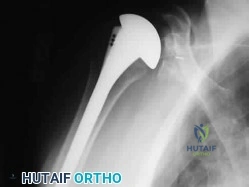
A 78-year-old female with severe, long-standing rheumatoid arthritis sustains a comminuted distal humerus fracture. A linked (semi-constrained) total elbow arthroplasty (TEA) is planned. What is the primary biomechanical advantage of utilizing a linked prosthesis over an unlinked prosthesis in this scenario?
Explanation
A linked (semi-constrained) TEA features a mechanical linkage between the humeral and ulnar components, providing intrinsic stability. This is highly advantageous in patients with poor bone stock or deficient collateral ligaments (such as in severe rheumatoid arthritis or trauma), preventing postoperative dislocation.
Question 14
A 50-year-old diabetic female presents with an insidious onset of severe shoulder pain and progressive stiffness over the last 4 months. Which of the following physical examination findings is considered pathognomonic for idiopathic adhesive capsulitis?
Explanation
The hallmark and pathognomonic physical exam finding in adhesive capsulitis (frozen shoulder) is a marked reduction in passive external rotation with the patient's arm resting at their side. This specific restriction is primarily caused by contracture and thickening of the coracohumeral ligament and the rotator interval.
Question 15
A 40-year-old male experiences a seizure and subsequently presents with a locked posterior shoulder dislocation. Imaging reveals a 30% anterior articular surface defect (reverse Hill-Sachs lesion). Which of the following surgical procedures is most appropriate to prevent recurrent engagement of this specific defect?
Explanation
For a locked posterior dislocation with an anteromedial humeral head defect (reverse Hill-Sachs or McLaughlin lesion) involving between 20% and 40% of the articular surface, the modified McLaughlin procedure is indicated. This involves transferring the lesser tuberosity (with its attached subscapularis) into the defect to restore stability. The classic McLaughlin involves only the subscapularis tendon.
Question 16
A 32-year-old male sustains a simple transverse fracture of the olecranon and undergoes tension band wiring. Which of the following biomechanical principles correctly describes how this construct facilitates fracture healing?
Explanation
The tension band wiring technique is ideal for simple transverse olecranon fractures. It follows the biomechanical principle of placing the implant on the tension side (the posterior cortex). As the triceps pulls during elbow flexion, the tensile distraction forces are converted by the wire into dynamic compression forces across the anterior articular surface.
Question 17
A 45-year-old female presents after a fall with a complex elbow fracture. Advanced imaging identifies a coronal shear fracture that involves the capitellum and extends medially to include most of the trochlea. According to the modified Bryan and Morrey classification (McKee modification), what type of fracture is this?
Explanation
In the Bryan and Morrey classification for capitellum fractures: Type I is a large osseous piece (Hahn-Steinthal), Type II is a sleeve of cartilage with minimal bone (Kocher-Lorenz), and Type III is comminuted. McKee added the Type IV fracture, which is a massive coronal shear fracture encompassing the capitellum and extending medially to involve the majority of the trochlea.
Question 18
A 12-year-old elite baseball pitcher presents with medial elbow pain that worsens during the acceleration phase of throwing. Radiographs demonstrate widening of the medial epicondyle apophysis. Which muscle group is the primary deforming force contributing to this condition?
Explanation
'Little League Elbow' is a traction apophysitis of the medial epicondyle caused by repetitive valgus overload during throwing. The primary deforming forces pulling on the medial epicondyle apophysis are the flexor-pronator mass and the ulnar collateral ligament.
Question 19
A 60-year-old male with severe cubital tunnel syndrome presents with noticeable intrinsic muscle wasting in his hand. When asked to firmly pinch a piece of paper between his thumb and index finger, his thumb interphalangeal joint strongly flexes. This compensatory finding (Froment's sign) is driven by which of the following muscles?
Explanation
Froment's sign is positive when the patient compensates for a weak adductor pollicis (ulnar nerve innervation) by heavily recruiting the flexor pollicis longus (anterior interosseous nerve/median nerve innervation) to maintain pinch grip, resulting in hyperflexion of the thumb interphalangeal joint.
Question 20
During a submuscular anterior transposition of the ulnar nerve, the surgeon must diligently release all potential sites of compression. One such structure is the Arcade of Struthers. Which of the following accurately describes the anatomical location of this structure?
Explanation
The Arcade of Struthers is a fascial band extending from the medial head of the triceps to the medial intermuscular septum. It is located approximately 8 cm proximal to the medial epicondyle and is a critical potential site of ulnar nerve compression, especially if not released during anterior transposition.
Question 21
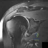
A 42-year-old man presents with acute, severe, unrelenting right shoulder pain that began 3 weeks ago without any antecedent trauma. The pain lasted for 10 days and has since significantly improved, but he now notices profound weakness when trying to elevate his arm or externally rotate it. Physical examination reveals prominent atrophy of the supraspinatus and infraspinatus fossae. MRI of the shoulder reveals no rotator cuff tears, but shows diffuse T2 hyperintensity and edema within the supraspinatus and infraspinatus muscles. What is the most likely diagnosis?
Explanation
Parsonage-Turner syndrome (idiopathic brachial neuritis) typically presents with an acute phase of severe shoulder girdle pain followed days to weeks later by painless weakness and muscle atrophy. The suprascapular nerve and anterior interosseous nerve are most commonly affected. MRI will show neurogenic edema (T2 hyperintensity) in the acutely denervated muscles without a structural tendon tear. Spinoglenoid notch entrapment would spare the supraspinatus.
Question 22
Scapular notching is a well-recognized complication following Reverse Total Shoulder Arthroplasty (RTSA). Based on modern biomechanical principles and implant design modifications, which of the following component positioning strategies is most effective in minimizing the risk of inferior scapular notching?
Explanation
Inferior scapular notching occurs due to mechanical impingement of the humeral component against the scapular neck during adduction. Inferior translation (placing the baseplate low on the glenoid) and inferior tilt of the glenosphere help to clear the inferior scapular pillar, thereby reducing the incidence of notching. Lateralization of the center of rotation and using a smaller humeral neck-shaft angle (e.g., 135-145 degrees) also decrease notching.
Question 23
A 28-year-old competitive weightlifter feels a sudden 'pop' and tearing sensation in his anterior chest wall while performing a heavy bench press. Examination reveals loss of the normal anterior axillary fold contour and weakness with internal rotation. In an acute pectoralis major rupture, which head is structurally most vulnerable and typically ruptures first?
Explanation
The sternal head of the pectoralis major undergoes a 180-degree twist before inserting on the humerus, causing its fibers to insert deep and proximal to the clavicular head fibers. During a bench press (arm in extension and abduction), the inferior fibers of the sternal head are under maximal tension and stretch, making them mechanically vulnerable and typically the first to rupture.
Question 24
A 35-year-old overhead athlete complains of chronic, vague posterior shoulder pain and numbness over the lateral deltoid. Physical examination demonstrates point tenderness at the posterior aspect of the shoulder, inferior to the teres minor and lateral to the long head of the triceps. Which neurovascular bundle is most likely entrapped in this anatomic space?
Explanation
The patient is describing Quadrilateral Space Syndrome. The quadrilateral space is bordered superiorly by the teres minor, inferiorly by the teres major, medially by the long head of the triceps, and laterally by the humeral shaft. Entrapment of the axillary nerve and posterior circumflex humeral artery in this space (often by fibrous bands) leads to posterior shoulder pain and deltoid paresthesias.
Question 25
A 60-year-old active laborer presents with a massive, retracted, and irreparable posterosuperior rotator cuff tear. He has significant weakness in external rotation and a positive external rotation lag sign, but intact anterior elevation. He wishes to avoid arthroplasty. Which of the following tendon transfers is biomechanically superior for restoring active external rotation and closely replicates the force vector of the infraspinatus?
Explanation
The lower trapezius transfer (often augmented with an Achilles tendon allograft) has gained popularity for massive irreparable posterosuperior cuff tears. Biomechanically, the lower trapezius vector matches the line of pull of the infraspinatus much more closely than the latissimus dorsi, making it superior for restoring active external rotation and minimizing out-of-phase muscle retraining.
Question 26
A 45-year-old man requires operative repair of a complete acute distal biceps tendon rupture. The surgeon must choose between a single-incision anterior approach and a two-incision approach. Compared to the single-incision technique, the two-incision approach has a historically higher risk of which of the following complications?
Explanation
The two-incision approach (modified Boyd-Anderson) was developed to limit radial nerve injury, but dissection between the radius and ulna significantly increases the risk of heterotopic ossification and radioulnar synostosis compared to a single anterior incision. The single-incision approach has a higher rate of lateral antebrachial cutaneous nerve (LABCN) neuropraxia due to anterior retraction.
Question 27
A 30-year-old patient presents with a 'terrible triad' injury of the elbow following a fall onto an outstretched hand. The injury involves an elbow dislocation, a radial head fracture, and a coronoid process fracture. According to standard surgical protocols, what is the most widely accepted sequence of structural repair to restore elbow stability?
Explanation
The standard inside-out surgical sequence for a terrible triad injury of the elbow is: 1. Fixation of the coronoid fracture (or reattachment of the anterior capsule), 2. Fixation or replacement of the radial head, 3. Repair of the lateral collateral ligament (LUCL) to the lateral epicondyle. The medial collateral ligament (MCL) is only repaired or a hinged external fixator applied if the elbow remains unstable in extension after the lateral side is secured.
Question 28
A 40-year-old female presents with a 6-month history of aching pain in her proximal lateral forearm. Physical examination reveals no wrist or finger drop, but she has focal tenderness 4 cm distal to the lateral epicondyle. Pain is exacerbated by resisted extension of the middle finger with the elbow extended. In Radial Tunnel Syndrome, what is the most common anatomic site of nerve compression?
Explanation
The patient's presentation is classic for Radial Tunnel Syndrome (compression of the posterior interosseous nerve without significant motor loss). While compression can occur at the fibrous bands over the radiocapitellar joint, the Leash of Henry, or the ECRB margin, the most frequent site of PIN compression is the proximal tendinous arch of the supinator, known as the Arcade of Frohse.
Question 29
During elbow arthroscopy, establishing standard portals requires precise knowledge of regional neurovascular anatomy. When creating the standard anteromedial portal, which neural structure is at the greatest risk of injury due to its close proximity (often 1-2 mm) to the arthroscopic trocar?
Explanation
The medial antebrachial cutaneous nerve (MABCN) is the structure at greatest risk during the establishment of the anteromedial portal in elbow arthroscopy, often passing within 1-2 mm of the portal site. The ulnar nerve is protected posterior to the medial epicondyle (unless subluxated), and the median nerve is generally located further laterally, safely away from the portal if established properly.
Question 30
A 24-year-old motorcyclist is involved in a high-speed collision and presents with massive soft tissue swelling around the shoulder girdle. AP chest radiograph shows severe lateral displacement of the scapula relative to the spinous processes and a widely displaced clavicle fracture. Examination shows a pulseless, flail upper extremity. In the setting of this suspected scapulothoracic dissociation, what is the most reliable prognostic indicator for long-term functional recovery of the limb?
Explanation
Scapulothoracic dissociation is a devastating, high-energy injury characterized by lateral displacement of the scapula and is highly associated with severe vascular (subclavian/axillary artery) and neurological injuries. While vascular injury is an acute limb-threatening emergency, the long-term functional outcome of the limb is almost entirely dependent on the severity of the brachial plexus injury (often complete avulsion), which dictates whether the limb will function or remain a flail, insensate appendage.
Question 31
An 18-year-old male presents to the emergency department after a football tackle complaining of severe pain at the base of his neck. He is experiencing mild stridor and a choking sensation. Clinical exam and CT imaging reveal a posterior displacement of the medial clavicle. Given the patient's age and clinical presentation, what is the most accurate anatomic description of this injury?
Explanation
The medial clavicular epiphysis is the last physis in the body to fuse, typically closing between 22 and 25 years of age. Therefore, what appears clinically and radiographically as a 'sternoclavicular joint dislocation' in a patient under age 22 is almost always a Salter-Harris type I or II physeal fracture of the medial clavicle, with the epiphysis remaining in the sternoclavicular joint.
Question 32
A 21-year-old collegiate baseball pitcher undergoes Medial Ulnar Collateral Ligament (MUCL) reconstruction (Tommy John surgery). The surgeon must anatomically restore the primary restraint to valgus stress at the elbow. Which bundle of the MUCL is being reconstructed, and what is its role in elbow kinematics?
Explanation
The anterior bundle of the MUCL originates on the anteroinferior surface of the medial epicondyle and inserts on the sublime tubercle of the ulna. It is the primary restraint to valgus stress at the elbow between 30 and 120 degrees of flexion. The posterior bundle is a secondary restraint, and the transverse (Cooper's) ligament contributes little to elbow stability.
Question 33
A 48-year-old man presents with painful swelling at the posterior aspect of both elbows and inability to actively extend against resistance. He reports a sudden 'tearing' sensation bilaterally while attempting to break a fall. MRI confirms bilateral triceps tendon ruptures. Which of the following systemic medical conditions is classically most associated with bilateral spontaneous triceps tendon ruptures?
Explanation
While unilateral triceps ruptures are generally traumatic (e.g., weightlifting), bilateral simultaneous triceps tendon ruptures are rare and highly associated with underlying systemic conditions that weaken the tendon. The classic board-tested association for bilateral spontaneous triceps rupture is secondary hyperparathyroidism (renal osteodystrophy), which causes pathologic changes at the bone-tendon junction. Other risks include chronic corticosteroid use, anabolic steroids, and fluoroquinolones.
Question 34
A 52-year-old female with poorly controlled type 1 diabetes presents with severe restriction of active and passive range of motion of her right shoulder, consistent with adhesive capsulitis (frozen shoulder). Pathophysiologically, the joint capsule in this condition demonstrates dense fibroblastic proliferation. Which of the following cytokines/growth factors is most heavily implicated in driving the fibrogenic cascade in adhesive capsulitis?
Explanation
Adhesive capsulitis is characterized by chronic inflammation leading to dense capsular fibrosis. Cytokine profiling of the capsule in patients with frozen shoulder shows significantly elevated levels of Transforming Growth Factor-beta (TGF-beta) and Platelet-Derived Growth Factor (PDGF). TGF-beta is considered the primary driver of the fibrotic response, stimulating fibroblasts to produce type I and type III collagen.
Question 35
A 34-year-old female sustains a coronal shear fracture of the distal humerus. Imaging demonstrates that the fracture involves both the capitellum and the lateral half of the trochlea in a single contiguous articular fragment. According to the Dubberley classification, this represents a Type 3 injury. What specific radiographic feature differentiates a Dubberley Type 3B from a Type 3A injury?
Explanation
The Dubberley classification of coronal shear fractures divides them by extent: Type 1 involves the capitellum only, Type 2 includes the capitellum and the lateral trochlear ridge, and Type 3 involves the capitellum and trochlea as a single piece. The modifier A or B is added based on the absence (A) or presence (B) of posterior condylar comminution, which is critical because Type B injuries lack posterior cortical buttressing and often require specialized plating.
Question 36
A 28-year-old male describes a sensation of his elbow 'clicking and giving way' when he tries to push himself up from an armchair. Physical exam reveals apprehension and a clunk with a supination, valgus, and axial compression load applied to the elbow as it is moved from extension to flexion. What is the primary ligamentous restraint that is deficient, and what is its anatomic insertion on the ulna?
Explanation
The patient is presenting with Posterolateral Rotatory Instability (PLRI) of the elbow. The primary deficient structure is the Lateral Ulnar Collateral Ligament (LUCL). The LUCL originates from the lateral epicondyle, blends with the annular ligament, and inserts on the supinator crest of the proximal ulna. Its incompetence allows the radial head and proximal ulna to subluxate posterolaterally away from the humerus.
Question 37
A 26-year-old man suffered a severe pan-brachial plexus avulsion injury 2 years ago and has no functional recovery of shoulder or elbow musculature, though he has undergone successful distal nerve transfers to restore rudimentary hand grip. He is scheduled for a shoulder arthrodesis to provide a stable proximal platform to aid in hand-to-mouth function. According to modern recommendations, what is the ideal position for glenohumeral fusion?
Explanation
The currently recommended, standard functional position for shoulder arthrodesis is approximately 30 degrees of flexion, 30 degrees of abduction, and 30 degrees of internal rotation (the '30-30-30' rule). This position allows the hand to reach the midline and mouth for feeding while enabling the arm to rest comfortably at the side when relaxed. Older recommendations (Rowe position) used more internal rotation and less abduction, but the 30-30-30 position optimizes modern activities of daily living.
Question 38
A 13-year-old elite Little League pitcher presents with progressive, activity-related right proximal humerus pain over the past 6 weeks. He denies any acute trauma. Plain radiographs reveal widening and irregularity of the proximal humeral physis compared to the contralateral side. What is the most appropriate initial management?
Explanation
The diagnosis is 'Little League Shoulder', which is an overuse injury resulting in a stress fracture of the proximal humeral physis (Salter-Harris type I). The standard of care is complete cessation of throwing for approximately 3 months (until radiographic and clinical resolution). Physical therapy should focus on core, periscapular, and lower extremity strengthening to correct kinetic chain deficits before initiating a structured return-to-throwing program.
Question 39
A 27-year-old professional volleyball attacker complains of subtle posterior shoulder pain and a subjective decrease in hitting power. Physical exam reveals normal active elevation, 5/5 strength in shoulder abduction, but notable weakness (3/5) in external rotation with the arm at the side. MRI of the shoulder is ordered. Based on the physical exam findings, where is the isolated neural compression most likely located, and what is the classic associated pathologic finding?
Explanation
The patient exhibits isolated weakness in external rotation (infraspinatus) with preserved abduction (supraspinatus). This indicates suprascapular nerve entrapment distal to the branches supplying the supraspinatus, specifically at the spinoglenoid notch. In overhead athletes, this is classically associated with a paralabral cyst extending from a posterior superior labral tear (SLAP lesion).
Question 40
A 45-year-old man falls from a ladder and sustains a highly comminuted, unsalvageable radial head fracture (Mason III). During evaluation, he is also noted to have intense pain at the wrist and extreme instability of the distal radioulnar joint (DRUJ), indicative of an Essex-Lopresti injury. If the surgeon simply excises the radial head without replacing it, what is the most profound long-term biomechanical consequence?
Explanation
An Essex-Lopresti injury involves a radial head fracture, rupture of the interosseous membrane (IOM), and disruption of the DRUJ. The radial head and IOM are the primary stabilizers against proximal migration of the radius. If the radial head is excised and not replaced in the setting of IOM disruption, the radius will migrate proximally. This results in significant positive ulnar variance, leading to severe ulnocarpal impaction syndrome and chronic wrist pain. Radial head arthroplasty is strictly required to maintain length.
Question 41
A 45-year-old female falls on her outstretched hand. Radiographs confirm an elbow dislocation, a comminuted radial head fracture, and a Type II coronoid fracture.
According to standard surgical protocols for this 'terrible triad' injury, what is the most appropriate sequence of repair to restore elbow stability?
According to standard surgical protocols for this 'terrible triad' injury, what is the most appropriate sequence of repair to restore elbow stability?
Explanation
The classic 'terrible triad' of the elbow includes an elbow dislocation, radial head fracture, and coronoid fracture. The widely accepted surgical protocol described by Pugh and McKee recommends repairing structures from deep to superficial, typically starting anteriorly: 1) Coronoid fixation or anterior capsule repair to restore the anterior buttress, 2) Radial head ORIF or arthroplasty, 3) LUCL repair to the lateral epicondyle, and 4) MCL repair or application of a hinged external fixator if the elbow remains unstable after the first three steps.
Question 42
When performing a lateral ulnar collateral ligament (LUCL) reconstruction for posterolateral rotatory instability (PLRI) of the elbow, accurate graft placement is critical. Where is the precise isometric origin of the LUCL on the lateral epicondyle to ensure stability and uniform tension throughout the elbow's range of motion?
Explanation
The isometric point for the origin of the lateral ulnar collateral ligament (LUCL) on the humerus is located at the center of the capitellum's axis of rotation. Placing the humeral tunnel at this exact point ensures that the reconstructed ligament maintains consistent tension and provides stability across the full arc of elbow flexion and extension.
Question 43
A 38-year-old bodybuilder sustains a distal biceps tendon rupture and undergoes a two-incision (Mayo) repair. Postoperatively, he presents with an inability to extend his fingers and thumb, though wrist extension is preserved but deviates radially. Sensation is intact. Which technical error most likely caused this complication?
Explanation
The patient has a posterior interosseous nerve (PIN) palsy, evidenced by the loss of finger/thumb extension and radial deviation with wrist extension (ECRL is intact via the radial nerve proper, but ECU is out). In the two-incision distal biceps repair, the PIN is at highest risk of injury during the posterolateral approach if retractors are placed too vigorously around the radial neck. Supinating the forearm moves the PIN further away from the surgical field during this step.
Question 44
A 29-year-old elite volleyball player presents with insidious onset of shoulder weakness. Physical examination reveals isolated atrophy of the infraspinatus fossa with normal bulk of the supraspinatus. Weakness is noted in external rotation. An MRI demonstrates a paralabral cyst. Where is the cyst most likely located, and what is the typical associated labral pathology?
Explanation
Isolated infraspinatus atrophy indicates compression of the suprascapular nerve distal to the innervation of the supraspinatus, which occurs at the spinoglenoid notch. Paralabral cysts in the spinoglenoid notch are classically associated with posterior or posterosuperior labral tears. Fluid is pumped through the labral defect into the notch, forming a cyst that compresses the suprascapular nerve.
Question 45
A 22-year-old male is involved in a high-speed motorcycle collision. On examination, he has massive swelling over the shoulder girdle and a complete lack of motor and sensory function in his left upper extremity. Radiographs demonstrate marked lateral displacement of the scapula. Which radiographic parameter is most strongly predictive of a major vascular injury in this condition?
Explanation
This patient has scapulothoracic dissociation, a devastating injury involving closed disruption of the scapulothoracic articulation. The normal scapulothoracic ratio (index) is 1.0. A scapulothoracic ratio greater than 1.07 indicates significant lateral displacement of the scapula and correlates strongly with severe brachial plexus injuries and major vascular trauma (subclavian or axillary artery tears) requiring emergent surgical exploration.
Question 46
A 65-year-old female with severe rheumatoid arthritis undergoes a linked (semi-constrained) total elbow arthroplasty (TEA).
Three years later, she develops rapid early polyethylene wear and aseptic loosening of the components. What is the most common iatrogenic cause of early bushing wear and failure in a linked TEA?
Three years later, she develops rapid early polyethylene wear and aseptic loosening of the components. What is the most common iatrogenic cause of early bushing wear and failure in a linked TEA?
Explanation
In a linked or semi-constrained total elbow arthroplasty, the prosthesis acts as a hinge that allows some varus/valgus toggle. If the humeral component is placed with its axis of rotation malaligned compared to the natural anatomic axis of the elbow, it creates a kinematic mismatch. This generates excessive stress on the polyethylene bushings during flexion and extension, leading to accelerated wear, osteolysis, and early aseptic loosening.
Question 47
A 45-year-old man receives a radial head replacement for a highly comminuted radial head fracture. Two years postoperatively, he complains of progressive lateral elbow pain and stiffness. Radiographs demonstrate widening of the lateral ulnohumeral joint space and erosive changes of the capitellum. What is the most likely cause of these findings?
Explanation
Using a radial head implant that is too thick 'overstuffs' the radiocapitellar joint. This causes increased contact pressures on the capitellum, leading to early cartilage wear, osteonecrosis, or capitellar erosion. It also artificially widens the lateral ulnohumeral joint space on radiographs and restricts elbow flexion and forearm rotation.
Question 48
A 32-year-old male weightlifter felt a sudden 'pop' and tearing sensation in his anterior axilla while performing a heavy bench press. Examination reveals loss of the anterior axillary fold contour. Operative repair is planned. Which of the following best describes the anatomic insertion of the most commonly ruptured segment of the pectoralis major tendon?
Explanation
Most pectoralis major ruptures occur at or near the humeral insertion and preferentially involve the sternal head. The pectoralis major tendon is bilaminar. As the sternal head courses laterally, it twists 180 degrees so that its fibers insert deep and proximal to the clavicular head. The clavicular head does not twist and inserts distally and superficial.
Question 49
During a Latarjet procedure for recurrent anterior shoulder instability, the coracoid process is transferred to the anterior glenoid neck. Postoperatively, the patient is unable to actively flex his elbow and reports decreased sensation over the lateral forearm. Which of the following surgical steps posed the greatest risk to the injured structure?
Explanation
The patient has a musculocutaneous nerve injury (loss of biceps/brachialis motor function and lateral antebrachial cutaneous sensation). The musculocutaneous nerve enters the deep surface of the coracobrachialis (part of the conjoint tendon) approximately 5-8 cm distal to the coracoid tip. Vigorous medial or inferior retraction of the conjoint tendon during the Latarjet procedure places this nerve at high risk of a traction neuropraxia.
Question 50
A 22-year-old male with recurrent anterior shoulder dislocations undergoes arthroscopic stabilization. Imaging reveals 20% glenoid bone loss and an engaging Hill-Sachs lesion. The surgeon performs a Bankart repair and a Remplissage procedure. Which anatomic structure is tenodesed into the Hill-Sachs defect, and what clinical restriction is most commonly observed postoperatively?
Explanation
The Remplissage procedure involves capsulotenodesis of the posterior capsule and the infraspinatus tendon into the Hill-Sachs defect. This effectively makes the defect extra-articular and prevents it from engaging the anterior glenoid rim. Because the posterior structures are tethered, the most common postoperative restriction is a mild to moderate decrease in external rotation.
Question 51
A 28-year-old offensive lineman presents with vague posterior shoulder pain that worsens during blocking maneuvers. The surgeon performs a Kim test to evaluate for a posterior labral tear. Which of the following accurately describes the correct execution of the Kim test?
Explanation
The Kim test evaluates for a posteroinferior labral tear. With the patient seated, the examiner holds the patient's elbow and the lateral aspect of the proximal humerus. The arm is abducted to 90 degrees, and a strong axial load is applied. The arm is then elevated 45 degrees diagonally upward while the examiner simultaneously applies a downward and posterior force to the proximal humerus. A sudden onset of posterior shoulder pain implies a posteroinferior labral tear.
Question 52
A 35-year-old female falls on an outstretched hand and sustains an isolated capitellar fracture.
According to the Dubberley classification of capitellum fractures, what finding distinguishes a Type 3 fracture from a Type 1 or 2, and what is its primary surgical implication?
According to the Dubberley classification of capitellum fractures, what finding distinguishes a Type 3 fracture from a Type 1 or 2, and what is its primary surgical implication?
Explanation
The Dubberley classification for capitellum and trochlea fractures is based on the presence of posterior comminution. Type 1 involves the capitellum, Type 2 involves the capitellum and trochlea as a single piece. A Type 3 fracture is distinguished by the presence of posterior comminution, which destroys the posterior buttress. This implies that simple anterior-to-posterior screw fixation will fail due to lack of posterior support, often necessitating bone grafting or posterior buttress plating.
Question 53
A 45-year-old male loses his footing and falls directly onto a flexed elbow. He is unable to actively extend his elbow against gravity, and a palpable gap is felt posteriorly. During operative repair of this distal triceps tendon rupture, understanding the native footprint is crucial. Which of the following describes the normal anatomic insertion of the triceps tendon on the olecranon?
Explanation
Anatomic studies demonstrate that the triceps tendon does not insert precisely at the tip of the olecranon. The footprint is broad and dome-shaped, beginning approximately 1.1 to 1.4 cm distal to the tip of the olecranon. Repairing the tendon to the absolute tip can lead to prominent hardware and abnormal kinematics.
Question 54
A 12-year-old competitive baseball pitcher presents with medial elbow pain that worsens during pitching. Radiographs show widening and irregularity of the medial epicondyle apophysis. What is the primary pathophysiologic mechanism causing 'Little League Elbow' in this patient population?
Explanation
'Little League Elbow' represents a spectrum of medial elbow injuries in overhead throwers, most commonly medial epicondyle apophysitis in skeletally immature patients. The pitching motion, particularly during the late cocking and early acceleration phases, generates tremendous valgus stress on the elbow. This results in tension-overload on the medial structures (causing apophysitis or avulsion) and compression on the lateral structures (radiocapitellar joint, potentially leading to osteochondritis dissecans of the capitellum).
Question 55
A 40-year-old female complains of a vague, aching pain in her proximal volar forearm. She reports numbness in the radial three and a half digits. Phalen's test and Tinel's sign at the carpal tunnel are negative. She experiences significant pain reproduction upon resisted pronation of the forearm while her elbow is held in full extension. Which structure is most likely causing the nerve compression?
Explanation
This patient has Pronator Syndrome, a proximal median neuropathy. The provocative maneuvers can help isolate the site of compression. Resisted forearm pronation with the elbow extended tests the two heads of the pronator teres. Resisted elbow flexion in supination isolates the bicipital aponeurosis (lacertus fibrosus). Resisted flexion of the PIP joint of the middle finger implicates the FDS fibrous arch.
Question 56
A 6-year-old child presents after a fall from monkey bars. Radiographs reveal a fracture of the proximal third of the ulnar diaphysis with an anterior dislocation of the radial head.
According to the Bado classification, what type of Monteggia lesion is this, and what is the classically described mechanism of injury?
According to the Bado classification, what type of Monteggia lesion is this, and what is the classically described mechanism of injury?
Explanation
The Bado classification categorizes Monteggia fracture-dislocations based on the direction of radial head dislocation. Type I (most common) is an anterior radial head dislocation with an anteriorly angulated ulnar fracture. The classically described mechanism for a Type I lesion is an excessive forced hyperpronation injury (or hyperextension).
Question 57
A 45-year-old male presents with sudden inability to make an 'OK' sign with his thumb and index finger following an episode of viral neuritis. He has normal sensation throughout the hand and forearm. Which of the following muscle groups is innervated by the specific nerve involved in this condition?
Explanation
The inability to make the 'OK' sign implies weakness of the flexor pollicis longus (FPL) and flexor digitorum profundus (FDP) to the index finger, resulting in a pinch using the pads of the digits (Mannerfelt's sign). This is characteristic of Anterior Interosseous Nerve (AIN) syndrome. The AIN is a pure motor branch of the median nerve that innervates the FPL, the radial half of the FDP (index and middle fingers), and the pronator quadratus.
Question 58
A 25-year-old female presents with severe, painful crepitus and snapping at the superomedial border of her scapula during active arm elevation. Extensive non-operative management has failed to provide relief. If an arthroscopic excision of the superomedial angle of the scapula is performed, which muscle's insertion must be carefully elevated and subsequently securely repaired?
Explanation
Snapping scapula syndrome is often caused by friction between the superomedial angle of the scapula and the underlying rib cage. Surgical treatment involves resecting the superomedial angle of the scapula and bursectomy. The levator scapulae muscle inserts onto the superomedial angle of the scapula. During the resection, this insertion is elevated and must be repaired back to the scapular border to prevent functional deficits.
Question 59
A 30-year-old male with a devastating global brachial plexus injury (C5, C6, C7 root avulsions) is undergoing a shoulder arthrodesis to stabilize his shoulder and allow better positioning for his remaining hand/elbow function. According to current biomechanical and functional literature, what is the optimal position for glenohumeral arthrodesis?
Explanation
The optimal position for shoulder arthrodesis is a classic 'Rule of 30s': approximately 30° of abduction, 30° of forward flexion, and 30° of internal rotation. This position allows the hand to reach the mouth (crucial for feeding and hygiene) and minimizes winging of the scapula while keeping the arm close enough to the body to pass through doorways.
Question 60
A 35-year-old male sustains a closed, extra-articular fracture of the distal third of the humeral shaft (Holstein-Lewis fracture). On initial presentation in the emergency department, examination reveals a complete radial nerve palsy. According to current orthopaedic guidelines, what is the most appropriate initial management for the nerve palsy in this specific scenario?
Explanation
In the setting of a closed humeral shaft fracture, even a Holstein-Lewis type, presenting with a primary radial nerve palsy, the standard of care remains initial observation of the nerve and conservative fracture management (e.g., coaptation splint transitioning to a functional brace). Most palsies are neuropraxias that will resolve spontaneously. Immediate exploration is indicated for open fractures, severe vascular compromise, or if the nerve palsy develops *after* closed reduction attempts (secondary palsy).
Question 61
A 12-year-old boy presents after falling onto an outstretched hand. He was found to have an elbow dislocation, which was closed reduced in the emergency department. Post-reduction, the elbow joint appears congruent, but the medial epicondyle is no longer visible in its anatomic position. A radiograph reveals a bone fragment within the joint space.
Which of the following is the most appropriate management for this patient?
Which of the following is the most appropriate management for this patient?
Explanation
An incarcerated medial epicondyle fragment within the elbow joint after reduction of a dislocation is an absolute indication for operative intervention. The fragment is typically tethered by the flexor-pronator mass and the medial collateral ligament. Attempting to mobilize the elbow with an intra-articular fragment will lead to rapid cartilage destruction and mechanical block. Open reduction and internal fixation (ORIF) allows for joint clearance, anatomic restoration of the flexor-pronator origin, and stabilization of the MCL.
Question 62
During open reduction and internal fixation of a highly comminuted capitellum fracture, excessive posterior soft tissue stripping is performed. The patient subsequently develops avascular necrosis (AVN) of the capitellum. Which of the following best describes the anatomical basis for this complication?
Explanation
The capitellum is primarily supplied by penetrating end-vessels that enter the bone posteriorly through the capsular attachments. Because the articular surface covers the anterior, inferior, and lateral aspects of the capitellum, there is no direct anterior vascular supply. Aggressive posterior dissection or stripping of the posterior soft tissues during surgical approaches (such as an extended lateral approach) significantly increases the risk of avascular necrosis.
Question 63
A 35-year-old male presents with sudden-onset, severe right shoulder pain that awakened him from sleep. The acute severe pain lasted for 5 days and then gradually subsided, but was immediately followed by profound weakness in shoulder elevation and external rotation. An MRI of the cervical spine and shoulder is unremarkable.
What is the most likely diagnosis?
What is the most likely diagnosis?
Explanation
Parsonage-Turner Syndrome (acute brachial neuritis) classically presents with severe, unrelenting, acute shoulder pain (often worse at night) lasting days to weeks, followed by patchy, profound weakness and muscle atrophy (most commonly affecting the suprascapular, axillary, or long thoracic nerves) as the pain subsides. It is often preceded by a viral illness or vaccination. MRI is typically normal initially but may show denervation edema in the affected muscles later. Treatment is primarily conservative with NSAIDs, steroids, and physical therapy.
Question 64
A professional baseball pitcher presents with vague posterior shoulder pain during the late cocking phase of throwing. Physical examination reveals an internal rotation deficit of 25 degrees compared to the non-dominant shoulder, while external rotation is increased by 10 degrees. Which of the following pathomechanical processes is most directly responsible for this glenohumeral internal rotation deficit (GIRD)?
Explanation
Glenohumeral internal rotation deficit (GIRD) in overhead throwers is primarily driven by contracture and thickening of the posteroinferior capsule. This contracture shifts the glenohumeral contact point posterosuperiorly during the late cocking phase (abduction and maximal external rotation), leading to increased shear stress on the superior labrum (peel-back mechanism) and internal impingement of the articular-sided rotator cuff against the posterosuperior glenoid.
Question 65
A 68-year-old female with advanced rheumatoid arthritis undergoes a primary linked semi-constrained total elbow arthroplasty (TEA).
Following comprehensive postoperative rehabilitation, what specific lifelong functional limitation must she be advised to adhere to in order to minimize the risk of aseptic loosening?
Following comprehensive postoperative rehabilitation, what specific lifelong functional limitation must she be advised to adhere to in order to minimize the risk of aseptic loosening?
Explanation
Patients who undergo total elbow arthroplasty (TEA) are traditionally placed on lifelong lifting restrictions to prevent accelerated polyethylene wear, bushing failure, and aseptic loosening, which remain the most common long-term complications. The standard recommendation is a lifting limit of 5 to 10 pounds for a single event and 1 to 2 pounds for repetitive lifting.
Question 66
A 28-year-old manual laborer complains of a painful grating sensation and audible popping at the superomedial border of his right scapula with overhead activities. Nonoperative management, including targeted periscapular strengthening and corticosteroid injections, has failed after 6 months. If surgical excision of the inflamed bursae is performed, which of the following bursae are specifically targeted in this region?
Explanation
Snapping scapula syndrome (scapulothoracic crepitus) often involves inflammation of the bursae located at the superomedial angle of the scapula. The two primary bursae in this specific anatomic location that become pathologic and require excision in refractory soft-tissue cases are the supraserratus bursa (located between the subscapularis and serratus anterior) and the infraserratus bursa (located between the serratus anterior and the chest wall).
Question 67
A 40-year-old female presents with a highly comminuted, un-reconstructable radial head fracture and distal radioulnar joint (DRUJ) instability after a high-energy fall.
What is the primary reason why simple radial head excision is strictly contraindicated in this specific injury pattern?
What is the primary reason why simple radial head excision is strictly contraindicated in this specific injury pattern?
Explanation
This patient has an Essex-Lopresti injury, characterized by a radial head fracture, rupture of the interosseous membrane (IOM), and disruption of the DRUJ. The interosseous membrane and the radial head act as the primary and secondary longitudinal stabilizers of the forearm. If the radial head is excised in the setting of an IOM tear, the radius will migrate proximally, resulting in ulnar-positive variance, chronic DRUJ pain, and functional impairment. Radial head arthroplasty is mandatory in this scenario.
Question 68
During the surgical management of a 'Terrible Triad' injury of the elbow, the lateral ulnar collateral ligament (LUCL) is repaired to the humerus using suture anchors.
To ensure isometric function of the LUCL throughout the arc of elbow flexion and extension, where must the humeral anchor be placed?
To ensure isometric function of the LUCL throughout the arc of elbow flexion and extension, where must the humeral anchor be placed?
Explanation
The isometric point for the origin of the lateral collateral ligament complex (specifically the LUCL) is located at the center of the axis of rotation of the capitellum on the lateral epicondyle. Repairing the ligament at this exact center of rotation ensures that the ligament maintains consistent tension throughout the entire arc of elbow flexion and extension, minimizing the risk of recurrent instability or severe stiffness.
Question 69
A 32-year-old elite volleyball player presents with progressive, isolated weakness in external rotation of the shoulder. He denies any acute trauma but reports deep posterior shoulder pain. Clinical examination reveals profound atrophy of the infraspinatus fossa but normal bulk and strength of the supraspinatus. Which of the following intra-articular pathologies is most commonly associated with this specific nerve entrapment syndrome?
Explanation
Isolated infraspinatus weakness with atrophy suggests compression of the suprascapular nerve at the spinoglenoid notch (after it has already supplied motor branches to the supraspinatus). The most common cause in an overhead athlete is a paralabral ganglion cyst. These cysts are highly associated with posterior or posterosuperior labral tears, which act as a one-way valve allowing synovial fluid to form the cyst in the spinoglenoid notch.
Question 70
A 13-year-old male baseball pitcher complains of right shoulder pain that worsens when throwing hard. He has localized tenderness over the lateral aspect of the proximal humerus. Radiographs reveal widening of the proximal humeral physis compared to the contralateral side.
What is the recommended initial management for this condition?
What is the recommended initial management for this condition?
Explanation
This clinical picture describes 'Little League Shoulder', which is a proximal humeral epiphysiolysis (a stress fracture through the physis, Salter-Harris Type I) caused by repetitive rotational torque during the throwing motion. The gold standard treatment is absolute rest from throwing, typically for a minimum of 3 months, until clinical symptoms resolve and radiographs demonstrate physeal healing. Surgery is almost never indicated.
Question 71
A 30-year-old male presents with severe chest wall bruising, loss of the anterior axillary fold, and weakness in internal rotation and adduction after feeling a "pop" while performing a heavy bench press.
During surgical repair of the ruptured pectoralis major tendon, the surgeon isolates the torn sternocostal head. Relative to the clavicular head footprint on the humerus, where is the anatomic footprint of the sternocostal head located?
During surgical repair of the ruptured pectoralis major tendon, the surgeon isolates the torn sternocostal head. Relative to the clavicular head footprint on the humerus, where is the anatomic footprint of the sternocostal head located?
Explanation
The pectoralis major tendon undergoes a 180-degree twist as it inserts onto the lateral lip of the bicipital groove of the humerus. Due to this twist, the sternocostal head (which originates inferiorly on the chest) crosses deep to the clavicular head and inserts deep and proximal (superior) on the humerus. Pectoralis major ruptures most commonly occur at the sternocostal head insertion during maximal eccentric contraction (e.g., bench pressing).
Question 72
A 22-year-old collegiate baseball pitcher is undergoing evaluation for medial elbow pain.
The examiner places the patient's shoulder in 90 degrees of abduction and external rotation, rapidly extends the elbow from full flexion to 30 degrees, and applies a valgus torque. The patient reports a sharp pain specifically between 120 and 70 degrees of elbow flexion. This clinical test (Moving Valgus Stress Test) evaluates the integrity of which specific structure?
The examiner places the patient's shoulder in 90 degrees of abduction and external rotation, rapidly extends the elbow from full flexion to 30 degrees, and applies a valgus torque. The patient reports a sharp pain specifically between 120 and 70 degrees of elbow flexion. This clinical test (Moving Valgus Stress Test) evaluates the integrity of which specific structure?
Explanation
The Moving Valgus Stress Test is highly sensitive and specific for insufficiency of the anterior bundle of the ulnar collateral ligament (UCL), which is the primary restraint to valgus stress at the elbow. A positive test is indicated by medial elbow pain reproduced at the shear range of 120 to 70 degrees of elbow flexion as the elbow is rapidly extended with applied valgus torque.
Question 73
A patient presents with poorly localized shoulder pain and paresthesias over the lateral deltoid that worsen when the arm is abducted and externally rotated. An MRI reveals fibrous bands compressing structures within the quadrilateral space. Which of the following correctly defines the anatomic borders of the quadrilateral space?
Explanation
The boundaries of the quadrilateral space are the teres minor (superiorly), teres major (inferiorly), the long head of the triceps (medially), and the surgical neck of the humerus (laterally). Quadrilateral space syndrome results from compression of the axillary nerve and the posterior humeral circumflex artery within this space, often by anomalous fibrous bands.
Question 74
A 65-year-old sedentary male with a massive, irreparable rotator cuff tear and severe biceps tendinopathy is scheduled for surgery.
The surgeon discusses performing a biceps tenotomy versus a biceps tenodesis. According to highest-level current evidence (systematic reviews and meta-analyses), what is the primary expected difference in clinical outcome between these two procedures in this demographic?
The surgeon discusses performing a biceps tenotomy versus a biceps tenodesis. According to highest-level current evidence (systematic reviews and meta-analyses), what is the primary expected difference in clinical outcome between these two procedures in this demographic?
Explanation
Current evidence demonstrates no significant difference in objective functional shoulder scores (Constant, ASES) or objective strength between biceps tenotomy and tenodesis, particularly in older, less active patients. However, tenotomy is consistently associated with a higher incidence of cosmetic 'Popeye' deformity (up to 20-30%) and subjective biceps cramping compared to tenodesis. Tenodesis requires slightly longer operative/rehab time but lowers the risk of deformity.
Question 75
A 45-year-old mechanic presents with an inability to actively extend his thumb and the fingers at the metacarpophalangeal (MCP) joints.
Wrist extension is preserved but distinctly deviates radially. Sensation in the hand and forearm is entirely normal. Which of the following structures is the most frequent site of compression causing this exact clinical picture?
Wrist extension is preserved but distinctly deviates radially. Sensation in the hand and forearm is entirely normal. Which of the following structures is the most frequent site of compression causing this exact clinical picture?
Explanation
This is a classic presentation of Posterior Interosseous Nerve (PIN) syndrome. The PIN is a purely motor nerve after it branches from the radial nerve proper. Compression classically occurs at the Arcade of Frohse (proximal edge of the superficial head of the supinator). It leads to paralysis of the EDC, EIP, EDM, EPL, EPB, and APL. Wrist extension is preserved (but deviates radially) because the extensor carpi radialis longus (ECRL) and brevis (ECRB) are typically innervated by the radial nerve proximal to the bifurcation or arcade.
Question 76
A 6-year-old girl falls off monkey bars and presents with elbow swelling and forearm deformity. Radiographs demonstrate an anterior dislocation of the radial head and a fracture of the ulnar diaphysis with anterior apex angulation. Which of the following is the standard initial treatment principle for achieving and maintaining reduction of the radial head in this specific fracture-dislocation (Bado Type I)?
Explanation
This describes a Bado Type I Monteggia fracture-dislocation (anterior dislocation of radial head, anterior angulation of ulnar shaft). The cardinal principle of treating Monteggia fractures is that anatomic restoration of ulnar length and alignment will almost always spontaneously reduce the radial head. If the radial head remains dislocated after anatomic ulnar fixation, one must suspect soft-tissue interposition (such as the annular ligament or joint capsule) requiring open reduction.
Question 77
A 35-year-old male suffered a severe traumatic brain injury and an ipsilateral elbow fracture-dislocation 8 months ago.
He now presents with a rigid elbow contracture secondary to massive heterotopic ossification (HO). Before proceeding with surgical excision of the HO and capsular release, which of the following criteria is considered the most reliable indicator that the HO is "mature" enough to minimize the risk of massive postoperative recurrence?
He now presents with a rigid elbow contracture secondary to massive heterotopic ossification (HO). Before proceeding with surgical excision of the HO and capsular release, which of the following criteria is considered the most reliable indicator that the HO is "mature" enough to minimize the risk of massive postoperative recurrence?
Explanation
Timing of heterotopic ossification (HO) excision is critical to prevent recurrence. Historically, surgeons waited for normalization of serum alkaline phosphatase or bone scan activity, but recent literature confirms that the most reliable practical indicator for maturation is radiographic appearance: the HO should have sharp, distinct cortical margins and a well-defined trabecular pattern (typically takes 6 months). Fluffy or cloud-like margins indicate active, immature bone formation.
Question 78
A 45-year-old male weightlifter sustains an acute distal biceps tendon rupture. He undergoes operative repair via a single-incision anterior approach using cortical button fixation.
Postoperatively, he complains of an area of numbness and tingling extending down the radial (lateral) aspect of his forearm. Which nerve is most likely injured, and what specific surgical maneuver places it at highest risk?
Postoperatively, he complains of an area of numbness and tingling extending down the radial (lateral) aspect of his forearm. Which nerve is most likely injured, and what specific surgical maneuver places it at highest risk?
Explanation
The Lateral Antebrachial Cutaneous Nerve (LABCN), which is the terminal sensory branch of the musculocutaneous nerve, exits lateral to the biceps tendon in the distal arm. It is highly susceptible to neuropraxia during a single-incision anterior approach to the distal biceps due to aggressive lateral retraction of the superficial soft tissues. It provides sensation to the radial/lateral aspect of the forearm.
Question 79
A 24-year-old motorcyclist is brought to the trauma bay after a high-speed collision. He presents with massive swelling over the shoulder girdle and a pulseless, flail upper extremity. Radiographs show significant lateral displacement of the scapula relative to the thoracic spine and an intact clavicle. Which of the following associated injuries is the most critical determinant of the long-term functional salvageability of this limb?
Explanation
This patient has a scapulothoracic dissociation, a severe injury caused by a massive lateral traction force resulting in complete disruption of the scapulothoracic articulation. It is highly associated with severe vascular (subclavian artery) and neurologic injuries. While vascular injury can often be repaired with a bypass graft, complete avulsion of the brachial plexus roots is a devastating, irreversible injury that leaves the arm completely flail and insensate. The severity of the brachial plexus injury is the primary prognostic factor for limb salvage, often ultimately necessitating early forequarter amputation if the plexus is avulsed.
Question 80
A 21-year-old collegiate javelin thrower presents with chronic posterior shoulder pain.
His pain is highly localized to the late cocking phase of his throwing motion. Arthroscopic evaluation reveals fraying of the posterosuperior labrum and an articular-sided, partial-thickness tear of the posterior supraspinatus and anterior infraspinatus tendons. Which of the following best describes the exact mechanism of 'internal impingement' occurring in this athlete?
His pain is highly localized to the late cocking phase of his throwing motion. Arthroscopic evaluation reveals fraying of the posterosuperior labrum and an articular-sided, partial-thickness tear of the posterior supraspinatus and anterior infraspinatus tendons. Which of the following best describes the exact mechanism of 'internal impingement' occurring in this athlete?
Explanation
Internal impingement is a pathologic condition seen almost exclusively in overhead athletes during the late cocking phase of throwing (extreme abduction and external rotation). In this position, the greater tuberosity rotates posteriorly and abuts against the posterosuperior glenoid labrum. This pinches the articular-sided fibers of the supraspinatus and infraspinatus tendons between the bone and the labrum, leading to 'kissing lesions': articular-sided rotator cuff tears and posterosuperior labral fraying/tearing.
Question 81
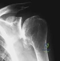
A 65-year-old female sustains a 4-part proximal humerus fracture. Which of the following radiographic findings is the strongest predictor of ensuing humeral head ischemia according to Hertel criteria?
Explanation
According to Hertel et al., a metaphyseal calcar length of < 8 mm attached to the articular segment, a disrupted medial hinge, and a basicervical fracture pattern are the most accurate predictors of ischemia. Of these, a calcar length < 8 mm has the highest positive predictive value for avascular necrosis.
Question 82
A 35-year-old overhead athlete presents with painless weakness in external rotation of the shoulder. Examination reveals atrophy isolated to the infraspinatus fossa. MRI demonstrates a paralabral cyst. At which of the following anatomic locations is the nerve compression most likely occurring?
Explanation
A paralabral cyst at the spinoglenoid notch compresses the suprascapular nerve after it has innervated the supraspinatus. This results in isolated infraspinatus atrophy and isolated weakness in external rotation without affecting the supraspinatus.
Question 83
A 40-year-old male sustains a 'terrible triad' injury of the elbow. During surgical reconstruction, what is the generally recommended sequence of repair to restore elbow stability?
Explanation
The standard surgical algorithm for a terrible triad injury begins deep and moves superficial: fixation or replacement of the coronoid first, followed by the radial head, and then repair of the lateral collateral ligament (LCL) complex. The medial collateral ligament (MCL) is only repaired if the elbow remains grossly unstable after these initial steps.
Question 84
A 28-year-old mechanic complains of right shoulder pain and weakness 4 months after a radical neck dissection. Examination reveals lateral winging of the scapula, with the scapula translated laterally and rotated downward. Which muscle-nerve combination is deficient, and what is the preferred salvage tendon transfer?
Explanation
Lateral winging with a downwardly rotated scapula following neck surgery suggests trapezius palsy secondary to a spinal accessory nerve injury. The Eden-Lange procedure, which transfers the levator scapulae and rhomboids, is the preferred reconstructive option for this condition.
Question 85
During the surgical reconstruction of a chronic type V acromioclavicular (AC) joint separation, the surgeon reconstructs the coracoclavicular (CC) ligaments. The native conoid ligament inserts on the clavicle at what distance from the distal clavicle, and what is its primary biomechanical role?
Explanation
The conoid ligament inserts approximately 4.5 cm medial to the distal clavicle and primarily resists superior translation of the clavicle. The trapezoid ligament inserts more distally (approximately 2.5 cm) and primarily resists axial compression.
Question 86
A 24-year-old rugby player undergoes a Latarjet procedure for recurrent anterior shoulder instability with 25% glenoid bone loss. Postoperatively, he exhibits profound weakness in elbow flexion and decreased sensation over the lateral aspect of his forearm. Which nerve was most likely injured during coracoid retraction?
Explanation
The musculocutaneous nerve enters the coracobrachialis muscle 3 to 8 cm distal to the coracoid tip and is at high risk during inferior retraction of the conjoint tendon. Injury causes biceps and brachialis weakness as well as lateral antebrachial cutaneous sensory loss.
Question 87
A 19-year-old collegiate baseball pitcher undergoes ulnar collateral ligament (UCL) reconstruction using a palmaris longus autograft. Which bundle of the native UCL is the primary restraint to valgus stress between 30 and 120 degrees of flexion, and where does it insert?
Explanation
The anterior bundle of the UCL is the primary restraint to valgus stress from 30 to 120 degrees of elbow flexion. It originates on the medial epicondyle and inserts on the sublime tubercle located on the anteromedial coronoid facet.
Question 88
A 30-year-old weightlifter feels a pop in his anterior chest while performing a bench press. MRI confirms a complete rupture of the sternal head of the pectoralis major. Compared to the clavicular head, how does the sternal head normally insert onto the humerus?
Explanation
The pectoralis major tendon undergoes a 180-degree twist before its insertion. As a result, the sternal head inserts deep, distal, and posterior to the clavicular head on the lateral lip of the bicipital groove.
Question 89
In planning a reverse total shoulder arthroplasty (rTSA), the surgeon decides to use a lateralized glenosphere rather than a standard medialized Grammont design. What is the primary biomechanical advantage of lateralizing the center of rotation on the glenoid side?
Explanation
A lateralized glenosphere moves the humerus laterally away from the scapular neck, significantly decreasing the risk of inferior scapular notching and improving external rotation by tensioning the remaining posterior rotator cuff. However, it increases shear forces at the glenoid baseplate interface.
Question 90
A 35-year-old female falls on an outstretched hand and sustains a coronal shear fracture of the distal humerus involving the capitellum and extending medially to include the lateral ridge of the trochlea. According to the Bryan and Morrey classification, what type of fracture is this?
Explanation
A Type IV coronal shear fracture, added by McKee to the Bryan and Morrey classification, involves the capitellum and extends medially to include the lateral ridge of the trochlea (the capitellotrochlear shear fracture). Type I involves primarily the capitellum with significant subchondral bone, whereas Type II is mostly an articular cartilage shell.
Question 91
A 45-year-old male develops severe heterotopic ossification (HO) and elbow stiffness following a distal humerus fracture treated with ORIF 6 months ago. His ROM is currently 60 to 90 degrees. What is the optimal timing and prerequisite for surgical excision of the HO?
Explanation
Current guidelines recommend excising heterotopic ossification when mature trabecular bone is seen on radiographs and clinical range of motion has plateaued, typically around 6 months post-injury. Waiting for a normal alkaline phosphatase level or a 'cold' bone scan is no longer considered necessary and can unnecessarily delay beneficial surgery.
Question 92
A 60-year-old male with a massive, irreparable posterosuperior rotator cuff tear and intact subscapularis undergoes a latissimus dorsi tendon transfer. During harvest, the primary neurovascular pedicle must be protected. What is the major arterial supply to the latissimus dorsi muscle?
Explanation
The primary blood supply to the latissimus dorsi is the thoracodorsal artery, which is a terminal branch of the subscapular artery. Protecting this pedicle and the accompanying thoracodorsal nerve is critical during mobilization for the transfer.
Question 93
A surgeon is performing a two-incision distal biceps repair (modified Boyd-Anderson approach). To minimize the risk of proximal radioulnar synostosis, which technical step is most critical during the procedure?
Explanation
In a two-incision distal biceps repair, heterotopic ossification and proximal radioulnar synostosis are major risks. This complication is minimized by strictly avoiding any exposure or subperiosteal elevation of the ulna when creating the posterolateral window.
Question 94
A 42-year-old man presents with anterior shoulder pain after a fall. On examination, the examiner places the patient's palm on his contralateral shoulder and attempts to lift the hand off the shoulder while the patient resists. Which specific portion of the rotator cuff is best isolated by this test?
Explanation
This describes the 'Bear Hug' test, which has been shown to be highly sensitive and specific for identifying tears of the superior portion of the subscapularis. The lift-off test is generally more specific for evaluating the inferior portion of the subscapularis.
None
Previous ChapterElbow Osteoarthritis Board Review: Interactive MCQ Case Stu…
Next Chapter Decode Upper Extremity Nerves: Drake RL ET Anatomy

Medically Verified Content by
Prof. Dr. Mohammed Hutaif Clinic
Consultant Orthopedic & Spine Surgeon