Total Wrist Arthrodesis: The Dorsal Compression Plate Technique
Key Takeaway
Total wrist arthrodesis utilizing a dorsal compression plate is a highly reliable salvage procedure for end-stage radiocarpal and midcarpal arthritis. This technique provides rigid internal fixation, promoting high fusion rates while maintaining optimal wrist alignment. By combining meticulous joint preparation, autologous bone grafting, and dynamic compression principles, surgeons can achieve a stable, pain-free wrist, significantly improving patient grip strength and overall upper extremity function.
Introduction to Total Wrist Arthrodesis
Total wrist arthrodesis remains the gold standard salvage procedure for end-stage pancarpal arthritis, severe carpal instability, and complex paralytic deformities of the upper extremity. When joint-preserving procedures or partial wrist fusions are no longer viable, a solid radiocarpal and intercarpal fusion provides a stable, pain-free foundation that reliably restores grip strength and functional utility to the hand.
The Compression Plate Technique, utilizing a dorsal dynamic compression plate (DCP) combined with autologous corticocancellous bone grafting, is a biomechanically superior method. By applying the tension band principle to the dorsal aspect of the wrist, this technique neutralizes flexion forces and provides rigid internal fixation, allowing for high union rates and predictable clinical outcomes.
Indications and Contraindications
Primary Indications
The decision to proceed with a total wrist arthrodesis should be based on a thorough clinical and radiographic evaluation. Primary indications include:
* Post-Traumatic Osteoarthritis: Advanced Scapholunate Advanced Collapse (SLAC) or Scaphoid Nonunion Advanced Collapse (SNAC) wrists (Stage III or IV).
* Rheumatoid Arthritis: Severe articular destruction, carpal subluxation, or ulnar translation of the carpus where arthroplasty is contraindicated.
* Neuromuscular Disorders: Spastic hemiplegia or brachial plexus palsies requiring a stable wrist to facilitate finger function or hygiene.
* Failed Previous Surgery: Nonunion of partial wrist fusions, failed proximal row carpectomy (PRC), or failed total wrist arthroplasty.
* Primary Osteoarthritis: End-stage pancarpal primary osteoarthritis.
Contraindications
- Active Infection: Local or systemic infection must be eradicated prior to hardware implantation.
- Open Physis: In pediatric patients, arthrodesis crossing the distal radial physis will arrest growth.
- Inadequate Soft Tissue Envelope: Poor dorsal skin coverage may preclude the use of bulky dorsal hardware.
- Quadriplegia: In certain spinal cord injury patients, preserving wrist flexion/extension is critical for the tenodesis effect.
Biomechanical Principles and Preoperative Planning
The Tension Band Principle
The wrist is subjected to significant volar flexion forces during normal hand use. Placing a dynamic compression plate on the dorsal surface acts as a tension band. When the hand attempts to flex, the dorsal plate is placed under tension, which in turn converts the deforming forces into compressive forces across the volar radiocarpal and intercarpal articulations. This dynamic compression is critical for primary bone healing.
Position of Fusion
The optimal position for wrist arthrodesis balances grip strength with activities of daily living (ADLs).
* Extension: 10 to 15 degrees of extension is generally preferred to maximize the mechanical advantage of the extrinsic finger flexors.
* Deviation: Slight ulnar deviation (0 to 5 degrees) helps align the visual axis of the hand with the forearm.
* Third Metacarpal Axis: The plate is typically aligned along the longitudinal axis of the radius, crossing the capitate, and anchoring into the third metacarpal.
💡 Clinical Pearl: The DRUJ Consideration
A critical element of preoperative planning is evaluating the Distal Radioulnar Joint (DRUJ). Total wrist arthrodesis alters carpal kinematics. If the DRUJ is arthritic or impinged, concurrent procedures such as a Darrach procedure, Suave-Kapandji procedure, or ulnar head arthroplasty must be planned.
Surgical Anatomy and Patient Positioning
Positioning
- The patient is placed supine on the operating table with the operative arm extended on a radiolucent hand table.
- A well-padded pneumatic tourniquet is applied to the proximal arm.
- The ipsilateral iliac crest is prepped and draped simultaneously for autologous bone graft harvest.
- Intravenous antibiotics are administered 30 minutes prior to tourniquet inflation.
Surgical Anatomy
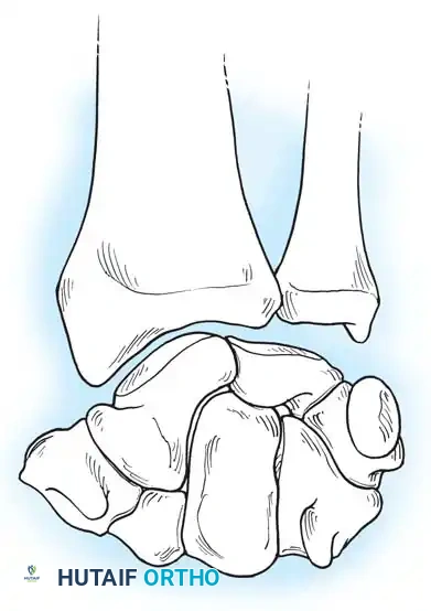
The dorsal approach to the wrist requires navigating the extensor retinaculum and the six extensor compartments. The primary surgical window is developed between the third compartment (containing the Extensor Pollicis Longus - EPL) and the fourth compartment (containing the Extensor Digitorum Communis - EDC and Extensor Indicis Proprius - EIP). Lister's tubercle serves as the primary osseous landmark for this interval.
Detailed Surgical Technique: The Compression Plate Method
1. Incision and Extensor Compartment Management
- Make a 10- to 15-cm longitudinal dorsal incision centered over the radiocarpal joint, extending from the distal third of the radius to the midshaft of the third metacarpal.
- Dissect through the subcutaneous tissue, taking care to identify and protect the dorsal sensory branches of the radial and ulnar nerves.
- Expose the extensor retinaculum. Incise the retinaculum over the third extensor compartment.
- Mobilize and transpose the EPL tendon radially.
- Elevate the fourth compartment subperiosteally and retract the finger extensors (EDC and EIP) medially (ulnarly).
- Retract the radial wrist extensors (ECRL and ECRB) laterally (radially).
2. Capsular Exposure and Joint Preparation
- Open the dorsal wrist capsule using an H-shaped incision. This creates robust proximal and distal capsular flaps that will later be utilized to cover the plate and protect the extensor tendons from hardware attrition.
- Expose the radiocarpal and intercarpal joints fully.
- Using a combination of sharp osteotomes, rongeurs, and a high-speed burr, meticulously denude the articular cartilage from the radiocarpal joint, the midcarpal joint (specifically the capitolunate and scaphocapitate articulations), and the carpometacarpal (CMC) joint of the third ray.
- Continue decortication until healthy, bleeding subchondral bone is exposed.
- Fill the resulting intercarpal and radiocarpal gaps with cancellous bone harvested from the iliac crest.
⚠️ Surgical Warning: Preserving the CMC Joints
While the third CMC joint is routinely fused to provide a distal anchor for the plate, avoid violating the CMC joints of the fourth and fifth rays. Preserving ulnar CMC mobility is crucial for maintaining the transverse metacarpal arch and allowing the hand to cup around objects.
3. The Radial Styloid-Capitate Lag Screw
A defining step in this specific technique (Technique 69-40) is the prevention of ulnar translation of the carpus, which can lead to devastating DRUJ impingement.
* Prior to plate application, place a 3.5-mm cortex lag screw directed from the radial styloid obliquely into the body of the capitate.
* As this screw is tightened, it pulls the carpal mass radially against the radial styloid.
* This maneuver effectively clears the ulnar side of the carpus away from the distal ulna, avoiding impingement of the DRUJ and ensuring unhindered forearm pronation and supination.
4. Autologous Bone Grafting
- Prepare a dorsal rectangular bed (trough) spanning from the distal radius, across the denuded carpus, to the base of the third metacarpal.
- Harvest a flat, rectangular corticocancellous bone graft from the inner table of the iliac crest.
- Inlay this corticocancellous graft directly into the prepared dorsal bed. The cortical surface of the graft should sit flush with the dorsal cortex of the radius and metacarpal, providing a continuous, flat surface for the compression plate.
5. Plate Application and Dynamic Compression
- Select a seven-hole or eight-hole, 3.5-mm dynamic compression plate (DCP). (Note: Modern pre-contoured wrist fusion plates may also be used, but the fundamental AO principles remain identical).
- Contour the plate to accommodate the desired 10 to 15 degrees of wrist extension.
- Place the plate directly over the inlaid corticocancellous graft, aligning it with the longitudinal axis of the radius and the third metacarpal.
- Compression Sequence:
- Insert the first screw into the capitate to anchor the plate centrally to the carpus.
- Insert the second screw into the distal radius, utilizing the eccentric drilling technique in the dynamic compression hole to compress the radiocarpal joint against the graft.
- Attach the distal end of the plate to the third metacarpal (and occasionally the second metacarpal if a specialized plate is used) with two or three screws.
- Complete the proximal fixation by placing three or four additional screws into the radial shaft.
💡 Clinical Pearl: Metacarpal Fixation
When placing screws into the third metacarpal, ensure they are bicortical for maximum pull-out strength, but avoid excessive length that could irritate the volar intrinsic muscles or flexor tendons.
Closure and Soft Tissue Management
Meticulous soft tissue closure is paramount to prevent postoperative tendon ruptures.
* Obtain meticulous hemostasis following tourniquet deflation.
* Close the H-shaped dorsal capsule over the plate using heavy absorbable sutures. This capsular layer acts as a vital barrier between the rigid hardware and the gliding extensor tendons.
* Leave the EPL transposed subcutaneously to prevent attrition against the plate.
* Place a closed suction drain deep to the subcutaneous tissue if significant oozing is present.
* Close the extensor retinaculum loosely, ensuring no bowstringing of the tendons occurs.
* Close the skin with non-absorbable sutures or surgical staples.
* Apply a sterile, non-adherent dressing followed by a bulky compression dressing supported by a volar and dorsal sugar-tong splint.
Postoperative Care and Rehabilitation Protocol
The postoperative protocol is designed to protect the rigid fixation while preventing stiffness in the adjacent unoperated joints.
Phase I: Immediate Postoperative (Days 1 to 14)
- Elevation: The hand must be kept strictly elevated above heart level to minimize edema.
- Early Motion: Active and passive range of motion of the fingers, thumb, elbow, and shoulder is encouraged starting on the first postoperative day. This prevents tendon adhesions and reduces swelling.
- Wound Check: At 10 to 14 days, the initial bandage is changed, the splint is removed, and sutures/staples are extracted.
Phase II: Intermediate Immobilization (Weeks 2 to 6)
- Following suture removal, the patient is transitioned to an above-elbow cast (Muenster style or long-arm cast) for 4 to 6 weeks.
- Rationale: Above-elbow immobilization is critical in the early healing phase to neutralize pronation and supination forces. Forearm rotation generates significant torsional stress across the radiocarpal fusion mass, which can lead to hardware failure or nonunion if not controlled.
Phase III: Late Immobilization and Weaning (Weeks 6 to 12+)
- At the 4- to 6-week mark, the above-elbow cast is removed. Radiographs are obtained to assess early trabecular bridging.
- The patient is transitioned to a short-arm cast or a rigid custom thermoplastic splint.
- Immobilization is continued until solid bony arthrodesis is evident both clinically (absence of pain with stress) and radiographically (bridging trabeculae across the radiocarpal and intercarpal joints).
- Once fusion is confirmed, progressive strengthening and functional use of the hand are initiated under the guidance of a certified hand therapist.
Hardware Removal
Plate removal is generally optional and is not routinely recommended unless the patient develops symptomatic hardware prominence, cold intolerance, or extensor tendon tenosynovitis. If required, hardware removal should be delayed until at least 12 to 18 months postoperatively to ensure complete remodeling of the fusion mass.
Complications and Pitfalls
While highly successful, total wrist arthrodesis carries potential complications that the orthopedic surgeon must be prepared to manage:
- Nonunion (Pseudarthrosis): Occurs in 2-5% of cases. Risk factors include smoking, inadequate joint decortication, failure to use bone graft, and insufficient rigid fixation. Treatment requires revision plating and supplemental bone grafting.
- Extensor Tendon Irritation/Rupture: The most common hardware-related complication. Failure to close the capsule over the plate or leaving screws excessively long dorsally can lead to EPL or EDC rupture.
- DRUJ Impingement: As noted, failure to place the radial styloid-capitate lag screw can allow the carpus to settle ulnarly, impinging the distal ulna. This presents as painful, restricted forearm rotation and may necessitate a secondary Darrach or Sauve-Kapandji procedure.
- Carpometacarpal Osteoarthritis: Fusing the wrist increases the lever arm and stress on the unfused CMC joints. Preserving the mobility of the 4th and 5th CMC joints during surgery helps mitigate this risk.
- Infection: Deep surgical site infections require prompt irrigation, debridement, and potentially hardware removal if the fusion mass is stable or if the infection cannot be suppressed with antibiotics.
By adhering strictly to the biomechanical principles of dynamic compression, ensuring meticulous joint preparation, and respecting the soft tissue envelope, the dorsal compression plate technique provides an exceptionally reliable solution for end-stage wrist pathology.
You Might Also Like