Diagnosing SC Joint Osteoarthritis: What You Need to Know

Key Takeaway
This topic focuses on Diagnosing SC Joint Osteoarthritis: What You Need to Know, SC joint osteoarthritis is the most common condition affecting the sternoclavicular joint, often seen in individuals over 60 years old. It involves degenerative changes, which can be asymptomatic or cause pain and a palpable protuberance. Higher risk factors for sc joint osteoarthritis include postmenopausal women, chronic SC joint instability, and manual laborers.
A 52-year-old active manual laborer presents with a 6-month history of localized right sternoclavicular (SC) joint pain. It is exacerbated by overhead reaching and heavy lifting. He reports a constant "grinding" sensation. Clinical examination reveals point tenderness over the SC joint and reproducible crepitus during shoulder abduction. How would you investigate this patient to confirm your diagnosis and rule out mimics?
Candidate: I would start with plain radiographs, specifically AP and Serendipity views, to look for joint space narrowing, subchondral sclerosis, and osteophytes. I would follow up with a CT scan to accurately assess the degree of joint destruction and evaluate for posterior osteophytes. To confirm the SC joint is the pain generator, I would perform a fluoroscopy-guided diagnostic anesthetic injection. I would also order an MRI if I suspected disc pathology or to rule out inflammatory arthritis or infection.
Candidates often forget the "Serendipity view" (40-degree cephalic tilt), which is the gold standard for SC joint assessment on plain films. Failing to emphasize the diagnostic injection as the "gold standard" for confirming the source of pain—thereby distinguishing it from referred pain (e.g., cervical spine or cardiac)—is a major oversight.
A structured approach: 1. Radiographic assessment: AP and Serendipity views (cephalic tilt) to assess morphology. 2. Cross-sectional imaging: CT is the gold standard for bony anatomy, especially to map the relationship of the medial clavicle to the retrosternal neurovascular structures. 3. Soft tissue assessment: MRI to evaluate for intra-articular disc tears or synovitis. 4. Confirmation: Diagnostic intra-articular injection (local anesthetic) to correlate imaging findings with the patient's chief complaint, ensuring the pain is not referred from the neck or thoracic viscera.
The patient has failed 6 months of non-operative management. You are planning a medial clavicle resection. Looking at the surgical site anatomy, what are the primary neurovascular concerns, and how do you protect them during the resection?
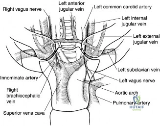
Candidate: The primary concerns are the great vessels—the brachiocephalic vein, the subclavian vessels, the vagus and phrenic nerves, and the thoracic duct on the left side. During the resection, I would use blunt, subperiosteal dissection to elevate the posterior capsule off the clavicle and use a malleable retractor or a Doyen elevator to shield the posterior structures before performing the osteotomy.
Candidates often miss the thoracic duct, which is a specific danger on the left side, or focus only on the blood vessels while ignoring the nerves (phrenic/recurrent laryngeal). Omitting the specific step of placing a posterior protective instrument (e.g., Doyen elevator) is a critical safety failure.
I prioritize patient safety by: 1. Anatomical awareness: Acknowledging the "Danger Zone" behind the manubrium and medial clavicle (brachiocephalic/subclavian vessels, vagus/phrenic nerves, and the thoracic duct). 2. Surgical Technique: Use of a subperiosteal plane to dissect posteriorly. 3. Mechanical Shielding: Utilizing a broad-based, blunt retractor (like a Doyen) to create a definitive barrier between the posterior cortex of the clavicle and the underlying mediastinal structures. 4. Instrumentation: Controlled usage of an oscillating saw rather than a burr to prevent "plunge" injuries.
How much of the medial clavicle should you resect, and what are the functional implications of over-resection vs. under-resection?
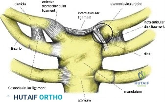
Candidate: I aim to resect 1.5 to 2.0 cm of the medial clavicle. Under-resection is a risk because it can lead to persistent osteophyte impingement or recurrence of the patient's original symptoms. Over-resection, particularly beyond 2 cm, risks iatrogenic instability by damaging the costoclavicular ligament attachments.
Failing to mention the costoclavicular (rhomboid) ligament. This is the key stabilizer; if the resection takes this out, the clavicle loses its vertical support, potentially causing significant functional shoulder pain and instability.
The standard resection is 1.5–2.0 cm. - Under-resection: Causes residual bony contact during shoulder motion, failing to alleviate the impingement/mechanical symptoms. - Over-resection: Risk of destabilizing the clavicle. The costoclavicular ligament, which anchors the medial clavicle to the first rib, must be preserved. Resecting medially to the ligament attachment compromises the fulcrum of the entire shoulder girdle, leading to symptomatic superior migration of the clavicle.
