Isolated Gastrocnemius Contracture & Foot Ulcers: Pathogenesis, Diagnosis, and Management
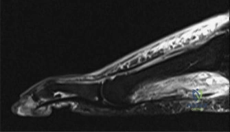
Key Takeaway
Isolated gastrocnemius contracture (IGC) significantly contributes to recurrent foot ulcers by limiting ankle dorsiflexion, increasing plantar forefoot pressures. This unperceived microtrauma, especially in neuropathic patients, initiates ulceration. Diagnosis is primarily via the Silfverskiöld test, differentiating IGC from global equinus by observing ankle dorsiflexion improvement with knee flexion.
A 58-year-old patient with long-standing Type 2 diabetes presents with a recurrent, non-healing ulcer beneath the second metatarsal head. They have been managed for 6 months with offloading and wound care. You suspect an isolated gastrocnemius contracture (IGC). Describe the clinical test you would perform and how you interpret the results.
Candidate: I would perform the Silfverskiöld test. With the patient supine, I assess passive ankle dorsiflexion with the knee extended, then again with the knee flexed to 90 degrees. If dorsiflexion is limited with the knee extended but improves significantly upon knee flexion, it confirms an isolated gastrocnemius contracture.
Failing to mention the degree of improvement. A borderline candidate forgets that the test is a binary differentiator: improvement indicates the gastrocnemius is the culprit; lack of improvement suggests a global equinus (soleus) contracture. They also often fail to mention the goal of testing (ruling out fixed osseous equinus).
The candidate defines the Silfverskiöld test clearly. They specify: 1) Passive dorsiflexion with the knee extended (measuring the triceps surae complex), 2) Passive dorsiflexion with the knee flexed (isolating the soleus). A positive test (IGC) shows restricted dorsiflexion with the knee extended that improves by >10 degrees upon flexion. They add that if the deformity remains fixed in both positions, it indicates a global equinus, likely involving the soleus or bony impingement, which changes the surgical plan.
You have decided to proceed with a surgical release for this patient. Look at the provided intraoperative image of a proximal gastrocnemius recession. Can you describe the landmarks for the incision and the critical structures you must protect?
Candidate: I use a longitudinal incision 2-3 cm medial to the midline of the posterior calf. The main structure at risk is the sural nerve, which is usually more lateral, and the small saphenous vein. I need to incise the gastrocnemius aponeurosis while preserving the underlying soleus fascia.
Ignoring the soleus fascia. The candidate fails to emphasize that the soleus fascia acts as the "safety net" protecting the neurovascular bundle (tibial nerve and posterior tibial vessels) lying deep to the soleus. They also fail to mention the exact level (10-15 cm proximal to the calcaneal insertion).
The candidate systematically outlines: 1) Incision: Posteromedial longitudinal, 2-3 cm medial to the midline. 2) Structures at risk: Sural nerve (lateral) and small saphenous vein (variable, often medial). 3) Surgical Plane: Identify the transition zone between muscle and aponeurosis. 4) Technical Crux: Transverse release of the gastrocnemius aponeurosis while strictly leaving the underlying soleus fascia intact. This protects the deep neurovascular bundle. They note the endpoint is achieving 5-10 degrees of passive dorsiflexion.
The surgery went well, and you achieved an excellent intraoperative release. Look at this intraoperative view. What do you see, and what is your immediate post-operative plan to ensure the patient doesn't develop complications?
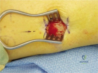
Candidate: The image shows the gap created in the gastrocnemius aponeurosis, with the soleus fascia clearly intact underneath. Post-operatively, I would place the patient in a short-leg splint or cast in slight dorsiflexion and keep them non-weight-bearing for 2-4 weeks to allow the soft tissues to heal before starting physical therapy.
Neglecting the "diabetic" context of the case. The candidate fails to mention glycemic control, vascular optimization, or the heightened vigilance required for wound dehiscence in a diabetic patient. They also ignore the potential for over-lengthening, which leads to a calcaneal gait.
The candidate confirms the visual evidence of the successful aponeurosis gap. They outline a structured post-operative protocol: 1) Immobilization: Splint in slight dorsiflexion to maintain length. 2) Loading: NWB for 2-4 weeks given the patient's diabetic/neuropathic status to prevent wound complications. 3) Rehab: Controlled transition to boot, followed by progressive ROM and strengthening. 4) Vigilance: Explicitly mention that because this is a diabetic foot, they will monitor for wound dehiscence and infection aggressively, and plan for custom orthotics/footwear long-term to prevent recurrent ulceration.
