The Elmslie-Trillat Operation: Comprehensive Surgical Guide

Key Takeaway
The Elmslie-Trillat operation is a highly specialized orthopedic procedure designed to realign the extensor mechanism by transferring the tibial tuberosity anteriorly and medially. It is primarily indicated for patients with recurrent patellar dislocations complicated by grade 3 or 4 chondromalacia. By optimizing patellofemoral tracking and reducing articular contact stress, this osteotomy provides a robust biomechanical solution for severe patellofemoral dysfunction.
Comprehensive Introduction and Patho-Epidemiology
The Elmslie-Trillat operation stands as a monumental achievement in the surgical armamentarium for addressing complex patellofemoral instability, particularly when superimposed upon degenerative joint disease. Originally conceptualized in the early 20th century by R.C. Elmslie and later popularized and refined by A. Trillat, the procedure was initially designed as a purely medializing transfer of the tibial tuberosity to correct extensor mechanism malalignment. However, as the biomechanical understanding of the patellofemoral joint expanded, it became evident that purely medializing the tuberosity in the presence of existing chondral damage often exacerbated articular wear. This realization catalyzed the evolution of the procedure through the seminal contributions of Brown, Fulkerson, and others, transforming it into a highly precise anteromedial transfer.
The patho-epidemiology of patellofemoral instability is multifactorial, involving a complex interplay of osseous dysplasia, soft tissue incompetence, and dynamic muscular imbalances. Patients presenting with recurrent lateral patellar dislocations frequently exhibit a lateralized tibial tubercle, quantified radiographically by an elevated Tibial Tubercle-Trochlear Groove (TT-TG) distance. This lateralization generates a pathological lateral force vector during quadriceps contraction, driving the patella out of the trochlear groove. Over time, recurrent subluxation and dislocation events lead to repetitive shear trauma across the articular cartilage, culminating in progressive chondromalacia, predominantly affecting the lateral patellar facet and the lateral trochlea.
Epidemiologically, the cohort requiring an Elmslie-Trillat procedure typically consists of young to middle-aged adults who have exhausted conservative management and, frequently, have failed isolated soft-tissue stabilizing procedures such as Medial Patellofemoral Ligament (MPFL) reconstruction. Performing an isolated MPFL reconstruction in a patient with a TT-TG distance exceeding 20 mm inevitably places supra-physiologic tension on the graft, leading to high failure rates. Therefore, the Elmslie-Trillat operation addresses the structural root cause of the instability. By physically altering the insertion vector of the patellar tendon, the procedure normalizes the Q-angle and eliminates the lateral subluxation vector.
Furthermore, the modern iteration of this procedure incorporates an anteriorization component, which is paramount for patients with concomitant Outerbridge Grade 3 or 4 chondromalacia. Anteriorization of the tibial tuberosity elevates the entire extensor mechanism, effectively increasing the moment arm of the quadriceps. This biomechanical shift significantly decreases the joint reaction forces across the patellofemoral articulation, providing essential decompression to the compromised cartilage. This dual biomechanical advantage—realignment and decompression—makes the Elmslie-Trillat procedure an invaluable, joint-preserving technique for orthopedic surgeons managing structurally compromised knees that are not yet candidates for arthroplasty.
Detailed Surgical Anatomy and Biomechanics
Anatomy of the Extensor Mechanism
A profound comprehension of the extensor mechanism anatomy is non-negotiable for the safe and effective execution of the Elmslie-Trillat operation. The extensor mechanism is a complex functional unit comprising the quadriceps muscle group, the quadriceps tendon, the patella, the patellar ligament (tendon), and the tibial tuberosity. The patella, the largest sesamoid bone in the human body, is embedded within this continuum and articulates with the femoral trochlea. Its primary anatomical function is to displace the extensor mechanism anteriorly, away from the axis of rotation of the knee, thereby acting as a fulcrum that enhances the mechanical advantage of the quadriceps.
The tibial tuberosity serves as the distal anchor for this mechanism. Its anatomical position relative to the trochlear groove dictates the terminal vector of the quadriceps pull. The lateral and medial retinacula provide secondary static stabilization, while the MPFL acts as the primary restraint to lateral patellar translation from 0 to 30 degrees of knee flexion. Surgically, the anterior compartment of the lower leg is of critical importance. The anterior tibial artery and the deep peroneal nerve course through this compartment, lying in perilous proximity to the posterolateral cortex of the tibia. When executing the osteotomy, the saw blade's exit point must be meticulously controlled to prevent catastrophic neurovascular injury.
Biomechanics of the Patellofemoral Joint
The biomechanics of the patellofemoral joint are characterized by massive, dynamic force transmissions. During normal gait, joint reaction forces across the patellofemoral articulation approximate 0.5 times body weight. However, during deep knee flexion activities such as squatting or ascending stairs, these forces can escalate to 7 or 8 times body weight. The distribution of these immense forces depends entirely on the congruence of the patella within the trochlea and the alignment of the extensor mechanism.
The Q-angle (Quadriceps angle) is a traditional clinical measurement of this alignment, formed by a line drawn from the Anterior Superior Iliac Spine (ASIS) to the center of the patella, intersecting with a line drawn from the center of the patella to the tibial tubercle. A hyper-physiologic Q-angle indicates a lateralized tibial tubercle, which exponentially increases the lateral vector forces on the patella during flexion. This lateral vector not only predisposes the patella to dislocation but also concentrates the immense joint reaction forces onto a smaller surface area of the lateral patellar facet, accelerating cartilaginous delamination and focal osteoarthritis.
Mechanics of Tibial Tuberosity Transfer
The Elmslie-Trillat operation, particularly as modified by Fulkerson, manipulates these biomechanics through a calculated anteromedial transfer. The medialization component directly addresses the abnormal Q-angle. By physically translating the tibial tuberosity medially, the TT-TG distance is normalized (typically to less than 15 mm). This realignment shifts the resultant force vector centrally, ensuring that the patella engages the deepest portion of the trochlear groove during early flexion, thereby restoring inherent osseous stability and protecting the MPFL from excessive strain.
The anteriorization component relies on the Maquet principle. By elevating the tibial tuberosity anteriorly, the patellar tendon is displaced forward. This geometric alteration increases the moment arm of the quadriceps mechanism, meaning less quadriceps force is required to generate the same extension torque. Consequently, the compressive joint reaction forces across the patellofemoral articulation are dramatically reduced. Mechanical testing demonstrates that a flat or slightly oblique osteotomy plane (as utilized in the modern Elmslie-Trillat/Fulkerson technique) possesses significantly higher mean load-to-failure and total energy-to-failure rates compared to steep, traditional oblique osteotomies. This structural superiority minimizes the risk of postoperative stress fractures while maximizing cartilaginous decompression.
Exhaustive Indications and Contraindications
Patient selection is universally recognized as the most critical determinant of success in tibial tuberosity osteotomies. The Elmslie-Trillat procedure is a highly specific, structural salvage operation; it is not a prophylactic intervention for generalized anterior knee pain or hypermobility. The surgeon must meticulously correlate the patient's clinical history, physical examination findings, and advanced imaging metrics to justify the surgical alteration of the extensor mechanism.
Primary indications center on the triad of recurrent instability, structural malalignment, and degenerative change. The classic candidate is a patient with recurrent lateral patellar dislocations who demonstrates a pathologically lateralized tibial tubercle (TT-TG > 20 mm) and concurrent Outerbridge Grade 3 or 4 chondromalacia of the patella or trochlea. In these cases, soft tissue procedures alone will fail due to the hostile biomechanical environment. Another profound indication is the presence of patella alta (an abnormally high-riding patella) combined with instability. When the Insall-Salvati index exceeds 1.2, the patella fails to engage the stabilizing bony confines of the trochlea during early flexion. In such scenarios, a modified Elmslie-Trillat incorporating a distal transfer is mandatory to physically draw the patella down into the functional groove.
Conversely, the contraindications are absolute and must be respected to avoid catastrophic outcomes. Performing this osteotomy in a skeletally immature patient with open physes is strictly prohibited. Disruption of the proximal tibial physis or the tibial tubercle apophysis will inevitably lead to premature growth arrest, resulting in a severe genu recurvatum deformity. Furthermore, this procedure is generally contraindicated in high-demand, competitive athletes (e.g., professional soccer or basketball players) due to the permanent alteration of extensor mechanism kinematics and the inherent, albeit low, risk of late stress fractures under extreme physiological loads.
| Parameter | Indications for Elmslie-Trillat Operation | Absolute and Relative Contraindications |
|---|---|---|
| Instability Profile | Recurrent lateral patellar dislocations failing conservative therapy. | First-time dislocation without structural bony malalignment. |
| Anatomical Alignment | TT-TG distance > 20 mm (Pathologic lateralization). | Normal TT-TG distance (< 15 mm) with isolated soft tissue laxity. |
| Cartilage Status | Outerbridge Grade 3 or 4 chondromalacia (lateral or distal facets). | Diffuse, pan-patellofemoral osteoarthritis; severe medial facet arthritis. |
| Patellar Height | Patella Alta (Insall-Salvati > 1.2) - requires distalization component. | Patella Baja (Insall-Salvati < 0.8) - distalization strictly prohibited. |
| Skeletal Maturity | Closed proximal tibial and tibial tubercle physes. | ABSOLUTE: Open physes (risk of genu recurvatum). |
| Patient Demographics | Young to middle-aged adults with structural salvage needs. | High-demand competitive athletes; patients with isolated medial tibiofemoral OA. |
Pre-Operative Planning, Templating, and Patient Positioning
Advanced Radiographic Evaluation and Templating
Meticulous preoperative planning is the bedrock of a successful Elmslie-Trillat procedure. The surgeon must quantify the exact degree of medial, anterior, and potentially distal transfer required before making the initial incision. The standard radiographic knee series must include a weight-bearing anteroposterior (AP) view, a true lateral view taken at exactly 30 degrees of knee flexion, and an axial view (Merchant or Skyline). The AP view assesses the overall tibiofemoral alignment and rules out concomitant tibiofemoral osteoarthritis.
The true lateral radiograph is critical for assessing patellar height. The Insall-Salvati ratio (the ratio of the patellar tendon length to the diagonal length of the patella) is calculated. An index greater than 1.2 indicates patella alta, necessitating a distal transfer component. Furthermore, Blumensaat’s line (the roof of the intercondylar notch) is identified. In a normal knee at 30 degrees of flexion, the inferior pole of the patella should intersect this line. This serves as a crucial intraoperative reference point to prevent iatrogenic patella baja during distal transfers.
Axial Computed Tomography (CT) imaging is the undisputed gold standard for quantifying the lateralization of the tibial tubercle. The TT-TG distance is measured by superimposing an axial slice through the deepest cartilaginous point of the femoral trochlea over an axial slice through the center of the patellar tendon insertion on the tibial tubercle. A TT-TG distance of 10-15 mm is considered normal, 15-20 mm is borderline, and greater than 20 mm is highly pathologic, providing a definitive mandate for medialization. Magnetic Resonance Imaging (MRI) is also routinely employed to map the exact location and severity of chondral defects, which dictates the necessity and extent of the anteriorization (elevation) component of the osteotomy.
Patient Positioning and Operating Room Setup
The patient is placed supine on a standard radiolucent operating table. A high thigh tourniquet is applied to the operative limb to ensure a bloodless surgical field, which is essential for precise identification of the fascial planes and neurovascular structures. The surgical limb is prepped and draped free in a manner that allows for unrestricted, full range of motion from 0 to 120 degrees of flexion. This mobility is absolutely critical, as the surgeon must dynamically assess patellar tracking and extensor mechanism tension intraoperatively prior to definitive hardware fixation.
A lateral post or a bump may be placed under the ipsilateral hip to prevent the natural tendency of the limb to externally rotate, ensuring the anterior aspect of the tibia faces directly upward. The fluoroscopy unit (C-arm) must be positioned on the contralateral side of the table, draped sterilely, and verified for unhindered access to obtain perfect lateral intraoperative radiographs. Prophylactic intravenous antibiotics are administered 30 minutes prior to tourniquet inflation, and a multimodal pain management protocol, often including a regional adductor canal block, is initiated by the anesthesia team.
Step-by-Step Surgical Approach and Fixation Technique
Surgical Approach and Soft Tissue Dissection
The operation commences with a longitudinal incision placed just lateral to the anterior tibial crest. The incision typically extends from the inferior pole of the patella to approximately 6 to 8 centimeters distal to the tibial tuberosity. Full-thickness fasciocutaneous flaps are meticulously elevated to preserve the delicate vascular supply to the anterior skin, minimizing the risk of postoperative wound necrosis. The medial border of the patellar tendon is identified and cleared of overlying bursa and fat pad, ensuring the entire insertion footprint on the tibial tuberosity is visualized.
Attention is then directed to the lateral aspect of the tibia. The anterior compartment fascia is incised longitudinally, approximately 1 centimeter lateral to the tibial crest. The anterior compartment musculature (primarily the tibialis anterior) is sharply and bluntly elevated off the lateral face of the tibia. This subperiosteal dissection must be performed with extreme care, utilizing a blunt Hohmann retractor to protect the anterior tibial artery and the deep peroneal nerve, which reside in the depths of this compartment and are highly vulnerable as the oscillating saw blade exits the posterolateral cortex.
Guide Pin Placement and Osteotomy Execution
To achieve a perfectly planar and mathematically precise cut, specialized drill guides, such as those popularized by Fulkerson, are highly recommended. Steinmann pins are drilled through the guide to establish the cutting plane. The pins are angled from anteromedial (entering just deep to the medial border of the patellar tendon) and directed posterolaterally. The angle of these pins dictates the ratio of medialization to anteriorization.
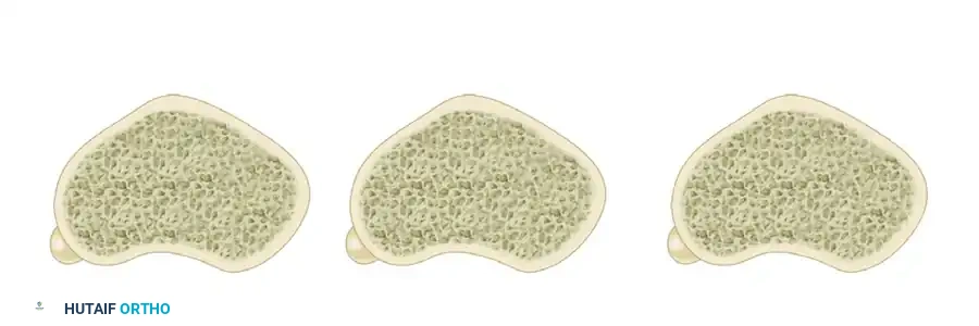
The depth of the osteotomy cut is a critical intraoperative decision. A deeper cut (originating further posterior on the medial tibia) allows for a larger bone block and more pronounced anterior transfer. However, this must be meticulously balanced against the risk of creating a massive stress riser. A cut that is too deep compromises the structural integrity of the proximal tibia, exponentially increasing the risk of late postoperative fractures. The oscillating saw is used to complete the osteotomy along the trajectory of the guide pins. Copious cold saline irrigation is mandatory during the saw cut to prevent thermal necrosis of the bone, which could lead to catastrophic nonunion. The distal end of the osteotomy must not end abruptly; it should be tapered or "feathered" out through the anterior cortex to dissipate stress forces.
Osteotomy Trajectory and Biomechanical Adjustments
The specific angle of the osteotomy directly influences the final three-dimensional position of the tibial tuberosity. Understanding these cross-sectional variations is paramount for tailoring the procedure to the patient's specific pathology.
Transverse Osteotomy
A purely transverse cut (parallel to the coronal plane) allows for direct medialization of the tuberosity. While this effectively corrects the TT-TG distance and normalizes the Q-angle, it does not inherently elevate (anteriorize) or depress the origin of the patellar tendon. This trajectory is suitable for patients with isolated instability and pristine articular cartilage, but it fails to decompress the joint in the setting of chondromalacia.

Depressing Oblique Osteotomy
An oblique osteotomy angled steeply from anterolateral to posteromedial can inadvertently depress the tuberosity as it is shifted medially. This posteriorization effectively shortens the moment arm of the quadriceps, thereby increasing patellofemoral contact pressures. This trajectory is strictly contraindicated in patients with any degree of chondromalacia, as it will rapidly accelerate cartilaginous destruction and exacerbate anterior knee pain.
Elevating Oblique Osteotomy (Fulkerson/Elmslie-Trillat Principle)
An oblique osteotomy directed from anteromedial to posterolateral is the modern gold standard. As the tuberosity block is shifted medially along this inclined plane, it is simultaneously elevated anteriorly. This trajectory beautifully achieves the dual goals of the operation: correcting the lateral subluxation vector while decompressing grade 3 or 4 chondromalacia by increasing the extensor mechanism's moment arm.
Management of Concomitant Patella Alta (Distal Transfer)
If preoperative templating reveals an Insall-Salvati index greater than 1.2, a distal transfer must be incorporated into the procedure. Unlike a purely hinged medialization where the distal periosteum is left intact, a distal transfer requires the tuberosity bone block to be completely detached. Once mobilized, approximately 5 to 10 millimeters of bone is carefully resected from the distal tip of the tuberosity block, or from the distal recipient bed.
This resection creates a structural void, allowing the entire bone block, along with the attached patellar tendon, to be translated distally. Before definitive fixation, this distalization must be rigorously checked. Intraoperative fluoroscopy is utilized to reference Blumensaat's line with the knee held at exactly 30 degrees of flexion. The inferior pole of the patella must not be drawn distal to this line. Overzealous distalization creates an iatrogenic patella baja, a debilitating condition characterized by severe anterior knee pain, restricted flexion, and accelerated global patellofemoral arthrosis.
Reduction and Rigid Internal Fixation
Once the tuberosity is translated to the meticulously calculated anteromedial (and potentially distal) position, it is temporarily secured to the tibial bed using two heavy Kirschner wires. The tourniquet is deflated, and hemostasis is achieved. The surgeon then takes the knee through a full, dynamic range of motion. Patellar tracking is directly visualized; the patella should engage the trochlea centrally and smoothly without any lateral subluxation or excessive medial tightness.
Upon confirmation of perfect tracking, definitive rigid internal fixation is achieved. Two or three 4.5 mm cortical lag screws are directed from anterior to posterior, perpendicular to the osteotomy plane. The lag technique is critical: the near cortex (tuberosity block) is overdrilled to the outer diameter of the screw threads, ensuring that tightening the screw aggressively compresses the osteotomy surfaces together. The screw heads must be carefully countersunk into the anterior cortex to prevent symptomatic hardware prominence beneath the thin anterior skin. Bicortical purchase (engaging the posterior tibial cortex) is absolutely mandatory to provide the rigid stability required for early rehabilitation and to prevent catastrophic hardware pullout.
Complications, Incidence Rates, and Salvage Management
While the Elmslie-Trillat operation yields 86% to 90% good to excellent results in properly selected patients, the complication profile is not insignificant and must be deeply respected by the operating surgeon. The procedure inherently creates a structural weakness in the proximal tibia, and deviations from meticulous surgical technique can lead to severe morbidity.
The most feared complication is a proximal tibial stress fracture, which occurs in approximately 1% to 3% of cases. These fractures can occur catastrophically months after clinical and radiographic healing appear complete, often during a sudden eccentric quadriceps contraction. The primary mechanism is the creation of a massive stress riser due to an excessively deep osteotomy cut or a failure to taper the distal end of the osteotomy smoothly into the anterior cortex. Management of a displaced stress fracture requires immediate open reduction and internal fixation (ORIF) with heavy proximal tibial locking plates, significantly compromising the patient's long-term functional outcome.
Over-medialization is a technical error that leads to iatrogenic medial patellar instability or dramatically increased contact pressures on the medial patellar facet. This occurs when the surgeon fails to accurately measure the TT-TG distance or overcompensates during the transfer. The resulting medial-sided arthritis is rapidly progressive and highly symptomatic. Salvage management often requires a complex revision osteotomy to lateralize the tubercle back to an anatomic position.
Hardware prominence is the most frequent minor complication, occurring in up to 30% of patients. Due to the lack of subcutaneous fat over the anterior tibia, the heads of the 4.5 mm cortical screws frequently become palpable and painful, particularly during kneeling. While this does not compromise the structural integrity of the repair, it routinely necessitates a secondary, minor surgical procedure for hardware removal once the osteotomy has fully and definitively united (typically after 9 to 12 months).
| Complication | Estimated Incidence | Etiology / Mechanism | Prevention Strategy | Salvage Management |
|---|---|---|---|---|
| Proximal Tibial Fracture | 1% - 3% | Deep osteotomy cut; sharp distal cortical step-off (stress riser). | Taper distal cut; avoid violating posterior cortex; restrict early high-impact loading. | ORIF with heavy proximal tibial locking plates; prolonged non-weight bearing. |
| Over-Medialization | 2% - 5% | Excessive medial transfer; failure to accurately measure TT-TG. | Precise preoperative CT templating; intraoperative dynamic tracking assessment. | Revision osteotomy to lateralize the tubercle; medial facet chondroplasty. |
| Patella Baja | < 2% | Overzealous distal transfer; failure to reference Blumensaat's line. | Intraoperative fluoroscopy at 30° flexion; limit distal transfer to < 10mm. | Extremely difficult; may require revision proximal transfer or patellar tendon lengthening. |
| Nonunion / Delayed Union | 1% - 2% | Thermal necrosis during saw cut; inadequate rigid compression (lag technique). | Copious cold irrigation during osteotomy; strict bicortical lag screw fixation. | Revision fixation with bone grafting; use of bone stimulators. |
| Symptomatic Hardware | 15% - 30% | Prominent screw heads beneath thin anterior tibial skin. | Aggressive countersinking of screw heads; placing screws slightly oblique. | Outpatient hardware removal after confirmed radiographic bony union (> 9 months). |
Phased Post-Operative Rehabilitation Protocols
A strict, scientifically phased rehabilitation protocol is essential to protect the healing osteotomy while simultaneously restoring extensor mechanism function and preventing debilitating arthrofibrosis. The protocol must balance the mechanical vulnerability of the bone cut with the biological necessity of early motion.
Phase I: Maximum Protection (Weeks 0-4)
The primary goals of Phase I are to protect the osteotomy site, control postoperative effusion, and initiate early passive motion. Weight-bearing status is heavily dependent on the surgeon's assessment of bone quality and fixation rigidity. Generally, patients are restricted to toe-touch weight-bearing (TTWB) or partial weight-bearing (up to 25% body weight) with bilateral crutches. The knee is locked in full extension in a hinged knee brace during all ambulation and during sleep to prevent accidental flexion under load.
Range of motion (ROM) therapy is initiated immediately to prevent capsular adhesions and arthrofibrosis. Passive and active-assisted ROM is permitted from 0 to 90 degrees. However, active open kinetic chain knee extension (e.g., performing a straight leg raise or kicking out against gravity) is strictly and absolutely prohibited. Active extension places massive shear forces directly across the osteotomy site via the patellar tendon, risking catastrophic avulsion of the tuberosity block and hardware failure.
Phase II: Moderate Protection (Weeks 4-8)
Progression to Phase II is contingent upon radiographic evidence of early callus formation and clinical absence of pain at the osteotomy site. Weight-bearing is progressively advanced to full weight-
