Reverse Radial Forearm Flap (RRFF): Anatomy, Indications & Hand Reconstruction
Key Takeaway
The Reverse Radial Forearm Flap (RRFF) is a workhorse pedicled flap for medium to large soft tissue defects of the hand, wrist, and distal forearm. It leverages retrograde blood flow through the radial artery to provide pliable, reliable coverage for exposed vital structures, preventing infection and preserving function following trauma, oncological resection, or chronic ulcers.
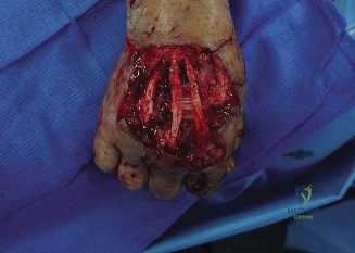
A 35-year-old male presents following a high-speed motorcycle accident with a complex soft tissue defect of the dorsal wrist exposing the distal radius hardware and extensor tendons. You are considering a reverse radial forearm flap (RRFF). What is your absolute non-negotiable pre-operative vascular requirement, and how do you clinically verify it?
Candidate: I need to ensure the hand is perfused by the ulnar artery. I would perform an Allen's test by occluding both the radial and ulnar arteries, having the patient clench their fist until it blanches, then releasing only the ulnar artery to see if the hand reperfuses quickly.
Failing to emphasize that this is an absolute contraindication. Many candidates describe the Allen's test vaguely or forget to state that a negative test (inadequate ulnar flow) means the procedure is contraindicated, as the radial artery will be sacrificed.
The absolute requirement is patent ulnar artery-based collateral circulation. I would confirm this with a clinical Allen's test, looking for rapid reperfusion within 5-7 seconds. If equivocal, I would proceed to Doppler ultrasound to quantify flow in the palmar and dorsal carpal arches. Without proof of adequate ulnar dominance, this flap is strictly contraindicated to prevent catastrophic hand ischemia.
You have confirmed vascular safety. Please describe the planning and marking of this flap, specifically regarding the pedicle and donor site.
Candidate: I would mark the radial artery course using Doppler. I'll design an elliptical skin paddle centered over the artery. I need to make sure the distal margin is at least 2cm away from the wrist joint to preserve the carpal arches.
Missing the detail about the "pivot point" or the importance of the perforators. Failing to mention the harvest of the venae comitantes or the necessity of preserving the paratenon for donor site grafting.
I would use Doppler to map the radial artery. The flap is designed as an ellipse over the mid-forearm. Crucially: The distal margin must be 2-3 cm proximal to the radial styloid to preserve the distal perforators and prevent aesthetic morbidity at the wrist. I would mark the superficial radial nerve if a sensate flap is requested. During elevation, I prioritize preserving the paratenon over the flexor tendons, as this is essential for a high-quality skin graft take on the donor site.
Post-operatively, the nursing staff calls you because the flap appears dusky and has sluggish capillary refill. What are your immediate steps?
Candidate: I would immediately assess the flap for venous congestion or arterial insufficiency. I'd check the dressing for constriction and elevate the arm. If it doesn't improve, I might need to take the patient back to the theater.
Providing a passive management plan. Examiners want to see a structured "ABC" approach for surgical complications: physical release of obstruction, then re-exploration.
My approach is systematic: 1. Exclusion of external factors: Remove all dressings and splints to ensure no pedicle kinking or compression. 2. Physical assessment: Assess color, turgor, and Doppler signal. 3. Immediate maneuvers: Optimize venous return through elevation. 4. Re-exploration: If no rapid improvement or if the Doppler signal is absent, I would take the patient to the OR for urgent revision of the pedicle/venous outflow, as early intervention is the only way to salvage the flap.
