Subtalar Arthrodesis: Comprehensive Surgical Technique and Clinical Protocols
Key Takeaway
Subtalar arthrodesis is a highly effective procedure for addressing end-stage arthritis, severe hindfoot deformity, and subtalar instability. This comprehensive guide details the lateral surgical approach, meticulous joint preparation, and rigid internal fixation techniques required to achieve optimal outcomes. Key steps include precise deformity correction to 5-10 degrees of hindfoot valgus, preservation of the calcaneocuboid ligaments, and strategic screw placement to maximize compression and restore the talar declination angle.
INTRODUCTION TO SUBTALAR ARTHRODESIS
Subtalar arthrodesis (talocalcaneal fusion) is a cornerstone procedure in reconstructive foot and ankle surgery. Designed to eliminate painful motion and correct complex hindfoot deformities, it is highly effective for restoring a plantigrade, stable, and functional foot. When performed with meticulous attention to joint preparation, anatomical alignment, and rigid internal fixation, subtalar arthrodesis yields excellent long-term clinical outcomes.
This comprehensive guide expands upon the foundational principles of subtalar arthrodesis, providing orthopedic residents, fellows, and practicing consultants with an exhaustive, step-by-step technical masterclass.
CLINICAL INDICATIONS AND PATIENT SELECTION
The decision to proceed with a subtalar arthrodesis must be rooted in a thorough clinical and radiographic evaluation. The primary goal is to relieve pain originating from the talocalcaneal articulation while correcting any associated coronal or sagittal plane deformities.
Primary Indications
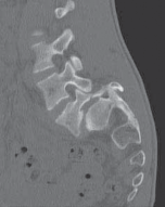
- Post-Traumatic Osteoarthritis: Most commonly secondary to calcaneal fractures (e.g., Sanders Type IV) or talar body/neck fractures resulting in articular incongruity or avascular necrosis (AVN).
- Primary Osteoarthritis: Idiopathic degeneration of the posterior, middle, or anterior facets of the subtalar joint.
- Inflammatory Arthropathy: Rheumatoid arthritis, psoriatic arthritis, or ankylosing spondylitis leading to erosive joint destruction and progressive valgus/varus deformity.
- Posterior Tibial Tendon Dysfunction (PTTD): Stage III adult-acquired flatfoot deformity characterized by a rigid, non-reducible hindfoot valgus.
- Tarsal Coalition: Symptomatic talocalcaneal coalitions that have failed conservative management or prior resection.
- Neuromuscular Deformity: Polio, Charcot-Marie-Tooth disease, or cerebral palsy resulting in rigid hindfoot instability.
💡 Clinical Pearl: The "Triple" vs. "Isolated" Arthrodesis
Before committing to an isolated subtalar arthrodesis, critically evaluate the transverse tarsal joints (talonavicular and calcaneocuboid). If severe subluxation exists—defined as more than one-third of the lateral surface of the calcaneus exposed and abutting the fibula—an isolated subtalar fusion will fail to correct the global deformity. In such cases, a triple arthrodesis or adjunctive medial column procedures are mandatory.
APPLIED BIOMECHANICS AND ANATOMY
A profound understanding of hindfoot biomechanics is essential for successful arthrodesis. The subtalar joint acts as a complex, multi-axial hinge that translates tibial rotation into foot pronation and supination.
- The Locking Mechanism: The position of the subtalar joint dictates the flexibility of the transverse tarsal joint. When the subtalar joint is everted (valgus), the axes of the talonavicular and calcaneocuboid joints become parallel, unlocking the midfoot and allowing flexibility for shock absorption. Conversely, when inverted (varus), the axes diverge, locking the midfoot to create a rigid lever for push-off.
- Surgical Implication: Fusing the subtalar joint in varus is a catastrophic technical error. It permanently locks the transverse tarsal joint, leading to a rigid, painful lateral border overload and rapid adjacent-segment degeneration. The target alignment is strictly 5 to 10 degrees of valgus.
PREOPERATIVE PREPARATION AND ANESTHESIA
Anesthesia and Pain Management
Optimal postoperative pain control begins in the preoperative holding area.
* Regional Anesthesia: A continuous or single-shot popliteal sciatic nerve block is highly recommended. This provides excellent intraoperative muscle relaxation and significantly reduces postoperative opioid consumption.
* Adjunctive Blocks: A saphenous nerve block may be added to cover the medial hindfoot, though the primary surgical insult is lateral.
Patient Positioning
- The patient is placed in the lateral decubitus position or supine with a large bump under the ipsilateral hip to internally rotate the leg, bringing the lateral hindfoot directly superior.
- A well-padded thigh tourniquet is applied to ensure a bloodless surgical field, which is critical for identifying the subtle anatomical planes of the sinus tarsi.
SURGICAL TECHNIQUE: STEP-BY-STEP
1. Incision and Superficial Dissection
- Incision Placement: Begin a straight lateral incision approximately 1 cm inferior to the tip of the fibular malleolus. Extend the incision distally and slightly anteriorly over the anterolateral border of the calcaneus and the cuboid.
- Neurovascular Protection: Meticulously protect the sural nerve and the peroneal tendons (peroneus longus and brevis) in the posterior and inferior aspects of the wound. Retract them gently using a Ragnell or Senn retractor.
2. Deep Exposure and the Sinus Tarsi
- Landmark Identification: Abduct and adduct the midfoot to dynamically locate the calcaneocuboid (CC) joint. Invert and evert the hindfoot to identify the subtalar joint line.
- Muscle Reflection: Locate the proximal tendinous origins of the extensor digitorum brevis (EDB) and extensor hallucis brevis (EHB). Using sharp dissection, elevate these muscle bellies from the sinus tarsi, working from proximal to distal until the CC joint is exposed.
- Ligamentous Preservation: Do not open the CC joint capsule. Use a small-blade scalpel (e.g., #15) merely to identify the joint line. It is imperative to preserve the dorsolateral calcaneocuboid ligament and the lateral calcaneocuboid ligament.
- Entering the Subtalar Joint: Enter the subtalar joint just superior to the lateral CC ligament. The CC ligaments, along with the bifurcate ligament and a portion of the short plantar ligament, are vital stabilizers of the transverse tarsal complex and must remain intact.
3. Joint Debridement and Preparation
- Soft Tissue Clearance: Identify the subtalar joint and excise the deep components of the inferior extensor retinaculum from the floor of the sinus tarsi. Release the talocalcaneal interosseous ligament, located medial and posterior to the cervical ligament.
- Retraction: Once the sinus tarsi is cleared of adipose and ligamentous tissue, place a right-angle retractor or a curved Hohmann retractor parallel to the posterior facet. This lifts the peroneal tendons laterally and posteriorly, maximizing exposure.
- Joint Distraction: Insert a lamina spreader into the depths of the sinus tarsi. Position it so that it rests on the inferior (plantar) aspect of the talar neck, just lateral to the middle and anterior facet components of the talocalcaneal articulation.
- Cartilage Removal: Depending on the degree of arthrosis, begin debridement with a curette or a sharp osteotome, starting anterolaterally over the talar side.
🚨 Surgical Warning: The Flexor Hallucis Longus (FHL)
As you debride the posterior facet, be acutely aware of the medial anatomy. Posteromedially, as the posterior facet curves downward (plantarward), the flexor hallucis longus (FHL) tendon is highly vulnerable to iatrogenic transection by an over-penetrating osteotome or curette. Always direct instruments away from the medial neurovascular bundle.
- Subchondral Bone Preparation: If the posterior facet is obscured, distract further with the lamina spreader. In cases of severe fibrosis or post-traumatic scarring, drill multiple 2.0 mm holes into both articular surfaces to facilitate distraction and subsequent debridement.
- Remove all eburnated, sclerotic subchondral bone down to healthy, bleeding cancellous bone. Use small, thin osteotomes (curved and straight) to "fish-scale" or "feather" the surfaces. This maximizes the surface area for osteogenesis without sacrificing critical calcaneal height.
4. Deformity Correction and Alignment
- Assessment of Apposition: Once the posterior, middle, and anterior facets are prepared, remove the lamina spreader and evaluate the position of the foot with the denuded surfaces apposed.
- Addressing Subluxation:
- Mild to Moderate: If subluxation is not severe (less than one-third of the lateral calcaneal surface is exposed), adequate soft tissue release, distraction, and manual translation will correct the deformity.
- Severe: If more than one-third of the lateral calcaneus abuts the fibula, isolated subtalar arthrodesis is insufficient. Additional procedures (e.g., lateral column lengthening, medial column fusion) are required.
- Target Alignment: Manually compress the joint. Place the calcaneus in 8 to 10 degrees of valgus relative to the tibial axis, ensuring the tibiotalar joint is held at 90 degrees (neutral dorsiflexion).
5. Bone Grafting Considerations
- Evaluate the apposition of the prepared surfaces. If achieving the mandatory 5 to 10 degrees of valgus creates a structural gap, structural autograft (e.g., iliac crest) or allograft is required to fill the void and prevent malunion or loss of height.
- In cases of avascular necrosis or severe sclerosis, the addition of orthobiologics (e.g., Demineralized Bone Matrix, Bone Marrow Aspirate Concentrate) may be beneficial to stimulate union.
6. Internal Fixation Techniques
Rigid internal fixation is paramount. While several techniques exist, large-diameter screw fixation is the gold standard.
- Temporary Fixation: Use one or two 0.062-inch Kirschner wires (K-wires) or threaded guidewires to provisionally hold the joint in the corrected alignment.
- Standard Fixation (Good Bone Stock):
- Utilize a 6.5-mm or 7.0-mm partially threaded cannulated (or noncannulated) cancellous screw.
- Trajectory: Insert the screw from the posterior aspect of the calcaneal tuberosity, directing it anteriorly, medially, and superiorly into the body and neck of the talus.
- Entry Point: Approximately 1 cm lateral to the midline of the calcaneal tuberosity, near the junction of the heel pad and the glabrous skin of the hindfoot.
- Exit Point: The optimal trajectory allows the screw to exit the talus just distal to the body-neck junction. This ensures the screw threads achieve maximum purchase in the dense superior cortex of the talar neck.
- Biomechanics of Screw Placement: If a large cannulated screw is used, ensure the threads rest entirely distal to the arthrodesis site (within the talus). This allows for dynamic interfragmentary compression. Furthermore, this specific trajectory slightly plantarflexes the talar head, improving the talar declination angle and decreasing the risk of anterior ankle impingement.
- Fixation in Osteopenic/Rheumatoid Bone:
- In patients with severe osteopenia (e.g., rheumatoid arthritis), standard screws may toggle or pull through the soft cancellous bone.
- Use a heavy washer on the head of the screw to distribute the compressive load over the posterior calcaneal cortex.
- Alternatively, large, smooth Steinmann pins can be utilized. They are inserted quickly under fluoroscopic guidance and provide stable, albeit non-compressive, neutralization.
💡 Clinical Pearl: Multi-Planar Assessment
Before final tightening, visually and fluoroscopically inspect the arthrodesis from the plantar, medial, and lateral aspects. Ensure there is no residual varus, no anterior translation of the calcaneus, and that the hardware does not penetrate the ankle or talonavicular joints.
7. Closure and Immediate Postoperative Care
- Deflate the tourniquet and obtain meticulous hemostasis. Hematoma formation in the sinus tarsi can lead to wound breakdown and deep infection.
- If significant postoperative bleeding is anticipated (e.g., extensive decortication), place a 1/8-inch closed-suction drain in the sinus tarsi, to be removed at 24 hours.
- Perform a supplementary ankle block with 0.5% bupivacaine (Marcaine) for immediate postoperative analgesia.
- Close the extensor retinaculum and subcutaneous tissues with absorbable sutures, followed by a non-absorbable monofilament for the skin.
- Apply a large, bulky Jones compression dressing reinforced with a posterior and U-shaped plaster splint, locking the ankle in neutral.
POSTOPERATIVE REHABILITATION PROTOCOL
The success of a subtalar arthrodesis relies heavily on strict adherence to postoperative weight-bearing restrictions to prevent hardware failure and nonunion.
- Phase I: Immediate Postoperative (Weeks 0-2)
- The procedure is typically performed on an outpatient basis, though patients with significant comorbidities may require a 1- to 2-day admission.
- Strict non-weight bearing (NWB). If the patient is frail or has an unstable gait, touch-down weight bearing (TDWB) with a walker or wheelchair use is mandated.
- Elevation of the limb above heart level is critical to minimize edema and protect the lateral incision.
- Phase II: Suture Removal and Casting (Weeks 2-6)
- At 14 to 17 days, the surgical dressing is removed. Once wound healing is confirmed, sutures are removed.
- The patient is transitioned into a well-molded, fiberglass short-leg cast.
- Strict NWB is maintained.
- Phase III: Transition to Weight Bearing (Weeks 6-12)
- At 6 to 8 weeks, radiographs are obtained to assess early trabecular bridging.
- If clinical and radiographic signs of healing are present, the patient is transitioned to a removable controlled ankle motion (CAM) walking boot.
- Progressive, partial weight-bearing is initiated, advancing to full weight-bearing as tolerated over the next 4 weeks.
- Phase IV: Maturation and Return to Activity (Weeks 12+)
- By 12 weeks, the arthrodesis has usually progressed to solid clinical fusion.
- A supportive compression stocking is applied to manage residual dependent edema.
- The patient transitions from the CAM boot to a supportive, stiff-soled athletic shoe or a shoe with a rocker-bottom modification to compensate for the loss of hindfoot motion.
- Physical therapy focuses on strengthening the Achilles tendon, peroneal musculature, and improving proprioception.
COMPLICATIONS AND PITFALLS
- Nonunion: Occurs in 5-10% of cases. Risk factors include smoking, diabetes, avascular necrosis of the talus, and inadequate joint debridement. Treatment requires revision arthrodesis with structural bone grafting and enhanced fixation.
- Malunion (Varus Deformity): The most devastating complication. Fusing the hindfoot in varus locks the transverse tarsal joint, causing severe lateral column overload, fifth metatarsal stress fractures, and rapid degeneration of the ankle joint.
- Sural Nerve Injury: Can occur during the initial incision or from aggressive retraction. Results in painful neuromas or lateral foot numbness.
- Hardware Prominence: The screw head at the posterior calcaneal tuberosity may cause irritation against footwear. Routine hardware removal is not indicated unless symptomatic after solid fusion is achieved (typically >1 year postoperatively).
You Might Also Like