Spinal Arthrodesis: Bone Graft Biology, Biomechanics, and Surgical Techniques
Key Takeaway
Spinal arthrodesis requires a profound understanding of bone graft biology, biomechanics, and meticulous surgical technique. This comprehensive guide explores the physiological cascade of graft incorporation, comparing autografts, allografts, and modern osteoinductive extenders like BMPs. It provides orthopedic surgeons with evidence-based protocols for graft selection, site preparation, and postoperative management to minimize pseudarthrosis and achieve robust, long-lasting spinal fusion across cervical, thoracic, and lumbar segments.
Introduction to Spinal Arthrodesis
Since the pioneering descriptions of spinal fusion by Hibbs and Albee in the early 20th century, arthrodesis of the spine has evolved into a cornerstone of orthopedic surgery. It is performed for a myriad of spinal pathologies, including tuberculosis and pyogenic infections, traumatic fractures, congenital and developmental deformities (e.g., scoliosis, kyphosis), arthritic degeneration, and intervertebral disc lesions.
Despite significant advancements in segmental instrumentation (rods, plates, pedicle screws) and a deeper understanding of the physiology of bone graft incorporation, pseudarthrosis (nonunion) remains a formidable clinical challenge. Achieving a solid arthrodesis is not merely a mechanical endeavor; it is a complex biological partnership between the graft material (autograft, allograft, or synthetic substitute) and the meticulously prepared recipient bed. Both elements must provide specific contributions to the fusion cascade. Furthermore, systemic factors—such as patient age, nutritional status, metabolic disorders, and the local mechanical environment—profoundly influence osteogenesis.
Biomechanics of Spinal Fusion
A successful spinal arthrodesis relies heavily on the biomechanical environment. Bone formation and remodeling are governed by Wolff’s Law, which dictates that bone adapts to the loads under which it is placed.
- Load-Sharing vs. Load-Bearing: Interbody grafts (anterior, transforaminal, or lateral) are subjected to compressive forces, which are highly osteogenic. Conversely, posterolateral fusions are subjected to tensile and shear forces, which are less favorable for bone formation.
- The Tension-Band Principle: Posterior instrumentation acts as a tension band, neutralizing flexion forces and converting them into compressive forces across the anterior column, thereby facilitating interbody graft incorporation.
- Micromotion vs. Macromotion: While absolute rigid fixation can lead to stress shielding and graft osteopenia, excessive macromotion disrupts the delicate fibrovascular stroma during the early phases of healing, leading to fibrous nonunion (pseudarthrosis). Optimal instrumentation provides stability while allowing physiological load-sharing (micromotion) to stimulate osteoblastic activity.
Clinical Pearl: The "race to fusion" is a competition between the biological incorporation of the bone graft and the fatigue life of the metallic implant. If the fusion mass does not consolidate before the implant reaches its fatigue limit, hardware failure (e.g., screw breakage, rod fracture) is inevitable.
Bone Grafts and Arthrodesis Healing: The Biological Cascade
Knowledge of the histological events and sequences of bone healing is paramount for the spine surgeon. Graft incorporation requires three fundamental properties:
1. Osteogenesis: The presence of surviving, viable osteoprogenitor cells within the graft (exclusive to fresh autograft or bone marrow aspirate).
2. Osteoinduction: The chemical stimulation of mesenchymal stem cells to differentiate into osteoblasts, mediated primarily by Bone Morphogenetic Proteins (BMPs).
3. Osteoconduction: The provision of a three-dimensional structural scaffold for vascular ingrowth and cellular migration.
Phases of Graft Incorporation
Research utilizing animal models (such as Boden et al.'s investigation into the coupling of membranous and enchondral ossification in intertransverse process fusions) has elucidated that spinal bone grafting undergoes three primary phases. These phases differ qualitatively from standard fracture healing, and different regions of the fusion mass heal at varying rates depending on their proximity to the decorticated host bone and vascular supply.
1. The Initial (Inflammatory) Phase
Following surgical decortication, a fracture hematoma forms. This hematoma is rapidly invaded by inflammatory cells (macrophages, neutrophils), leading to the formation of a fibrovascular stroma. At the decorticated surfaces, membranous bone formation initiates. During this phase, local expression of BMP-6, BMP-4, alkaline phosphatase, and osteonectin is significantly upregulated.
2. The Middle (Reparative) Phase
This phase is characterized by aggressive revascularization and the coupled resorption of necrotic graft bone by osteoclasts. Osteoprogenitor cells, recruited from the decorticated host surfaces and the autograft, differentiate into osteoblasts and chondroblasts.
* Enchondral Ossification: Occurs centrally within the fusion mass to bridge the gap between adjacent transverse processes.
* Gene Expression: Levels of osteocalcin and osteopontin peak, accompanied by a secondary surge in BMP-6 expression.
New bone matrix is deposited onto the trabecular scaffolding of the substrate, beginning at the areas of earliest vascular ingrowth (the decorticated host bone) and proceeding outward.
3. The Late (Remodeling) Phase
Beginning approximately at week 6, the cartilaginous intermediate is minimized. Marked remodeling occurs in accordance with Wolff’s Law, forming a peripheral cortical rim from which mature trabecular bone extends. Gene expression generally returns to baseline, with the exception of sustained BMP-6 activity.
Surgical Warning: Non-Steroidal Anti-Inflammatory Drugs (NSAIDs) and systemic corticosteroids profoundly inhibit the initial inflammatory phase and the subsequent prostaglandin-mediated pathways essential for osteoinduction. Their use should be strictly avoided for at least 6 to 12 weeks postoperatively to minimize the risk of pseudarthrosis.
Bone Graft Materials: Selection and Application
Autografts (The Gold Standard)
Autologous bone, most commonly harvested from the anterior or posterior iliac crest, remains the gold standard. It is the only material that inherently possesses all three properties: osteogenesis, osteoinduction, and osteoconduction. Local bone harvested during laminectomy is also highly valuable but often insufficient in volume for multilevel fusions.
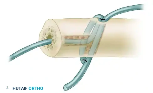
* Drawbacks: Iliac crest bone graft (ICBG) harvesting is associated with significant morbidity, including donor site pain, hematoma, infection, pelvic fracture, and lateral femoral cutaneous nerve injury.
Allografts
Due to autograft limitations, allografts (cadaveric bone) are extensively used. Allografts are highly osteoconductive but only weakly osteoinductive (due to the depletion of viable cells and degradation of proteins during processing).
- Cervical Spine: For single-level Anterior Cervical Discectomy and Fusion (ACDF), fresh-frozen and freeze-dried allografts yield acceptable results. However, allografts should not be used for multilevel cervical interbody fusions without supplemental internal fixation, as nonunion and subsidence rates are unacceptably high. Freeze-dried allograft is contraindicated for posterior cervical fusions.
- Thoracolumbar Spine: In adults, allograft bone should generally not be used as a standalone material for posterior thoracolumbar fusions due to high failure rates. Ethylene oxide-sterilized allografts are strictly contraindicated due to severe biological degradation.
- Structural Allografts: Anteriorly placed structural allografts (e.g., femoral ring allografts) supplemented with autologous bone provide acceptable fusion rates. However, high subsidence rates are noted in the lumbar spine unless supplemental segmental instrumentation is utilized to maintain deformity correction.
Bone Graft Extenders and Synthetics
Demineralized Bone Matrix (DBM)
DBM is produced via the acid extraction of allograft bone, leaving behind type I collagen, noncollagenous proteins, and osteoinductive growth factors (primarily BMPs).
* Properties: DBM is osteoinductive and osteoconductive but provides zero mechanical support.
* Efficacy: While animal models show promise, well-controlled human studies demonstrate variable efficacy. DBM is best utilized as a graft extender, mixed with local autograft or bone marrow aspirate.
Ceramics (Hydroxyapatite and Tricalcium Phosphate)
Available in block or granular forms, ceramics are purely osteoconductive scaffolds. They resorb at varying rates (Tricalcium phosphate resorbs faster than Hydroxyapatite). While useful in pediatric deformity surgery (e.g., adolescent idiopathic scoliosis) to expand autograft volume, their standalone role in adult degenerative spinal fusion remains limited.
Recombinant Human Bone Morphogenetic Proteins (rhBMPs)
The pioneering work by Urist led to the isolation of BMPs—low-molecular-weight noncollagenous glycoproteins belonging to the TGF-β superfamily. Recombinant technology has made rhBMP-2 and rhBMP-7 (OP-1) commercially available.
* rhBMP-2 (INFUSE): FDA-approved for Anterior Lumbar Interbody Fusion (ALIF) using a specific tapered titanium cage. Boden’s multicenter studies demonstrated 100% fusion rates at 6 months when rhBMP-2 was applied on an absorbable collagen sponge.
* rhBMP-7 (OP-1): Studies by Vaccaro and Johnsson have shown OP-1 to be safe and equivalent to autologous bone graft for single-level uninstrumented posterolateral lumbar fusions.
Pitfall: Off-label use of rhBMP-2 in the cervical spine (particularly anteriorly) has been associated with severe, life-threatening complications, including massive prevertebral soft tissue swelling, dysphagia, and airway compromise. Extreme caution and strict adherence to dosage guidelines are mandatory.
Step-by-Step Surgical Approach: Posterolateral Lumbar Fusion (PLF)
To translate the biology of graft healing into surgical success, meticulous technique is required. The following details the standard approach for a posterolateral lumbar arthrodesis.
1. Patient Positioning
- The patient is placed prone on a radiolucent spine table (e.g., Jackson frame).
- The abdomen must hang free to decrease intra-abdominal pressure, which in turn reduces epidural venous engorgement and intraoperative bleeding.
- All bony prominences are meticulously padded to prevent pressure necrosis and peripheral nerve palsies.
2. Surgical Exposure
- A midline longitudinal incision is made over the target levels.
- Subperiosteal dissection of the paraspinal musculature is performed bilaterally, exposing the spinous processes, laminae, facet joints, and transverse processes.
- Crucial Step: Exposure must extend to the tips of the transverse processes and the pars interarticularis. Meticulous hemostasis is maintained using bipolar electrocautery and hemostatic agents.
3. Site Preparation (Decortication)
- The success of the fusion relies entirely on the preparation of the recipient bed.
- Using a high-speed burr (e.g., matchstick or acorn tip) and sharp curettes, the dorsal surfaces of the transverse processes, the lateral aspect of the superior articular processes, and the pars interarticularis are aggressively decorticated.
- The goal is to expose bleeding cancellous bone, which provides the osteoprogenitor cells and vascular supply necessary for the inflammatory and reparative phases of graft healing.
4. Graft Preparation and Delivery
- If utilizing ICBG, the graft is harvested, morcellized into 2-3 mm fragments, and mixed with local bone obtained from the laminectomy/decompression.
- If using extenders (e.g., DBM or ceramics), they are hydrated with bone marrow aspirate or local blood and mixed thoroughly with the autograft.
- The graft material is packed tightly into the lateral gutters, spanning the decorticated transverse processes.
5. Instrumentation and Closure
- Pedicle screws and rods are placed to provide rigid segmental stabilization (the tension band), minimizing macromotion while allowing physiological load-sharing.
- The wound is irrigated copiously. A subfascial drain may be placed depending on the extent of dead space and bleeding.
- The fascia is closed in a watertight fashion using heavy, interrupted absorbable sutures to prevent muscle herniation and reduce infection risk.
Postoperative Protocols and Complication Management
Rehabilitation and Bracing
- Mobilization: Early mobilization is encouraged to prevent deep vein thrombosis (DVT) and pulmonary complications. Patients are typically mobilized on postoperative day one.
- Bracing: The use of a Thoracolumbosacral Orthosis (TLSO) or cervical collar depends on the rigidity of the internal fixation and the quality of the patient's bone (e.g., osteoporosis). Rigidly instrumented patients often do not require bracing, whereas uninstrumented or osteoporotic patients may require orthotic support for 6 to 12 weeks.
Radiographic Monitoring
- Serial radiographs (AP, lateral, and dynamic flexion-extension views) are obtained at 6 weeks, 3 months, 6 months, and 1 year.
- Signs of Fusion: Continuous trabecular bridging across the fusion segments, absence of radiolucent lines around the hardware, and less than 2 degrees of angular motion or 2 mm of translation on dynamic views.
- Signs of Pseudarthrosis: Hardware failure (broken screws, haloing around screw tracts), persistent radiolucent lines through the graft mass, and progressive deformity.
Management of Pseudarthrosis
If pseudarthrosis is diagnosed (typically after 1 year of failed conservative management and persistent axial pain), revision surgery is indicated.
* Revision Strategy: The surgical approach must be altered. If a posterior fusion failed, an anterior interbody approach (ALIF or LLIF) should be strongly considered to place the graft in a compressive, highly vascularized environment.
* Biological Enhancement: Revision surgery mandates the use of potent biological enhancers, such as fresh ICBG or rhBMP-2, to overcome the biologically depleted environment of the nonunion site.
Conclusion
Arthrodesis of the spine is a complex interplay of biomechanical stabilization and biological regeneration. The modern orthopedic surgeon must possess a profound understanding of the cellular cascade of bone healing, the distinct properties of various graft materials, and the meticulous surgical techniques required to prepare the host bed. By respecting the principles of osteogenesis, osteoinduction, and osteoconduction, and by carefully selecting the appropriate autograft, allograft, or biological extender, surgeons can optimize fusion rates, minimize complications, and achieve excellent long-term clinical outcomes for their patients.
===CONTENT===
You Might Also Like