Little League Elbow: Spectrum of Pathologies, Epidemiology, Anatomy & Biomechanics
Key Takeaway
Little League Elbow (LLE) encompasses medial elbow pathologies in young overhead throwing athletes, primarily medial epicondyle apophysitis. It's caused by repetitive valgus stress, applying tensile forces to the medial epicondyle physis and UCL during late cocking and early acceleration. Key risk factors include skeletal immaturity, high pitch counts, and poor mechanics.
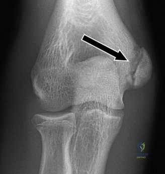
A 14-year-old competitive baseball pitcher presents with acute onset medial elbow pain after a high-velocity pitch. He reports a "pop" followed by immediate inability to continue throwing. He has tenderness over the medial epicondyle and mild ulnar nerve paraesthesia. The radiograph is shown below.
Candidate: The radiograph demonstrates a displaced medial epicondyle avulsion fracture. Given the mechanism and the neurovascular status, I would perform a thorough physical exam to assess for ulnar nerve deficit. Management would likely involve ORIF, especially since it is displaced and there are symptoms of nerve involvement.
Failing to mention the "coin-in-the-joint" sign (intra-articular entrapment), omitting the significance of the ulnar nerve status, or jumping immediately to "screw fixation" without discussing the importance of physeal preservation in a 14-year-old.
The candidate should identify the displaced medial epicondyle avulsion fracture and immediately highlight the risk of intra-articular entrapment (the "coin-in-the-joint" sign). They must emphasize a neurovascular assessment, specifically checking the ulnar nerve (motor/sensory). The perfect answer addresses: 1) Clinical status (instability/nerve), 2) Radiographic assessment (looking for fragments in the joint), and 3) Decision-making (surgical ORIF is indicated for displacement >2-3mm, ulnar nerve symptoms, or intra-articular entrapment), while acknowledging the need to respect the physis using techniques like tension band wiring or careful screw placement.
How do you explain the biomechanical rationale for this injury, and what is your plan for ensuring this patient returns safely to sport?
Candidate: It’s caused by repetitive valgus stress during the late cocking and acceleration phases of throwing. The flexor-pronator mass and UCL pull on the immature apophysis, causing it to fail. For return to sport, I’d use an interval throwing program after the fracture unites.
Ignoring the "kinetic chain." Candidates often focus only on the elbow, failing to mention the critical need to correct deficits in shoulder girdle strength, scapular dyskinesis, and core stability, which are often the root cause of the elbow overload.
Structure the answer into: 1) Biomechanics: Explain the high valgus torque in late cocking/early acceleration, the role of the UCL/FPM, and the "weakest link" being the cartilaginous physis. 2) Kinetic Chain: Acknowledge that elbow failure is often secondary to proximal deficits (e.g., shoulder/hip/core). 3) Rehabilitation: Describe a structured interval throwing program (ITP) that is phased, pain-free, and overseen by a physiotherapist, noting that return to play must wait for radiographic evidence of union and restoration of full, pain-free ROM.
