Management of Fracture Nonunions: A Surgical Guide
Key Takeaway
Fracture nonunion represents a complex orthopedic challenge requiring meticulous evaluation of mechanical stability and biological vitality. This comprehensive guide details the pathophysiology, Weber and Cech classification, and evidence-based surgical interventions for hypertrophic and atrophic nonunions. Emphasizing soft tissue management, bone grafting techniques, and rigid internal or external fixation, it provides orthopedic surgeons with a systematic approach to restoring osseous continuity, optimizing biomechanics, and ensuring successful functional recovery in complex nonunion cases.
INTRODUCTION TO DELAYED UNION AND NONUNION
Despite the multitude of studies investigating fracture healing, establishing a universal criterion for declaring a fracture a nonunion remains a clinical challenge. A diagnosis of nonunion is unjustified until clinical or radiographic evidence definitively demonstrates that the biological healing process has ceased and that osseous union is highly improbable without surgical intervention.
The U.S. Food and Drug Administration (FDA) panel established a widely accepted definition of nonunion: a fracture is considered a nonunion when a minimum of 9 months has elapsed since the injury, and the fracture shows no visible progressive signs of healing for 3 consecutive months. However, this criterion cannot be rigidly applied to every fracture pattern. For instance, a diaphyseal fracture of a long bone (e.g., tibial shaft) should generally not be considered a nonunion until at least 6 months post-injury, as union often requires extended time, particularly in the presence of local complications such as severe soft tissue stripping or infection. Conversely, an intracapsular fracture of the femoral neck may be defined as a nonunion after only 3 months due to the inherently precarious vascular supply and high mechanical shear forces.
Clinical Pearl: Do not wait 9 months to intervene if a fracture is clearly destined for nonunion. A femoral neck fracture with hardware failure or a tibial shaft fracture with a massive segmental defect and no callus at 4 months warrants early, definitive surgical intervention.
PATHOPHYSIOLOGY AND CONTRIBUTING FACTORS
The exact etiology of delayed union and nonunion is multifactorial, representing a failure of the delicate interplay between mechanical stability and biological osteogenesis. Both systemic and local factors profoundly influence the trajectory of fracture healing.
Systemic Factors
Systemic optimization is a mandatory prerequisite for successful nonunion surgery.
* Vitamin D Deficiency: Recent literature highlights the critical role of Vitamin D in bone metabolism. Patients presenting with unexplained nonunions despite adequate mechanical stabilization, or those with multiple low-energy fractures, frequently exhibit hypovitaminosis D. Routine assessment of serum 25-hydroxyvitamin D levels is strongly recommended for all nonunion patients, with aggressive supplementation initiated prior to surgical intervention.
* Tobacco Use: Smoking is a profound deterrent to osteogenesis. Nicotine induces peripheral vasoconstriction, while carbon monoxide competitively binds to hemoglobin, drastically reducing tissue oxygen tension in the cutaneous and subcutaneous tissues. This hypoxic environment impairs angiogenesis, decreases vascularization at the fracture site, and significantly increases the risk of deep infection and osteomyelitis. Complete smoking cessation is imperative.
* Nonsteroidal Anti-inflammatory Drugs (NSAIDs): The role of NSAIDs in fracture healing remains a subject of intense academic debate. Multiple animal models demonstrate that COX-2 inhibition suppresses endochondral ossification and delays fracture healing. While human studies are conflicting, the prevailing evidence-based consensus dictates that patients with delayed unions or established nonunions should strictly abstain from NSAIDs and systemic corticosteroids during their treatment course.
Local Factors
Nonunion of long bones is significantly more common under the following local conditions:
1. Open fractures with severe soft tissue compromise (Gustilo-Anderson Types II and III).
2. Presence of deep infection or osteomyelitis.
3. Segmental fracture patterns with impaired endosteal and periosteal blood supply (typically affecting the intercalary segment).
4. High-energy comminution.
5. Insecure or mechanically unstable fixation.
6. Insufficient duration of immobilization.
7. Ill-advised open reduction that excessively strips the periosteal blood supply.
8. Fracture distraction (iatrogenic via traction, external fixation, or poorly contoured plating).
9. Irradiated bone.
Surgical Warning: In the tibia, an intact fibula can act as a mechanical strut, preventing axial compression at the tibial fracture site and leading to a delayed or nonunion. Fibular osteotomy may be required to allow dynamic compression of the tibial nonunion.
PREOPERATIVE CONSIDERATIONS AND EVALUATION
Successful management of a nonunion requires a holistic evaluation of the host, the soft tissue envelope, and the mechanical environment.
Status of Soft Tissues and Neurovascular Structures
With contemporary techniques in orthobiologics and rigid fixation, definitive surgery can often be performed earlier, facilitating accelerated joint rehabilitation. However, the soft tissue envelope dictates the surgical approach.
* Soft Tissue Contractures: Unyielding scar tissue, particularly on the concave side of an angular deformity, poses a high risk for postoperative skin necrosis upon deformity correction. Deep scarring may preclude local bone grafting or bone transport. The necessity for rotational flaps or free tissue transfer (e.g., latissimus dorsi or anterolateral thigh flaps) must be anticipated and coordinated with a microsurgeon.
* Vascular Assessment: In patients with a history of high-energy trauma, vascular injury, or diminished peripheral pulses, advanced imaging (CT angiography or formal arteriography) is mandatory. A compromised vascular tree will doom any biological grafting procedure. Vascular reconstruction must precede or accompany nonunion takedown.
* Neurological Status: Neurological deficits must be meticulously documented. If a nerve is encased in scar tissue, neurolysis is indicated. In cases of severe bone loss requiring massive lengthening, the Ilizarov method of distraction osteogenesis is preferred to prevent acute traction neurapraxia. If the extremity is insensate and lacks motor function (e.g., complete sciatic or tibial nerve transection), amputation may be the most functional and practical choice.
Status of the Bones and Classification Systems
The biological viability of the fracture ends dictates the surgical strategy. The most universally applied classification is that of Weber and Cech, which divides nonunions into hypervascular (capable of biological reaction) and avascular (inert) types.
Hypervascular (Hypertrophic) Nonunions
These nonunions demonstrate robust uptake on bone scintigraphy, indicating a rich blood supply. They are biologically active but mechanically unstable.
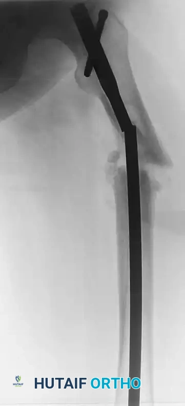
1. "Elephant Foot" Nonunions: Hypertrophic and rich in callus. They result from insecure fixation, inadequate immobilization, or premature weight-bearing in a reduced fracture with viable fragments.
2. "Horse Hoof" Nonunions: Mildly hypertrophic and poor in callus. They typically occur after unstable internal fixation (e.g., a loose plate) where micromotion exceeds the tolerance for woven bone formation.
3. Oligotrophic Nonunions: Not highly hypertrophic, but biologically viable. Often seen after major displacement or distraction without adequate reduction.
Avascular (Atrophic) Nonunions
These nonunions lack biological potential and require both mechanical stabilization and biological augmentation (bone grafting).
1. Torsion Wedge Nonunions: Characterized by an intermediate fragment with a unilateral blood supply that has healed to one main fragment but not the other.
2. Comminuted Nonunions: Characterized by one or more necrotic intermediate fragments. Radiographs show an absence of callus. Often results from plate breakage or catastrophic hardware failure.
3. Defect Nonunions: Characterized by the loss of a diaphyseal segment (e.g., post-debridement for open fractures or osteomyelitis). The fragment ends are viable, but the gap exceeds the biological capacity for spontaneous bridging.
4. Atrophic Nonunions: The end-stage result of missing intermediate fragments and interpositional scar tissue lacking osteogenic potential. The bone ends are osteoporotic, sclerotic, and biologically inert.
Paley Classification
Paley et al. introduced a highly practical classification based on bone loss and deformity, particularly useful for planning external fixation and deformity correction:
* Type A (Bone loss < 1 cm):
* Type A1: Mobile deformity.
* Type A2: Fixed deformity (A2-1: stiff without deformity; A2-2: stiff with fixed deformity).
* Type B (Bone loss > 1 cm):
* Type B1: Bony defect.
* Type B2: Loss of bone length.
* Type B3: Both bony defect and loss of length.
BIOMECHANICS OF NONUNION REPAIR
Understanding Perren’s Strain Theory is paramount. Strain is the relative change in gap length divided by the original gap length.
* Hypertrophic nonunions have adequate biology but excessive strain. The tissue in the fracture gap (fibrocartilage) cannot ossify because the mechanical deformation exceeds the tolerance of osteoblasts. The surgical goal is to decrease strain (increase stability) to allow the existing fibrocartilage to convert to bone.
* Atrophic nonunions lack both stability and biology. The surgical goal is to provide absolute stability while introducing osteoconductive, osteoinductive, and osteogenic elements (e.g., autograft) to stimulate healing.
GENERAL TREATMENT OF NONUNIONS: SURGICAL PRINCIPLES
1. Preparation and Positioning
- Optimization: Eradicate any active infection. If infection is suspected, hold preoperative antibiotics until deep intraoperative cultures are obtained.
- Positioning: Place the patient on a radiolucent table. Ensure unimpeded access for the C-arm fluoroscope in both AP and lateral planes. If autogenous bone grafting is planned, prep and drape the ipsilateral or contralateral iliac crest.
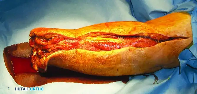
2. Surgical Approach and Soft Tissue Handling
- Utilize extensile, internervous approaches.
- Handle soft tissues with meticulous care. Avoid excessive periosteal stripping, which further devascularizes the bone ends.
- Excise all interpositional fibrous tissue down to healthy, bleeding bone (the "paprika sign").
3. Decortication and Preparation of the Nonunion Site
For atrophic and oligotrophic nonunions, osteoperiosteal decortication (as described by Judet) is a powerful technique.
* Using a sharp osteotome, elevate thin petals of cortical bone attached to the overlying periosteum and muscle around the circumference of the nonunion.
* This creates a vascularized, osteogenic envelope that bridges the nonunion site.
* Open the medullary canal on both sides of the nonunion using drill bits or curettes to re-establish endosteal blood flow and allow marrow elements to populate the fracture gap.
4. Bone Grafting Strategies
Bone grafting is mandatory for atrophic nonunions and defect nonunions.
* Autogenous Iliac Crest Bone Graft (ICBG): The gold standard. Provides osteoconductive scaffold, osteoinductive growth factors (BMPs), and osteogenic cells.
* Reamer-Irrigator-Aspirator (RIA): Harvests massive volumes of highly osteogenic autograft from the femoral or tibial medullary canal. Ideal for large defects, minimizing the morbidity associated with massive ICBG harvest.
* Orthobiologics: Demineralized bone matrix (DBM), bone morphogenetic proteins (rhBMP-2, rhBMP-7), and synthetic ceramics (tricalcium phosphate) can be used as graft extenders, though they do not replace the need for mechanical stability.
5. Stabilization of Fragments
Intramedullary Nailing (Exchange Nailing)
The treatment of choice for diaphyseal nonunions of the femur and tibia, particularly hypertrophic nonunions.
* Technique: Remove the existing hardware. Over-ream the canal by 1 to 2 mm larger than the previous nail. This generates autogenous bone graft (reamings) that deposits at the nonunion site and increases the working diameter for a larger, stiffer nail.
* Biomechanics: Provides load-sharing, relative stability, and excellent control of bending and torsional forces.
Compression Plating
Ideal for metaphyseal nonunions, atrophic nonunions requiring structural grafting, or cases where the medullary canal is obliterated.
* Technique: Apply a dynamic compression plate (DCP) or locking compression plate (LCP). For hypertrophic nonunions, rigid compression alone is often sufficient to induce union. For atrophic nonunions, the plate must bridge the grafted defect, often requiring a longer plate to distribute stress and prevent hardware failure.
External Fixation (Ilizarov / Taylor Spatial Frame)
Indicated for infected nonunions, nonunions with massive bone loss, or those with complex multiplanar deformities.
* Technique: Utilizes tensioned wires and half-pins attached to circular rings. Allows for gradual deformity correction, compression/distraction, and bone transport (distraction osteogenesis) to bridge massive segmental defects.
Pitfall: Failing to achieve absolute mechanical stability in an atrophic nonunion will result in rapid resorption of the bone graft and catastrophic failure of the reconstruction. Always prioritize rigid fixation.
MANAGEMENT OF INFECTED NONUNIONS
Infected nonunions represent the most formidable challenge in orthopedic trauma. The principles of management follow a staged approach:
1. Stage 1: Radical Debridement. Remove all necrotic bone, infected hardware, and devitalized soft tissue. "If it doesn't bleed, it must be excised." Stabilize the limb with a temporary external fixator or antibiotic-impregnated cement spacer (Masquelet technique).
2. Stage 2: Soft Tissue Coverage. Achieve a closed, healthy soft tissue envelope via local or free flaps.
3. Stage 3: Bone Reconstruction. Once infection is eradicated (confirmed by normal inflammatory markers and negative cultures), proceed with definitive reconstruction.
* Masquelet Technique: The cement spacer induces a highly vascularized pseudosynovial membrane. At 6-8 weeks, the spacer is removed, and the void is densely packed with autogenous bone graft (RIA or ICBG) within the induced membrane, followed by rigid internal fixation.
* Bone Transport: For defects > 5 cm, a corticotomy is performed in healthy bone, and the segment is gradually transported via an Ilizarov frame to dock at the nonunion site.
POSTOPERATIVE PROTOCOLS AND ADJUNCTIVE THERAPIES
Rehabilitation and Weight-Bearing
Postoperative protocols must be tailored to the fixation construct and the biological status of the nonunion.
* Intramedullary Nailing: Often allows for early, progressive weight-bearing, which dynamically compresses the nonunion site and stimulates osteogenesis.
* Plating/Grafting: Requires protected weight-bearing (toe-touch or partial) until radiographic evidence of graft incorporation and bridging callus is observed, typically at 8 to 12 weeks.
Biophysical Stimulation
- Low-Intensity Pulsed Ultrasound (LIPUS): FDA-approved for the treatment of established nonunions. LIPUS transmits mechanical energy through soft tissue to the fracture site, upregulating mechanoreceptors, increasing intracellular calcium, and stimulating COX-2 and PGE2 production, thereby accelerating endochondral ossification.
- Pulsed Electromagnetic Fields (PEMF): Induces weak electrical currents within the bone, stimulating osteoblast proliferation and extracellular matrix synthesis. Particularly useful as an adjunct in recalcitrant atrophic nonunions.
CONCLUSION
The successful eradication of a fracture nonunion demands a masterful understanding of bone biology, biomechanics, and advanced surgical techniques. By meticulously optimizing the host, respecting the soft tissue envelope, accurately classifying the nonunion, and applying the precise balance of rigid mechanical stability and biological augmentation, the orthopedic surgeon can reliably restore osseous continuity and functional capacity to the severely compromised limb.
You Might Also Like