Surgical Management of Chronic Posterior Hip Dislocation: A Comprehensive Guide
Key Takeaway
Chronic posterior hip dislocations present complex reconstructive challenges due to soft tissue contractures, acetabular bone loss, and high rates of osteonecrosis. Management depends on the chronicity of the injury, patient age, and femoral head viability. While open reduction and internal fixation may be attempted in injuries under three months old, total hip arthroplasty remains the gold standard for older, complex fracture-dislocations, requiring meticulous acetabular reconstruction.
Comprehensive Introduction and Patho-Epidemiology
Chronic or neglected posterior hip dislocations represent one of the most formidable and technically demanding challenges in the realm of orthopedic trauma and adult reconstructive surgery. By strict orthopedic definition, a dislocation is classified as "chronic" or "neglected" when it has remained unreduced for a period exceeding three weeks. However, in the context of resource-stratified healthcare systems or delayed presentations, these injuries frequently present months or even years following the initial traumatic event. The etiology is almost universally high-energy blunt force trauma, such as motor vehicle collisions or falls from significant heights, where an axial load is applied to a flexed, adducted, and internally rotated femur. This vector of force drives the femoral head posteriorly, often shearing off the posterior wall or column of the acetabulum, resulting in complex fracture-dislocation patterns such as those described in the Thompson-Epstein classification (Types IV and V) or the Pipkin classification.
The natural history of an unreduced posterior hip dislocation is characterized by a cascade of profound, inexorable pathoanatomical changes that severely complicate delayed surgical intervention. Immediately following the dislocation, the empty acetabulum begins to undergo physiological alterations. Within weeks, the acetabular fossa fills with a dense, adherent fibrocartilaginous scar tissue, while the native pulvinar undergoes massive hypertrophy. Concurrently, the articular cartilage of both the femoral head and the acetabulum, deprived of its essential synovial fluid nourishment and normal mechanical loading, undergoes rapid fibrillation, chondrolysis, and eventual full-thickness necrosis. The proximal femur, untethered from its anatomical center of rotation, migrates superiorly and posteriorly along the iliac wing, driven by the unopposed pull of the robust pelvifemoral musculature.
Epidemiologically, while acute hip dislocations are managed emergently in developed nations with a high rate of success, chronic presentations remain a significant burden in developing regions. The socio-economic impact is devastating, as these injuries predominantly affect young, economically active individuals, leading to profound disability, severe limb-length discrepancy, and intractable pain. The management of these complex injuries requires a highly individualized, multidisciplinary approach. The surgical decision-making matrix is dictated by a confluence of critical factors: the exact chronicity of the dislocation, the presence and morphology of associated acetabular or femoral head fractures, the biological viability of the femoral head, and the patient's physiological age and functional demands. The timeline of the dislocation remains the primary driver; injuries less than three months old with a biologically viable femoral head may occasionally be amenable to heroic open reduction and internal fixation (ORIF), whereas those exceeding this temporal threshold almost universally necessitate primary reconstructive arthroplasty due to established osteonecrosis and insurmountable joint contractures.
Detailed Surgical Anatomy and Biomechanics
A profound understanding of the altered biomechanics and distorted surgical anatomy is absolutely critical for meticulous preoperative planning and the avoidance of catastrophic intraoperative complications. When the femoral head remains dislocated posteriorly and superiorly over the outer table of the ilium for an extended duration, the surrounding soft tissue envelope undergoes severe adaptive shortening and fibrotic transformation. The pelvifemoral musculature—specifically the abductors (gluteus medius and minimus), the iliopsoas, the rectus femoris, and the adductor complex—develops rigid, unyielding contractures. This muscular shortening not only acts as a formidable physical barrier to anatomical reduction but also drastically alters the mechanical lever arms of the hip joint, resulting in a pronounced Trendelenburg gait and severe functional impairment even if reduction is achieved.
The neurovascular anatomy in the setting of a chronic posterior dislocation is highly precarious and demands the utmost surgical respect. The sciatic nerve, which normally courses posterior to the short external rotators, is frequently tethered, stretched, or draped directly over the displaced femoral neck or jagged posterior acetabular wall fragments. The superior migration of the femur places chronic, insidious tension on the sciatic nerve, significantly increasing the risk of iatrogenic neuropraxia or axonotmesis during surgical exposure or reduction maneuvers. Furthermore, the nerve is often encased in dense, organized hematoma and post-traumatic scar tissue, completely obliterating normal anatomical tissue planes. The vascular supply to the femoral head, primarily derived from the deep branch of the medial circumflex femoral artery (MCFA) and its terminal retinacular vessels, is invariably stretched, kinked, or frankly avulsed during the initial traumatic displacement. Consequently, the incidence of avascular necrosis (AVN) approaches 100% in dislocations left unreduced for several months, rendering the femoral head a biologically inert, structurally compromised segment of necrotic bone.
Biomechanically, the chronic superior migration of the hip center creates a highly disadvantageous mechanical environment. The "high hip center" significantly shortens the abductor lever arm while simultaneously lengthening the body weight lever arm. This biomechanical mismatch necessitates exponentially higher abductor muscle forces to maintain pelvic stability during the single-leg stance phase of gait, leading to rapid muscle fatigue and accelerated wear of any reconstructive components if the anatomical center of rotation is not restored. Furthermore, the empty acetabulum undergoes disuse dysplasia; the posterior wall, if fractured, often resorbs or heals in a severely malunited position, leaving a massive structural defect that compromises the containment and stability of any future arthroplasty components. Restoring the true anatomical center of rotation is the paramount biomechanical objective of any reconstructive intervention, necessitating complex bone grafting techniques and occasionally femoral shortening osteotomies to achieve a stable, functional articulation without placing undue tension on the compromised neurovascular structures.
Exhaustive Indications and Contraindications
The surgical management of chronic posterior hip dislocations is highly nuanced, requiring the surgeon to balance the desire for joint preservation against the grim biological reality of osteonecrosis and cartilage degradation. The decision-making process must be rigidly guided by the chronicity of the injury, advanced imaging findings, and the patient's physiological profile. Open Reduction and Internal Fixation (ORIF) is an aggressive, joint-preserving endeavor reserved strictly for a narrow subset of patients. Total Hip Arthroplasty (THA) has emerged as the definitive gold standard for the vast majority of chronic cases, offering predictable pain relief and functional restoration, albeit at the cost of high technical complexity.
Arthrodesis (hip fusion) and salvage subtrochanteric osteotomies represent niche procedures, typically reserved for specific patient demographics or resource-limited environments where advanced arthroplasty is either contraindicated or unavailable. Arthrodesis is particularly challenging in this cohort because achieving solid bony fusion across a necrotic, avascular femoral head is biologically unfavorable, often requiring massive autogenous bone grafting and rigid dual-plate fixation. Salvage osteotomies, based on the principles of the Schanz osteotomy, do not attempt to reduce the joint but rather aim to realign the mechanical axis, convert shearing forces into compressive forces, and equalize limb length by introducing a valgus angulation at the subtrochanteric level.
Below is a comprehensive matrix detailing the precise indications and absolute contraindications for the primary surgical modalities utilized in the management of chronic posterior hip dislocations.
| Surgical Modality | Primary Indications | Absolute Contraindications | Relative Contraindications |
|---|---|---|---|
| Open Reduction and Internal Fixation (ORIF) | - Dislocation chronicity < 3 months. - MRI-confirmed viability of the femoral head. - Young, highly active patients. - Reconstructable acetabular bone stock. - Minimal articular cartilage damage. |
- Dislocation chronicity > 3 months. - Established osteonecrosis (AVN) on MRI/bone scan. - Severe femoral head impaction fractures (Pipkin). - Advanced physiological age. |
- Moderate soft tissue contractures. - Delayed presentation between 8 to 12 weeks. - Pre-existing degenerative joint disease. |
| Total Hip Arthroplasty (THA) | - Dislocation chronicity > 3 months. - Established osteonecrosis (AVN). - Thompson-Epstein Type IV or V with severe comminution. - Older patients with lower functional demands. - Failed prior ORIF. |
- Active, untreated local or systemic infection. - Severe neuromuscular compromise (paraplegia). - Insufficient bone stock precluding even custom/cage reconstruction (rare). |
- Extremely young age (< 25 years) with high labor demands. - Active intravenous drug use. - Severe, uncorrectable abductor deficiency. |
| Arthrodesis (Hip Fusion) | - Young, heavy manual laborers. - Unilateral hip disease. - High functional demands where THA would fail prematurely. - Salvage for recurrent instability post-THA. |
- Contralateral hip degenerative joint disease. - Ipsilateral knee arthritis or instability. - Advanced lumbar spine degenerative disc disease. - Bilateral hip dislocations. |
- Established, extensive osteonecrosis (impairs fusion mass consolidation). - Female patients of childbearing age (relative to cultural/functional needs). |
| Salvage Subtrochanteric Osteotomy | - Late, unreduced dislocations in resource-stratified settings. - Severe joint contracture (adduction/internal rotation). - Debilitating limb-length inequality. - Unavailability of arthroplasty implants. |
- Viable, reducible joint < 3 months old. - Active infection. - Severe, intractable hip pain (osteotomy does not relieve articular pain). |
- Pre-existing severe ipsilateral knee deformity. - Inability to comply with postoperative altered biomechanics. |
Pre-Operative Planning, Templating, and Patient Positioning
A meticulous, exhaustive preoperative workup is the cornerstone of successful surgical intervention in chronic posterior hip dislocations. The margin for error is exceedingly narrow, and failure to anticipate anatomical distortions or bone stock deficiencies will inevitably lead to intraoperative catastrophes.
Radiographic Assessment and Advanced Imaging
Standard radiographic evaluation must begin with an anteroposterior (AP) view of the pelvis, centered over the symphysis pubis, to assess the degree of superior femoral migration and to evaluate the contralateral hip. Judet views (iliac and obturator obliques) are mandatory to delineate the integrity of the anterior and posterior columns, as well as the anterior and posterior walls of the acetabulum. However, plain radiography is vastly insufficient for definitive preoperative planning. A fine-cut Computed Tomography (CT) scan with three-dimensional (3D) surface-rendered reconstructions is the absolute gold standard. The CT scan allows the surgeon to precisely quantify the volume of the posterior wall defect, identify intra-articular incarcerated osteochondral fragments, and assess the true depth and bone quality of the native acetabulum. Furthermore, Magnetic Resonance Imaging (MRI) is critical if joint-preserving ORIF is being considered. MRI is highly sensitive for detecting early osteonecrosis; the absence of normal marrow signal in the femoral head dictates an immediate pivot from joint preservation to primary reconstructive arthroplasty.
Preoperative Skeletal Traction Protocols
If the head of the femur is displaced superiorly—which is nearly ubiquitous in chronic cases—preoperative skeletal traction is an indispensable and mandatory adjunct. Attempting an immediate, acute intraoperative reduction of a chronically migrated femur carries an unacceptably high risk of catastrophic sciatic nerve stretch injury and profound vascular compromise. The protocol involves the placement of a heavy, threaded Steinmann pin in either the distal femur or the proximal tibia, depending on the integrity of the ipsilateral knee ligaments. Traction is initiated at 10% to 15% of the patient's body weight. The traction vector must be meticulously controlled: initially longitudinal to overcome the proximal migration, combined with gradual, progressive abduction to gently coax the femoral head down to the level of the true anatomical acetabulum. This traction is typically maintained for 1 to 3 weeks, with serial AP pelvis radiographs obtained every 3 to 5 days to objectively monitor the descent of the femoral head. It is absolutely imperative that the patient's neurological status is monitored daily during this phase; any emerging signs of sciatic neuropraxia, such as a nascent foot drop or paresthesias in the common peroneal nerve distribution, mandate an immediate reduction in traction weight.
Digital Templating and Patient Positioning
Digital preoperative templating is essential, particularly for THA. The surgeon must template the acetabular component to the true anatomical floor, ignoring the false superior acetabulum created by the migrated femoral head. Templating will dictate the size of the required multi-hole hemispherical shell and anticipate the need for structural bone grafting or anti-protrusio cages. Regarding patient positioning, the lateral decubitus position is standard for the Kocher-Langenbeck approach. The patient must be rigidly secured with pelvic positioners (peg board or specialized clamps), ensuring the pelvis is perfectly orthogonal to the floor. The entire lower extremity must be prepped and draped free to allow for dynamic intraoperative manipulation, assessment of limb length, and evaluation of sciatic nerve tension during trial reduction.
Step-by-Step Surgical Approach and Fixation Technique
The surgical execution requires a masterful command of pelvic anatomy and a vast armamentarium of reconstructive techniques. The Kocher-Langenbeck approach serves as the universal workhorse for accessing the posterior column and the dislocated femoral head.
The Kocher-Langenbeck Approach and Neurolysis
The incision begins at the posterior superior iliac spine (PSIS), curves distally toward the greater trochanter, and extends longitudinally down the femoral shaft. The fascia lata is incised in line with the skin incision, and the gluteus maximus is bluntly split in line with its fibers. The most critical, non-negotiable step at this juncture is the immediate identification and meticulous neurolysis of the sciatic nerve. In chronic dislocations, the nerve is frequently displaced from its normal anatomical bed, encased in dense fibrotic scar tissue, and stretched tightly over the migrated femoral neck. The nerve must be traced from its exit at the greater sciatic notch, carefully freed from all tethering adhesions, and protected with vessel loops throughout the entirety of the procedure. The short external rotators (piriformis, obturator internus, and gemelli), if not already avulsed by the initial trauma, are tagged and released near their trochanteric insertion and reflected posteriorly to serve as an additional protective buffer for the sciatic nerve.
Joint-Preserving Open Reduction and Internal Fixation
If the strict criteria for ORIF are met, the surgeon proceeds with clearing the true acetabulum. The hypertrophied pulvinar, organized hematoma, and any capsular debris must be aggressively excised using a combination of curettes, rongeurs, and electrocautery until the pristine native articular cartilage is visualized. A heavy Schanz pin is driven into the greater trochanter to serve as a joystick. Utilizing manual longitudinal traction and internal rotation via the Schanz pin, the viable femoral head is gently levered back into the true acetabulum. Once reduced, any posterior wall fracture fragments must be anatomically reduced and provisionally held with Kirschner wires. Definitive fixation is achieved using a combination of interfragmentary lag screws and a contoured pelvic reconstruction plate (typically 3.5mm) spanning from the ischial tuberosity to the intact ilium. In cases of severe marginal comminution, spring plates may be utilized beneath the primary reconstruction plate to buttress the smaller osteochondral fragments and prevent posterior subluxation.
Total Hip Arthroplasty and Acetabular Reconstruction
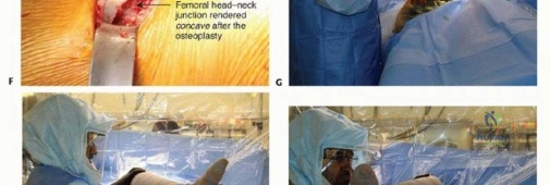
In the vast majority of chronic cases, THA is the mandated procedure. Following the exposure and neurolysis described above, the necrotic femoral head is excised in situ using an oscillating saw. This excised head is an invaluable resource; it must be preserved on the back table to serve as a structural autograft for posterior wall reconstruction. The true acetabulum is identified—often requiring the surgeon to look significantly inferior and medial to the false acetabulum. The native acetabulum is cleared of all fibrotic tissue. If a massive posterior wall defect exists, the native femoral head is fashioned with a saw to precisely fit the defect. This structural graft is temporarily secured with K-wires, and then definitively fixed to the intact ilium and ischium using multiple 4.0mm or 6.5mm cancellous lag screws. The acetabulum, now incorporating the structural graft, is concentrically reamed. A highly porous, multi-hole titanium hemispherical shell is impacted into the true acetabulum, and multiple screws are placed into the safe zones (posterosuperior and posteroinferior quadrants) to ensure rigid primary stability.
Subtrochanteric Shortening Osteotomy
In cases of extreme superior migration where preoperative skeletal traction was either unsuccessful or unavailable, attempting to reduce the femoral component into the true acetabulum will place catastrophic tension on the sciatic nerve. In these instances, a subtrochanteric shortening osteotomy is required. Following acetabular reconstruction, the femur is prepared, and a trial stem is inserted. The amount of required shortening is calculated by overlapping the trial stem with the proximal femur. A transverse or step-cut osteotomy is performed in the subtrochanteric region, excising a cylindrical segment of the femoral diaphysis (typically 2 to 4 centimeters). A modular, diaphyseal-engaging, fully porous-coated revision stem is then utilized to bypass the osteotomy site, providing rigid rotational and axial stability while allowing the hip to be reduced without neurovascular compromise.
Complications, Incidence Rates, and Salvage Management
The complication profile for the surgical management of chronic posterior hip dislocations is significantly higher than that of primary elective arthroplasty or acute trauma surgery. The extensive soft tissue dissection, the inherently compromised neurovascular structures, and the complex bone grafting required create a perfect storm for postoperative morbidity. Surgeons undertaking these procedures must be intimately familiar with the identification and salvage management of these severe complications.
| Complication | Estimated Incidence | Pathophysiology & Risk Factors | Salvage Management & Mitigation |
|---|---|---|---|
| Sciatic Nerve Palsy | 10% - 25% | Chronic tethering, intraoperative stretch during reduction, direct retractor injury, or postoperative hematoma. The peroneal division is most susceptible due to its lateral anatomical position and relative lack of connective tissue support. | Immediate removal of compressive hematomas. Application of an Ankle-Foot Orthosis (AFO) to prevent equinus contracture. Gabapentinoids for neuropathic pain. Most neuropraxias recover within 6-12 months; complete axonotmesis has a poor prognosis. |
| Avascular Necrosis (AVN) | 80% - 100% (if ORIF attempted late) | Disruption of the medial circumflex femoral artery (MCFA) and its retinacular branches during initial trauma. Prolonged dislocation exacerbates ischemia and cellular death. | If AVN develops post-ORIF, the patient will present with recurrent pain and segmental collapse. The definitive salvage is conversion to a Total Hip Arthroplasty (THA). |
| Heterotopic Ossification (HO) | 30% - 50% | Extensive soft tissue stripping, muscle trauma, and bone dust generation during reaming/grafting trigger osteoprogenitor cells in the surrounding musculature. | Prophylaxis is mandatory: Indomethacin (75mg SR daily for 3-6 weeks) or localized radiation (700 cGy single fraction). If severe, mature HO restricts motion (Brooker Class III/IV), surgical excision is performed after 12-18 months once bone scans confirm quiescence. |
| Aseptic Loosening / Graft Resorption | 15% - 30% (at 10 years) | Failure of structural autograft incorporation, poor initial cup fixation, or placement of the cup in the high "false" acetabulum leading to altered, excessive biomechanical forces. | Revision arthroplasty utilizing highly porous metal augments, cup-cage constructs, or custom triflange acetabular components to bridge massive uncontained pelvic discontinuities. |
| Postoperative Instability / Dislocation | 5% - 15% | Abductor insufficiency due to chronic contracture, failure to restore offset, or malpositioning of the acetabular component (inadequate anteversion/abduction). | Closed reduction and application of an abduction orthosis. If recurrent, revision surgery is required to optimize component position, increase head size, or utilize dual-mobility or constrained acetabular liners. |
Phased Post-Operative Rehabilitation Protocols
The postoperative rehabilitation regimen is as critical to the ultimate functional outcome as the surgical execution itself. Because these procedures involve extensive soft tissue releases, complex bone grafting, and frequently osteotomies, the rehabilitation protocol cannot follow standard primary arthroplasty guidelines. It must be highly customized, rigidly phased, and closely monitored by both the surgical team and specialized physical therapists.
Immediate Postoperative Phase and Prophylaxis (Weeks 0-6)
The primary goals in the immediate postoperative phase are the protection of the surgical reconstruction, the prevention of medical complications, and the initiation of safe, restricted mobility. Weight-bearing status is strictly dictated by the surgical procedure. For patients who underwent ORIF with posterior wall plating, or THA with structural autografting or subtrochanteric osteotomy, strict Toe-Touch Weight-Bearing (TTWB) or Non-Weight-Bearing (NWB) is enforced for the first 6 to 8 weeks to prevent catastrophic graft subsidence or fixation failure. Strict posterior hip precautions (no flexion past 90 degrees, no adduction across the midline, no internal rotation) are universally applied. In patients with profound preoperative abductor insufficiency or those deemed non-compliant, a rigid hip abduction orthosis is fitted before the patient leaves the operating room.
Medical prophylaxis during this phase is aggressive. Venous Thromboembolism (VTE) prophylaxis is mandatory due to the high-risk nature of pelvic surgery and restricted mobility. Chemical prophylaxis utilizing Low Molecular Weight Heparin (LMWH), direct oral anticoagulants (DOACs), or adjusted-dose Warfarin is continued for a minimum of 28 to 35 days. Simultaneously, Heterotopic Ossification (HO) prophylaxis is initiated immediately; if radiation therapy was not administered postoperatively, Indomethacin (75 mg sustained release daily) is prescribed for 3 to 6 weeks, provided there are no gastrointestinal or renal contraindications.
Intermediate Mobilization and Weight-Bearing Progression (Weeks 6-12)
At the 6-to-8-week mark, a critical clinical and radiographic evaluation is performed. AP and Judet radiographs are scrutinized for signs of structural graft incorporation, fracture consolidation, or early component subsidence. If radiographic healing is progressing satisfactorily, the patient is transitioned to a graduated weight-bearing protocol. Weight-bearing is advanced by 25% of body weight per week, utilizing crutches or a walker, until full weight-bearing is achieved. Physical therapy during this phase shifts focus toward active-assisted and active range of motion, carefully avoiding extremes of flexion and internal rotation. Isometrics for the quadriceps and hamstrings are intensified, and gentle, progressive strengthening of the abductor complex (gluteus medius) is initiated. Abductor rehabilitation is notoriously prolonged in these patients due to the chronic preoperative shortening and intraoperative manipulation.
Late Rehabilitation and Functional Restoration (Months 3-12)
Once full weight-bearing is achieved without pain, the late rehabilitation phase focuses on functional restoration, gait normalization, and the elimination of the Trendelenburg lurch. Patients engage in closed-kinetic-chain exercises, proprioceptive training, and advanced core stabilization. It is vital to counsel the patient that maximum medical improvement following the reconstruction of a chronic posterior hip dislocation may take up to 18 to 24 months. Persistent abductor weakness is common and may require the long-term use of a cane in the contralateral hand to optimize gait biomechanics and protect the reconstruction from excessive shear forces.
Summary of Landmark Literature and Clinical Guidelines
The evolution of surgical thought regarding chronic posterior hip dislocations has been shaped by several decades of rigorous clinical observation and landmark orthopedic literature. Historically, there was a strong impetus to perform heroic open reductions regardless of the timeline, driven by a desire to preserve the native joint. However, as longitudinal data emerged, the catastrophic rates of AVN and secondary osteoarthritis forced a paradigm shift in treatment algorithms.
The foundational principles of acetabular exposure and reconstruction were established by Letournel and Judet. Their exhaustive anatomical studies defined the Kocher-Langenbeck approach and the biomechanical necessity of restoring the posterior wall to prevent recurrent instability. Their work remains the bedrock upon which modern ORIF and complex THA for fracture-dislocations are based.
In the realm of chronic dislocations specifically, the landmark series by Garrett et al. provided sobering data on the natural history of delayed reductions. They definitively demonstrated that dislocations left unreduced for greater than three months have an AVN rate approaching 100%, rendering joint-preserving procedures futile and highly prone to early failure. This temporal threshold has since become the universally accepted dividing line in clinical guidelines between attempting ORIF versus proceeding directly to primary THA.
More recently, comprehensive reviews and surgical series by authors such as Ilyas & Rabbani have codified the techniques for complex arthroplasty in this cohort. Their work highlighted the critical importance of utilizing the excised, necrotic femoral head as a structural autograft to reconstruct the dysplastic, deficient posterior acetabulum. They also popularized the use of preoperative skeletal traction to mitigate the need for technically demanding subtrochanteric shortening osteotomies, thereby reducing operative time and lowering the incidence of catastrophic sciatic nerve palsies. Current clinical guidelines from major orthopedic bodies (such as the AAOS and OTA) strongly reflect this literature, advocating for a pragmatic, reconstructive approach in chronic cases, prioritizing the restoration of the anatomical hip center, rigid acetabular fixation, and the protection of the compromised neurovascular envelope to achieve durable, long-term functional outcomes.
