Correction of Cervicotrochanteric Malunion: Advanced Surgical Techniques and Outcomes
Key Takeaway
Cervicotrochanteric malunion presents a complex reconstructive challenge characterized by varus collapse, external rotation, and limb shortening. Successful surgical correction requires meticulous preoperative planning, precise osteotomy, and rigid internal fixation. This guide details the step-by-step surgical technique, from the lateral approach and fibrous tissue excision to deformity correction and pediatric considerations. Mastery of these principles is essential for restoring hip biomechanics and optimizing patient outcomes.
Comprehensive Introduction and Patho-Epidemiology
Cervicotrochanteric fractures, which occur at the critical anatomical junction between the base of the femoral neck and the intertrochanteric line, are inherently unstable and biomechanically precarious injuries. When these fractures are managed nonoperatively, or when internal fixation constructs fail due to poor bone quality, technical errors, or patient non-compliance, they frequently progress to a cervicotrochanteric malunion. This pathological state presents a formidable reconstructive challenge for the orthopaedic surgeon. The resulting deformity is rarely uniplanar; rather, it is characterized by a complex, multi-planar three-dimensional distortion encompassing severe varus collapse, marked external rotation of the distal femoral fragment, and significant limb shortening.
The epidemiology of cervicotrochanteric malunions follows a bimodal distribution, mirroring the incidence of the primary fractures. In the younger demographic, these malunions typically result from high-energy trauma, such as motor vehicle collisions or falls from significant heights, where severe comminution and initial displacement overwhelm the capacity of standard fixation constructs. In the elderly, osteoporotic population, malunions often follow low-energy falls where poor bone stock leads to hardware cut-out, varus settling, and eventual malunion. The profound alteration of the proximal femoral anatomy in these patients severely compromises the abductor lever arm. This biomechanical failure inevitably leads to a debilitating Trendelenburg gait, chronic hip and trochanteric pain, and accelerated degenerative joint disease of the hip due to altered joint reaction forces.
Surgical correction of a cervicotrochanteric malunion is technically demanding and requires a profound understanding of hip biomechanics, meticulous preoperative templating, and the precise execution of corrective osteotomies. The primary goal of intervention is not merely radiographic alignment, but the functional restoration of the abductor mechanism, equalization of limb length, and the re-establishment of normal joint kinematics. This comprehensive chapter details the evidence-based surgical management of cervicotrochanteric malunions, providing an exhaustive guide tailored for the postgraduate orthopaedic surgeon, fellow, and resident.
Detailed Surgical Anatomy and Biomechanics
Understanding the intricate pathoanatomy and biomechanics of a cervicotrochanteric malunion is the absolute foundation for executing a successful surgical correction. The proximal femur is subjected to some of the highest physiological loads in the human body, and the deformity seen in malunion is directly driven by the unopposed pull of powerful musculature crossing the hip joint during the vulnerable period of fracture healing.
Deforming Muscular Forces
The characteristic deformity of a cervicotrochanteric malunion is dictated by specific muscle groups acting on the uncoupled proximal and distal fragments.
* Proximal Fragment: The proximal fragment, comprising the femoral head and neck, is subjected to the robust forces of the short external rotators (piriformis, gemelli, obturator internus, and quadratus femoris) and the iliopsoas muscle. The iliopsoas, inserting on the lesser trochanter, exerts a profound force that pulls the proximal fragment into flexion, abduction, and external rotation.
* Distal Fragment: Conversely, the distal fragment (the femoral shaft) is acted upon by the massive adductor complex (adductor longus, brevis, and magnus), which pulls the shaft medially and proximally, resulting in the classic varus collapse and limb shortening. Simultaneously, the weight of the limb, combined with the pull of the gluteus maximus, contributes to the severe external rotation of the distal fragment relative to the proximal segment.
The Malunion Morphology and the Fibrous Wedge
Because of the severe external rotation of the distal fragment relative to the proximal fragment, the fractured surface of the greater trochanter rotates to face anteromedially rather than its native medial orientation. This rotational mismatch creates a highly specific and clinically vital morphological feature.
A hallmark of the cervicotrochanteric malunion is the presence of a distinct, wedge-shaped space between the anterior cortices of the two fragments. The base of this wedge is located anteriorly and is typically filled with dense, unyielding, and highly vascularized fibrous scar tissue. In stark contrast, the actual osseous union occurs predominantly along the posterior cortex, where the bone ends have been driven together by the deforming forces. Recognizing this specific pathoanatomy—the anterior fibrous wedge and the posterior osseous bridge—is the absolute key to successful intraoperative mobilization and reduction. Failure to recognize and completely excise this anterior fibrous block will render any attempt at anatomical reduction impossible.
Biomechanical Consequences of the Deformity
The varus collapse drastically decreases the neck-shaft angle (often falling below 100 degrees from a normal 130-135 degrees). This medializes the mechanical axis of the lower extremity and shortens the crucial abductor lever arm (the distance from the center of the femoral head to the tip of the greater trochanter). To maintain pelvic leveling during the single-leg stance phase of gait, the abductor muscles must generate exponentially higher forces, which increases the overall joint reaction force across the acetabulum. Over time, this mechanical disadvantage leads to abductor fatigue (Trendelenburg lurch) and asymmetric cartilage wear, culminating in secondary osteoarthritis.
Exhaustive Indications and Contraindications
The decision to proceed with the surgical correction of a cervicotrochanteric malunion must be carefully weighed against the patient's physiological age, bone quality, baseline functional status, and the presence of concurrent intra-articular pathology. Not all malunions require surgical correction; asymptomatic or minimally symptomatic malunions in low-demand patients may be managed conservatively with shoe lifts and physical therapy.
Patient Selection Criteria
Ideal candidates for corrective osteotomy are young to middle-aged adults with symptomatic malunions who possess good bone stock and minimal pre-existing degenerative changes in the hip joint. In older adults with significant osteopenia or established osteoarthritis, a corrective osteotomy is often bypassed in favor of a total hip arthroplasty (THA), which provides immediate pain relief and early mobilization.
Tabulated Indications and Contraindications
| Category | Specific Clinical Scenarios | Rationale / Considerations |
|---|---|---|
| Absolute Indications | Intractable pain, severe Trendelenburg gait, progressive varus collapse, impending hardware failure. | Deformity is causing mechanical failure of the joint and severe functional disability. Surgical intervention is required to prevent catastrophic failure or end-stage arthritis. |
| Relative Indications | Limb length discrepancy (LLD) > 2.0 cm, moderate restriction of internal rotation/abduction, cosmetic deformity in a young patient. | Symptoms may be managed with a shoe lift, but surgical correction offers a definitive biomechanical restoration. Depends heavily on patient expectations and physiological age. |
| Relative Contraindications | Advanced chronological age (>70 years), moderate to severe osteoarthritis, osteoporosis (T-score < -2.5), heavy smoking. | Poor bone stock increases the risk of fixation failure and nonunion. Osteoarthritis will not be cured by osteotomy; these patients are better served by Total Hip Arthroplasty (THA). |
| Absolute Contraindications | Active systemic or local infection, medically unfit for prolonged anesthesia, severe Charcot arthropathy, non-ambulatory baseline status. | High risk of perioperative mortality, catastrophic infection, or failure to achieve any functional benefit. |
Pre-Operative Planning, Templating, and Patient Positioning
A rigorous, exhaustive preoperative assessment is mandatory to quantify the three-dimensional deformity and to plan the precise geometry of the corrective osteotomy. The success of the procedure is largely dictated before the patient ever enters the operating theater.
Comprehensive Clinical Assessment
Patients typically present with a pronounced limp, a measurable limb length discrepancy (LLD), and restricted range of motion, particularly in internal rotation and abduction. The surgeon must differentiate between true LLD (measured from the anterior superior iliac spine to the medial malleolus) and apparent LLD (measured from the umbilicus to the medial malleolus), as pelvic obliquity from adductor contractures can confound measurements. A thorough neurovascular examination is essential; the planned surgical correction will involve significant lengthening and internal rotation of the limb, which places acute tension on the sciatic and femoral nerves. Pre-existing neuropathies must be documented to avoid confounding postoperative assessments.
Radiographic Imaging and 3D Templating
Standard two-dimensional radiographs are the starting point: an anteroposterior (AP) pelvis, a cross-table lateral of the affected hip, and full-length standing leg films are required to assess the current neck-shaft angle, femoral offset, and overall mechanical axis alignment. However, 2D imaging is insufficient for complex malunions.
A fine-cut Computed Tomography (CT) scan with 3D reconstructions is the gold standard for evaluating the rotational profile of the femur. The CT scan allows the surgeon to map the exact location and thickness of the posterior osseous bridge versus the anterior fibrous wedge.
Digital templating software must be utilized to simulate the osteotomy. The surgeon must calculate the exact wedge of bone or fibrous tissue to be resected to achieve the desired correction. Furthermore, templating determines the optimal angle of the fixation device (e.g., a 130-degree or 135-degree compression hip screw or cephalomedullary nail) required to restore the normal neck-shaft angle and mechanical axis.
Patient Positioning and Operating Room Setup
The patient is ideally placed supine on a radiolucent fracture table. This specialized positioning is critical as it allows for the application of marked, controlled longitudinal traction and facilitates unimpeded intraoperative fluoroscopy (image intensification) in both the AP and lateral planes. The perineal post must be heavily padded to prevent pudendal nerve neurapraxia. Alternatively, a flat radiolucent table with a sterile femoral distractor can be utilized. While a flat table allows for easier manipulation of the limb, a fracture table is generally preferred for the dynamic, hands-free control of traction and rotation during the critical phases of provisional fixation.
Step-by-Step Surgical Approach and Fixation Technique
The preferred surgical approach for the correction of a cervicotrochanteric malunion is a robust, curved lateral incision, utilizing the internervous plane between the tensor fasciae latae (TFL) and the gluteus medius. This is classically known as the Watson-Jones approach, which provides unparalleled access to the anterior capsule and the cervicotrochanteric junction.
The Watson-Jones Interval and Exposure
The incision begins approximately 2.5 cm posterior to the anterior superior iliac spine (ASIS), curving distally and posteriorly over the greater trochanter, and extending along the proximal 5 to 8 cm of the femoral shaft. The fascia lata is incised sharply in line with the skin incision.
The deep dissection identifies the interval between the TFL (supplied by the superior gluteal nerve) and the gluteus medius (also supplied by the superior gluteal nerve). While this is technically not a true internervous plane, it is an intermuscular plane that provides excellent, safe access. The surgeon must be acutely aware that the superior gluteal nerve and vessels cross the operative field approximately 3 to 5 cm proximal to the tip of the greater trochanter. Dissection proximal to this safe zone must be strictly avoided to prevent denervation of the abductor musculature, which would catastrophically and irreversibly worsen the patient's postoperative gait.
Once the interval is developed, the TFL is retracted anteriorly and the gluteus medius posteriorly. The anterior capsule of the hip is exposed. A robust T-shaped or H-shaped capsulotomy is performed to fully visualize the cervicotrochanteric junction, the base of the femoral neck, and the anterior articular surface of the femoral head.
Debridement and Excision of the Fibrous Wedge
With the joint exposed, the surgeon must identify the anterior wedge-shaped space characteristic of the malunion. Using a combination of heavy rongeurs, sharp curettes, and electrocautery, the surgeon meticulously excises the dense fibrous tissue filling this gap. This tissue must be excised completely down to healthy, bleeding normal bone. This step cannot be rushed; failure to clear this space entirely will leave a mechanical block that prevents the subsequent reduction maneuvers from closing the anterior gap.
Osteotomy and Deformity Correction
Once the anterior fibrous wedge is entirely cleared, the posterior osseous union is visualized. Using a sharp, broad osteotome or a cooled oscillating saw, the surgeon carefully divides the bony bridge posteriorly. Great care must be taken not to plunge posteriorly, which could endanger the sciatic nerve or the crucial medial femoral circumflex artery (MFCA) that supplies the femoral head.
Once the osteotomy is complete, the distal fragment must be completely mobilized and freed from the proximal fragment. The reduction maneuver is then executed, essentially reversing the mechanism of the initial deformity. Marked longitudinal traction is applied to the leg via the fracture table to overcome the shortened adductors. This is followed by controlled internal rotation of the distal fragment, which closes the anterior gap where the fibrous tissue was excised. Finally, abduction of the distal fragment restores the neck-shaft angle and re-establishes the abductor lever arm.
Soft Tissue Balancing: The Adductor Tenotomy
In chronic malunions, the adductor musculature becomes severely contracted and fibrotic. If marked traction and manual abduction fail to restore the neck-shaft angle, the adductors are acting as a rigid tether. In these instances, a percutaneous or open tenotomy of the adductor longus (and occasionally the adductor brevis) near their origin on the pubis is mandatory. This release is often the critical step required to obtain sufficient abduction of the distal fragment without placing undue, potentially catastrophic stress on the osteotomy site and the planned internal fixation.
Internal Fixation Strategies
Once the normal angle between the shaft and the neck has been restored, the reduction must be provisionally held with heavy, threaded Kirschner wires. The reduction is rigorously confirmed via image intensification in both AP and lateral planes.
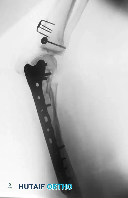
The fracture is typically fixed with a sliding compression hip screw (Dynamic Hip Screw - DHS) or a modern cephalomedullary nail. The guidewire is inserted centrally into the femoral head. It is imperative to achieve a tip-apex distance (TAD) of less than 25 mm, as described by Baumgaertner, to minimize the risk of hardware cut-out. The sliding mechanism of the compression screw allows for dynamic compression across the osteotomy site upon weight-bearing, promoting primary bone healing.
Pediatric Considerations and Physis Preservation
Cervicotrochanteric malunions in the pediatric population require highly specialized considerations due to the presence of open growth plates (the capital femoral physis and the greater trochanteric apophysis) and the unique biology of the developing hip. Standard adult implants are strictly contraindicated. A pediatric compression hip screw or a specialized pediatric locking proximal femoral plate is preferable.
The capital femoral physis must be meticulously avoided during guidewire and screw insertion. Penetration of the physis with large-diameter threaded implants will lead to premature physeal closure, resulting in severe, progressive leg length discrepancy and secondary deformities of the femoral head (coxa breva). If the child is nearing skeletal maturity, intentional epiphysiodesis may be acceptable, but in younger children, smooth pins or screws that stop short of the physis are mandatory.
Complications, Incidence Rates, and Salvage Management
The surgical correction of a cervicotrochanteric malunion is a massive undertaking fraught with potential complications. Managing patient expectations preoperatively is paramount, as the complication profile is significantly higher than that of primary fracture fixation.
Tabulated Complications and Salvage Strategies
| Complication | Estimated Incidence | Prevention Strategy | Salvage Management |
|---|---|---|---|
| Loss of Correction (Varus Collapse) | 10% - 15% | Meticulous templating, achieving TAD < 25mm, utilizing a 135-degree implant, strict adherence to weight-bearing restrictions. | Revision internal fixation with bone grafting; Valgus-producing subtrochanteric osteotomy; Total Hip Arthroplasty (THA) in older patients. |
| Avascular Necrosis (AVN) of Femoral Head | 5% - 10% | Careful protection of the posterior structures (Medial Femoral Circumflex Artery) during osteotomy; avoiding excessive traction. | Core decompression (if early pre-collapse); Total Hip Arthroplasty (THA) for late-stage collapse. |
| Nonunion of Osteotomy | 3% - 7% | Complete excision of fibrous tissue down to bleeding bone; dynamic compression; avoiding thermal necrosis with the saw. | Revision fixation with autologous bone grafting (iliac crest); optimization of patient biology (smoking cessation, Vitamin D). |
| Hardware Cut-Out | 4% - 8% | Central-central placement of the lag screw; avoiding superior quadrant placement; respecting poor bone density. | Removal of hardware and conversion to THA using a diaphyseal-engaging stem to bypass the compromised proximal bone. |
| Nerve Palsy (Sciatic / Superior Gluteal) | 1% - 3% | Avoiding dissection >5cm proximal to greater trochanter; padding the perineal post; avoiding excessive acute limb lengthening (>3cm). | Observation and supportive care (AFO for foot drop); neurolysis if entrapment is suspected; usually resolves within 3-6 months if neurapraxia. |
Prognosis and Long-Term Outcomes
In children, the results of corrective osteotomy are historically disappointing. Due to the rapid remodeling potential and the complex interplay of growth plates, only moderate improvement in position may be secured long-term, and recurrence of deformity is common. Because salvage procedures yield suboptimal results, the efficient and aggressive anatomical treatment of fresh cervicotrochanteric fractures in children is absolutely crucial to prevent malunion from occurring in the first place.
In young adults, the functional outcome is generally much improved following surgical correction. Patients typically experience significant relief of pain and an improvement in their Trendelenburg gait. However, the surgeon must counsel the patient preoperatively that rarely, if ever, is the absolute length of the limb restored completely to its pre-injury state. A residual mild limb length discrepancy (usually < 1.5 cm) or slight limitation in terminal hip rotation is to be expected and is considered an acceptable outcome.
Phased Post-Operative Rehabilitation Protocols
The postoperative rehabilitation protocol is a critical determinant of the final functional outcome. It must be meticulously tailored to the patient's age, bone quality, and the intraoperative assessment of the rigidity of the internal fixation achieved.
Pediatric Postoperative Protocol
If complete correction has been secured and the fracture was fixed internally in a child, the mechanical hold of the implants is often insufficient to withstand the robust, non-compliant activity levels characteristic of a pediatric patient. Therefore, supplemental external immobilization is mandatory. A one-and-a-half spica cast should be applied immediately postoperatively in the operating room. The hip should be immobilized in slight abduction and neutral rotation. This cast is typically worn for a minimum of 6 to 8 weeks. Serial radiographs are required every 2 to 3 weeks to monitor for loss of reduction within the cast. Once bridging callus is visualized, the cast is removed, and a phased return to weight-bearing and active range of motion is initiated under the guidance of a pediatric physical therapist.
Adult Postoperative Protocol: A Phased Approach
In young adults with good bone stock and rigid fixation utilizing a compression hip screw or cephalomedullary nail, a structured, three-phase rehabilitation program is employed.
- Phase 1: Maximum Protection (Weeks 0-6)
Patients are immediately mobilized but are strictly restricted to toe-touch weight-bearing (TTWB) or partial weight-bearing (PWB, max 20 lbs) using a walker or crutches. Early passive and active-assisted range of motion exercises are initiated to prevent capsular adhesions. Aggressive DVT prophylaxis (chemical and mechanical) is mandatory during this phase. Abductor strengthening is limited to isometric gluteal sets to avoid excessive sheer stress across the osteotomy site. - Phase 2: Progressive Loading (Weeks 6-12)
Progression to this phase is strictly contingent upon radiographic evidence of early bridging callus at the osteotomy site. Weight-bearing is gradually advanced by 25% of body weight per week. Active abductor strengthening (e.g., side-lying hip abduction, clamshells) is initiated. Stationary cycling with low resistance is excellent for restoring joint mobility and cartilage nutrition. - Phase 3: Functional Restoration (Months 3-6)
Once full weight-bearing is achieved without pain, and radiographs demonstrate solid osseous union, the focus shifts to gait retraining, proprioception, and advanced strengthening. Closed kinetic chain exercises (leg presses, mini-squats) are incorporated. The ultimate goal is the elimination of the Trendelenburg lurch and the return to pre-injury occupational and recreational activities.
Summary of Landmark Literature and Clinical Guidelines
The evolution of surgical techniques for proximal femoral malunions is deeply rooted in historical orthopaedic literature, which continues to guide modern clinical practice.
The surgical approach detailed in this chapter owes its foundation to the pioneering work of Sir Reginald Watson-Jones, who first described the anterolateral interval for hip access, minimizing trauma to the abductor mechanism. The biomechanical principles of deformity correction in the proximal femur are heavily influenced by the work of Friedrich Pauwels. Pauwels' theories on the conversion of shearing forces into compressive forces via valgus-producing osteotomies remain the biomechanical rationale for restoring the neck-shaft angle in these malunions.
Regarding internal fixation, Baumgaertner's landmark 1995 study on the Tip-Apex Distance (TAD) revolutionized the use of the sliding hip screw. His finding that a TAD of less than 25 mm is the single most important predictive factor for preventing hardware cut-out is a universally accepted clinical guideline that applies equally to acute fractures and corrective osteotomies.
Furthermore, when evaluating a patient with a proximal femoral malunion, the surgeon must maintain a high index of suspicion for concurrent intra-articular pathology, particularly if the patient has a history of high-energy trauma or hip dislocation. Literature reports have documented cases of femoral head avulsion fractures (Pipkin lesions) that have malunited directly to the acetabulum, creating a bony block to motion. In such complex scenarios, the guidelines established by Ganz regarding the surgical dislocation of the hip are paramount. The Ganz approach allows for a safe, 360-degree visualization of the femoral head while preserving the medial femoral circumflex artery, enabling meticulous surgical débridement of intra-articular malunions. Excellent functional results can be obtained after restoration of articular congruity, provided avascular necrosis has not already compromised the femoral head.
Modern clinical guidelines strongly advocate for joint-preserving osteotomies in symptomatic patients under the age of 50. However, in patients over the age of 65, or those with established osteoarthritis, the literature consistently demonstrates that Total Hip Arthroplasty (THA) provides superior, more predictable outcomes with lower revision rates compared to complex reconstructive osteotomies.
