Erb-Duchenne Brachial Plexus Birth Palsy: Clinical Diagnosis & Imaging in Newborns
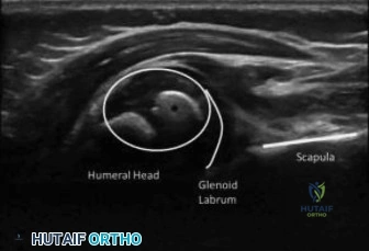
Key Takeaway
Erb-Duchenne brachial plexus birth palsy is diagnosed in newborns through a clinical exam noting 'waiter's tip' posture, flaccidity, and absent Moro/biceps reflexes. X-rays exclude fractures. MRI at 3 weeks assesses nerve root integrity, confirming the injury and guiding early management, essential for optimal outcomes.
A 3-week-old infant is referred for right-sided arm flaccidity following a difficult vaginal delivery complicated by shoulder dystocia. On examination, the arm is adducted, internally rotated, and pronated. Active Movement Scale (AMS) shows no anti-gravity movement at the shoulder or elbow. You suspect an Erb-Duchenne palsy. What are the key differential diagnoses you must exclude, and how do you differentiate them?
Candidate: I would consider birth-related fractures like a clavicle or proximal humerus fracture, which cause pseudoparalysis. I would also rule out septic arthritis of the shoulder, central nervous system injury/stroke, and congenital muscular torticollis. I differentiate these through clinical exam—checking for focal bony tenderness, crepitus, or systemic signs of infection—and confirm with plain radiographs to exclude fractures.
Failing to mention the "systemic" aspect of the examination. Candidates often focus purely on the limb. A poor candidate forgets to mention checking for Horner’s syndrome (cervical sympathetic chain) or respiratory distress (phrenic nerve involvement/C3-C5), which are essential for gauging the severity and extent of the plexus injury.
I would categorize the differential into: 1) Mechanical/Bony: Clavicle/Proximal humeral fractures (ruled out via X-ray and palpation for crepitus). 2) Infectious: Septic arthritis (systemic markers, fever, limited passive ROM due to pain). 3) Neurological: CNS/Stroke (look for contralateral deficits or altered mental status). 4) Soft Tissue: Torticollis (SCM mass). Crucially, I would highlight that in BPBP, the physical exam shows full passive ROM compared to the limited/guarded ROM in septic arthritis or fracture, and confirm the diagnosis with a focused plexus MRI to assess root integrity.
We are now at the 3-month follow-up mark. The infant still shows no antigravity biceps function. Based on the MRI image provided, discuss your management plan.
Candidate: At 3 months, the absence of biceps function is a clear indication for surgical exploration. The MRI shows T2 hyperintensity and thickening, indicating a neuroma-in-continuity. I would plan for surgical exploration to assess the nerve conductivity, potentially performing nerve grafting for the C5/C6 roots and considering an Oberlin nerve transfer to restore elbow flexion.
Suggesting that MRI findings alone dictate the surgery. A borderline candidate forgets that clinical findings—specifically the "biceps at 3 months" rule—are the gold standard indication. They also often fail to prioritize the specific nerves for transfer (e.g., confusing the medial pectoral nerve with other donors).
I would state that the "biceps at 3 months" rule is the primary driver for surgical intervention. The MRI findings of neuroma-in-continuity suggest a Sunderland Grade 3/4 injury, which will not achieve satisfactory spontaneous recovery. My plan: 1. Surgical Exploration: Micro-neurolysis and intraoperative nerve action potential (NAP) testing. 2. Reconstruction: Resection of the neuroma and interpositional sural nerve grafting. 3. Targeted Transfer: Perform an Oberlin transfer (medial pectoral nerve to musculocutaneous nerve branch) to ensure early, reliable reinnervation of the biceps, as grafting recovery is slow and often incomplete in proximal injuries.
The surgery was performed, including nerve grafting and an Oberlin transfer. Walk me through your postoperative rehabilitation protocol and how you objectively monitor progress.
Candidate: Postoperatively, the patient is immobilized for 4–6 weeks in an airplane splint. After that, I would start passive range of motion to prevent contractures. Long-term monitoring uses the Active Movement Scale (AMS) for motor function and the Mallet score for assessing shoulder function, tracked every 3 months.
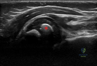
Neglecting the "secondary deformities" aspect. Examiners want to hear about the risk of glenohumeral dysplasia and internal rotation contractures. A candidate who only focuses on the nerve repair misses the necessity of long-term screening for posterior humeral head subluxation.
I would structure this into three phases: 1) Protection (0-6 wks): Immobilization to protect the coaptation. 2) Rehabilitation (6 wks-1 yr): Gentle passive ROM, moving to active-assisted as the nerves reinnervate. 3) Long-term Surveillance: Regular Mallet scoring and serial imaging to screen for glenohumeral dysplasia/retroversion. I would emphasize that even with successful nerve repair, we must remain vigilant for secondary contractures (e.g., subscapularis tightness), which may require secondary orthopaedic procedures like tendon transfers (Latissimus Dorsi/Teres Major) later in childhood.
