Ring Finger Fracture Management: CRPP Techniques, Surgical Anatomy & Biomechanics
Key Takeaway
Closed Reduction and Percutaneous Pin Fixation (CRPP) is a minimally invasive treatment for unstable or displaced ring finger fractures, including metacarpal and phalangeal types. It preserves soft tissue, reduces surgical time, and often allows earlier mobilization than open surgery. Success depends on meticulous technique, accurate reduction, and stable pin placement, critical for preserving hand function and preventing malunion.
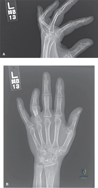
A 28-year-old amateur boxer presents to the ED following an altercation. He has pain and swelling over the ring finger metacarpal. On examination, he has a stable, closed injury. You obtain radiographs and suspect an unstable neck fracture. Explain the biomechanics of the deformity and the clinical assessment you would perform to rule out rotational malalignment.
Candidate: The fracture occurs at the neck, and the distal fragment tilts volarly because the interosseous muscles and flexor tendons pull it forward. I would check the finger cascade by asking the patient to make a fist to see if the fingers overlap, which would indicate rotational malalignment.
Failing to mention the extensor hood mechanism or the specific anatomical direction of the apex-dorsal angulation. Candidates often describe "overlapping" but miss the specific instruction to look for "scissoring" and convergence of the fingertips toward the scaphoid tubercle.
The fracture deformity is apex-dorsal angulation. This is driven by the intrinsic muscle pull (interossei) and the FDS/FDP tendons, which create a flexion moment at the MCP joint, causing the distal fragment to displace volarly. For assessment, I must check the digital cascade: when the patient makes a gentle fist, all fingertips must point toward the scaphoid tubercle. Rotational malalignment is detected by observing the nail bed planes—they should be parallel. Any 'scissoring' or overlapping of the digits is a clinical emergency indicating rotational malunion.
You have decided to proceed with closed reduction and percutaneous pin fixation (CRPP) for the patient's metacarpal fracture. Describe your technical approach and how you would ensure stability.
Candidate: I would use the Jahss maneuver to reduce the fracture by flexing the MCP joint to 90 degrees and applying pressure to the dorsal aspect. Then I would insert two K-wires percutaneously, ideally crossing them within the metacarpal canal to ensure stability.
Neglecting to mention the "danger" of the extensor tendons when placing pins, or failing to emphasize the need for fluoroscopic confirmation of bicortical purchase. Candidates also forget to mention the specific drill speed to avoid thermal necrosis.
Following reduction with the Jahss maneuver, I use retrograde intramedullary pinning. I insert 0.035" or 0.045" K-wires dorsally, just distal to the fracture site, taking care to avoid the extensor mechanism. I drill at a slow speed to prevent thermal bone necrosis. I obtain bicortical purchase in the proximal fragment, usually with two crossed wires. The stability is confirmed by ensuring the digital cascade is restored, fluoroscopic confirmation of alignment in AP/lateral planes, and checking for rotational stability before burying the pins.
What are the absolute contraindications for CRPP in phalangeal fractures, and how would you manage a PIP joint volar plate avulsion fracture?
Candidate: Absolute contraindications are infection, severe comminution that prevents stable reduction, and non-reducible articular fractures. For a volar plate avulsion, I would use a K-wire to block the joint from extending, keeping it in slight flexion.
Forgetting that "severe soft tissue loss" is a significant contraindication. Additionally, candidates often fail to specify the degree of PIP flexion (20-30 degrees) required for the blocking pin.
Absolute contraindications include gross contamination/infection, severe comminution that precludes closed reduction, and non-reducible articular fractures requiring open visualization. For a volar plate avulsion, I utilize a 'dorsal blocking pin' technique: a K-wire is placed across the PIP joint to block hyperextension while allowing for limited, protected motion. The joint is typically positioned in 20-30 degrees of flexion to allow the volar plate to heal anatomically against the proximal phalanx.
