

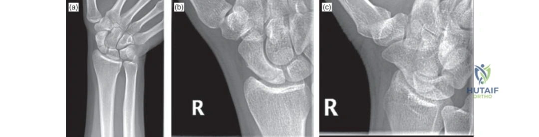
A 28-year-old rugby player presents with persistent wrist pain following a FOOSH injury 3 weeks ago. Initial X-rays were reported as normal, but he remains symptomatic. You are concerned about an occult scaphoid fracture. What is your systematic diagnostic approach, and why is this patient's presentation potentially high-risk?
Candidate: I would perform a thorough clinical exam, specifically looking for anatomical snuffbox and scaphoid tubercle tenderness. If suspected, I would request specialized scaphoid views. Since he is symptomatic after 3 weeks, I would order a CT scan or MRI. It is high-risk because the scaphoid has a retrograde blood supply, putting the proximal pole at risk of AVN if the fracture is missed or unstable.
Candidates often fail to mention the specific mechanism or the "time-dependency" of diagnostics. They may suggest a repeat X-ray in 2 weeks as the *initial* step, forgetting that at 3 weeks, a CT is more diagnostic. They also often fail to mention the impact of smoking or the functional consequences of SNAC wrist.
Start with the clinical assessment: palpation of the anatomical snuffbox, tubercle, and axial loading of the thumb. Acknowledge that standard films have poor sensitivity (often <70%). Given the 3-week delay, I would bypass repeat radiographs and proceed immediately to a fine-cut (0.625–1mm) CT scan to evaluate for fracture, displacement, or early nonunion. I would explain that the retrograde blood supply (primarily the dorsal carpal branch) makes the proximal pole susceptible to AVN. I would also screen for risk factors like smoking which impede healing.
The CT scan confirms a displaced scaphoid waist fracture with a 15-degree humpback deformity. You decide to proceed to surgery. Please justify your choice of surgical approach and describe the key anatomical intervals.
Candidate: I would choose a volar approach (Modified Henry's). This is better for waist fractures as it allows me to address the humpback deformity by manipulating the fragments. The interval is between the Flexor Carpi Radialis (FCR) tendon and the radial artery. I would be careful of the palmar cutaneous branch of the median nerve.
Failing to mention the "humpback deformity" correction. Simply saying "I fix it with a screw" ignores the biomechanical necessity of restoring length and correcting the flexion deformity of the proximal pole.
I would use a volar approach (Modified Henry's) because it provides excellent visualization of the waist and allows for reduction of the humpback deformity, often requiring a joystick maneuver or dorsal compression. The interval is between the FCR (retracted ulnarly) and the radial artery (retracted radially). I must mention the RSC (radioscaphocapitate) ligament, which may require a capsulotomy for access. Protection of the palmar cutaneous branch of the median nerve is essential. If the deformity is rigid, I would consider a bone graft to restore scaphoid length.
During the procedure, you are assessing the proximal pole. You observe it is sclerotic and fails to bleed upon curettage. How does this change your management plan?
Candidate: This suggests avascular necrosis. I would switch to a vascularized bone graft, likely a 1,2 ICSRA graft, to provide a blood supply to the proximal pole. A standard screw fixation alone will likely fail to achieve union.
Suggesting only a standard non-vascularized bone graft (like distal radius corticocancellous graft) without addressing the viability of the bone. In the presence of established AVN, vascularized tissue is generally required for biological success.
Intraoperative findings of sclerosis and lack of punctate bleeding confirm AVN. My priority shifts to revascularization. I would perform a vascularized bone graft—the 1,2 Intercompartmental Supraretinacular Artery (ICSRA) pedicled graft is the gold standard here. I would fix the scaphoid with a headless compression screw to provide stability, ensuring the graft is secured into the proximal pole to bridge the nonunion and provide biological support.
Detailed Chapters & Topics
Dive deeper into specialized chapters regarding mastering-the-management-of-scaphoid-fractures-understanding-classification-treatment-and-complications