Posterolateral Bone Grafting for Tibial Nonunion: A Comprehensive Surgical Guide
Key Takeaway
Posterolateral bone grafting is a highly effective, single-stage procedure indicated for complex tibial nonunions complicated by anterior infection, poor soft tissue envelopes, or extensive bone loss. By utilizing a pristine posterior muscular bed and avoiding the compromised anterior tissues, this technique achieves union rates of 80% to 97%. This guide details the precise surgical approach, interosseous membrane preservation, and structured postoperative rehabilitation protocols.
INTRODUCTION TO POSTEROLATERAL BONE GRAFTING
The management of tibial diaphyseal nonunions presents a formidable challenge to the orthopedic surgeon, primarily due to the precarious subcutaneous nature of the anteromedial tibial border. High-energy trauma often results in severe compromise of the anterior soft tissue envelope, rendering traditional anterior surgical approaches and plating techniques highly susceptible to wound breakdown, deep infection, and hardware exposure.
Posterolateral bone grafting has emerged as a highly reliable, single-stage, nondestructive salvage procedure specifically designed to bypass the hostile anterior environment. By utilizing the robust, highly vascularized, and typically unscarred posterior muscular bed, surgeons can deliver massive quantities of osteogenic autograft directly to the nonunion site. Historically recommended for tibial fractures complicated by active or quiescent anterior infection, or those with extensive segmental bone loss, this technique boasts reported union rates ranging from 80% to 97%. The average time to clinical and radiographic union spans 5 to 7 months.
The primary biomechanical and biological advantage of posterolateral grafting lies in its ability to stimulate profound osteogenesis without disrupting the fragile anterior soft tissues or destabilizing the existing fibrous nonunion. This comprehensive guide delineates the precise indications, surgical anatomy, operative technique, and postoperative rehabilitation protocols required to master this indispensable procedure.
INDICATIONS AND CONTRAINDICATIONS
Indications
The decision to proceed with a posterolateral bone graft is dictated by the condition of the host tissue and the morphology of the nonunion. Primary indications include:
* Infected Tibial Nonunions: Particularly those with a history of anterior soft tissue defects, chronic osteomyelitis, or active anterior draining sinuses, provided the posterior compartment remains pristine.
* Extensive Anterior Bone Loss: Cases where anterior cortical continuity is lost, and anterior grafting would lack sufficient soft tissue coverage.
* Failed Anterior Procedures: Patients who have undergone multiple failed anterior plating or grafting attempts, resulting in a scarred, avascular anterior bed.
* Compromised Soft Tissue Envelope: Severe soft tissue compromise (e.g., post-fasciotomy scarring, split-thickness skin grafts over the anterior tibia) that precludes safe anterior surgical incisions.
Contraindications
- Posterior Compartment Infection: Any history of deep infection within the posterior or lateral compartments of the leg is an absolute contraindication, as the procedure relies on a sterile, highly vascularized posterior muscle bed.
- Severe Vascular Compromise: Injury or occlusion of the posterior tibial artery. The posterior tibial artery is the primary blood supply to the posterior compartment and the graft bed; its absence significantly increases the risk of ischemic complications and graft failure.
- Inadequate Autograft Source: Patients with severe osteoporosis, prior extensive iliac crest harvests, or systemic conditions precluding the harvest of a large volume of high-quality corticocancellous autograft.
💡 Clinical Pearl: The "Nondestructive" Principle
The hallmark of the posterolateral approach is that it is a nondestructive procedure. You must not disturb the existing fibrous union or penetrate the interosseous membrane. The fibrous tissue provides inherent stability; disrupting it converts a stable nonunion into an unstable one, necessitating complex internal or external fixation.
PREOPERATIVE PLANNING AND IMAGING
Meticulous preoperative planning is critical for the success of posterolateral bone grafting.
- Radiographic Evaluation: Standard orthogonal (anteroposterior and lateral) radiographs of the entire tibia and fibula are mandatory to assess the nonunion's geometry, the extent of bone loss, and the presence of any retained hardware.
- Advanced Imaging: A computed tomography (CT) scan is highly recommended to precisely define the nonunion architecture, identify sequestra, and evaluate the quality of the posterior tibial cortex.
- Vascular Assessment: If there is any history of vascular injury or if peripheral pulses are asymmetric, a CT angiogram or formal angiography must be obtained to confirm the patency of the posterior tibial artery.
- Host Optimization: Optimization of host biology is non-negotiable. Smoking cessation must be strictly enforced, as nicotine profoundly inhibits angiogenesis and osteogenesis. Nutritional parameters (Albumin, Prealbumin, Vitamin D) should be optimized prior to surgical intervention.
SURGICAL ANATOMY
A profound understanding of the posterolateral anatomy of the leg is essential to safely navigate the intervals and protect critical neurovascular structures.
- The Surgical Interval: The approach exploits the internervous and intermuscular plane between the lateral compartment (peroneus longus and brevis, innervated by the superficial peroneal nerve) and the superficial/deep posterior compartments (gastrocnemius, soleus, flexor hallucis longus, and tibialis posterior, innervated by the tibial nerve).
- The Interosseous Membrane: This robust fibrous sheet connects the interosseous crests of the tibia and fibula. It serves as the anterior boundary of the dissection. Penetrating it risks injury to the anterior neurovascular bundle and violates the pristine posterior environment.
- The Neurovascular Bundle: The posterior tibial artery and tibial nerve descend through the deep posterior compartment, lying posterior to the tibialis posterior muscle. During the subperiosteal reflection of the deep posterior musculature from lateral to medial, these structures are safely retracted medially within the muscle belly.
SURGICAL TECHNIQUE: STEP-BY-STEP
1. Patient Positioning and Preparation
The patient is placed in the prone position on a radiolucent operating table. Ensure all bony prominences are meticulously padded. A thigh tourniquet is applied to the operative leg. The ipsilateral posterior iliac crest is prepped and draped simultaneously to allow for the harvest of a massive volume of corticocancellous autograft.
⚠️ Surgical Warning: Tourniquet Time
While a tourniquet provides a bloodless field, strict adherence to tourniquet time limits (maximum 120 minutes) is crucial to prevent ischemic injury to the posterior musculature, which will serve as the vascular bed for the bone graft.
2. The Incision
With the tourniquet inflated, make a longitudinal incision 1 to 2 cm posterior to the posterior border of the fibula and parallel to it. The length of the incision is dictated by the extent of the nonunion; it is imperative to expose at least 4 to 5 cm of healthy bone both proximal and distal to the nonunion site to ensure an adequate bed for graft incorporation.
3. Superficial Dissection and Fascial Incision
Divide the subcutaneous tissues sharply. Take care to identify and protect the sural nerve and lesser saphenous vein, which may cross the proximal or distal extents of the incision. Identify the deep fascial plane separating the gastrocnemius-soleus muscle group posteriorly from the peroneal muscles anteriorly. Incise the deep fascia longitudinally along this interval.
4. Deep Muscle Reflection
- Identify the Fibula: Retract the peroneal muscles anteriorly to expose the posterolateral aspect of the fibula.
- Reflect the Flexor Hallucis Longus (FHL) and Soleus: Using a periosteal elevator, subperiosteally reflect the fibular origins of the FHL and the soleus muscles from lateral to medial.
- Reflect the Tibialis Posterior: Continue the subperiosteal dissection medially, reflecting the origin of the posterior tibial tendon (tibialis posterior muscle) off the posterior aspect of the interosseous membrane.
5. Exposure of the Tibia and Neurovascular Protection
Continue the medial dissection to expose the posterior surface of the tibia. This requires elevating the remainder of the posterior tibial tendon origin and the FHL from the fibula and interosseous membrane.
🚨 Pitfall: Neurovascular Injury
As you dissect medially across the posterior tibia, you must retract the posterior tibial artery, posterior tibial vein, and tibial nerve medially. Do not attempt to isolate or expose them. They are safely enveloped within the retracted muscle mass. Overzealous dissection or the use of sharp retractors medially can result in catastrophic vascular injury.
6. Preparation of the Recipient Bed
The success of the graft depends entirely on the preparation of a bleeding, osteogenic bed.
* Preserve the Nonunion: Do not disturb the fibrous union at the fracture site. Do not penetrate the interosseous membrane, as this will allow the graft to migrate anteriorly into the compromised soft tissue envelope.
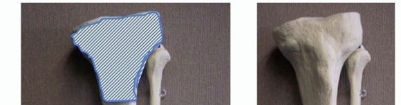
* Decortication: Using a sharp, curved osteotome, raise aggressive "shingle" shavings of the posterior tibial cortex for 4 to 5 cm above and below the nonunion. This decortication exposes the Haversian canals, promoting bleeding and the influx of osteoprogenitor cells.
* Fibular Preparation: Prepare the medial side of the fibula in a similar fashion, creating a continuous bleeding bed across the posterior tibia, interosseous membrane, and medial fibula.
7. Graft Harvesting and Placement
Simultaneous with the leg exposure (if a two-team approach is utilized), harvest a large volume of corticocancellous bone from the posterior iliac crest.
* Obtain small, matchstick-sized strips of corticocancellous bone and copious amounts of cancellous marrow.
* Pack the graft densely over the decorticated posterior tibial surfaces, across the intact interosseous membrane, and onto the prepared medial fibula. This creates a massive posterolateral bone block that will eventually consolidate into a robust synostosis.
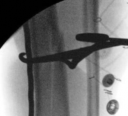
8. Radiographic Confirmation
Before closure, obtain an intraoperative radiograph. Crucially, the radiograph must be taken with the leg in 30 degrees of internal rotation. This specific angle profiles the posterolateral aspect of the tibia and fibula, allowing the surgeon to confirm that the graft mass is adequately positioned and spans the entirety of the nonunion site.
9. Closure
- Hemostasis and Drainage: Deflate the tourniquet and achieve meticulous hemostasis. Insert deep suction drains to prevent postoperative hematoma formation, which can lead to infection or compartment syndrome.
- Fascial Closure: Loosely close the deep fascia over the muscle mass with a few interrupted, absorbable sutures. The goal is merely to hold the graft in place; a tight fascial closure over the added volume of the bone graft will precipitate acute posterior compartment syndrome.
- Skin Closure: Close the subcutaneous tissue and skin in a routine, tension-free manner.
- Dressing: Apply a bulky, well-padded compression dressing to control edema.
POSTOPERATIVE CARE AND REHABILITATION
The postoperative protocol is as critical as the surgical execution. Because the procedure relies on the existing fibrous union for stability, the limb must be rigidly protected until the massive bone graft incorporates and consolidates.
Immediate Postoperative Phase (Days 1-3)
- Monitoring: Close monitoring for signs of compartment syndrome is mandatory, given the volume of graft placed within the posterior compartment.
- Drain Management: Suction drains are typically removed when output ceases or becomes negligible, usually within 1 to 2 days.
- Dressing Change: The initial bulky compression dressing is removed when acute swelling subsides, generally around day 3.
Phase I: Maximum Protection (Weeks 1-6)
- Once the initial swelling has decreased, a long-leg, strictly non-weight-bearing (NWB) cast is applied.
- The patient must remain NWB for a full 6 weeks. This period is critical for the initial vascularization of the bone graft and the prevention of shear forces that could disrupt the fragile early osteoid formation.
Phase II: Progressive Loading (Weeks 6 to Clinical Union)
- At the 6-week mark, the cast is removed, and clinical/radiographic evaluations are performed.
- A new long-leg cast is applied.
- The patient is transitioned to partial weight-bearing (PWB). Controlled axial loading during this phase stimulates Wolff's Law, promoting the consolidation and remodeling of the posterolateral graft mass. This cast is maintained until union is clinically evident (absence of pain on palpation or stress, and absence of gross motion).
Phase III: Maturation (Clinical Union to Radiographic Union)
- Once clinical union is achieved, the patient is transitioned to a short-leg, full weight-bearing (WB) cast.
- This cast is worn for an additional 6 to 9 weeks.
- Immobilization is discontinued only when union is apparent radiographically—defined as the presence of bridging trabeculae across the nonunion site on at least three out of four cortices on orthogonal views. The total average time to union is 5 to 7 months.
COMPLICATIONS AND MANAGEMENT
While highly successful, posterolateral bone grafting carries specific risks:
- Compartment Syndrome: The addition of a large volume of autograft into the posterior compartment increases intracompartmental pressure. Loose fascial closure and vigilant postoperative monitoring are imperative.
- Vascular Injury: Laceration or thrombosis of the posterior tibial artery during medial retraction can lead to graft failure or limb ischemia. Gentle, blunt retraction of the muscle belly is the primary preventative measure.
- Graft Resorption/Persistent Nonunion: Occurs in 3% to 20% of cases, often due to inadequate decortication, insufficient graft volume, premature weight-bearing, or patient non-compliance (e.g., smoking). Management may require a repeat grafting procedure or, if the anterior soft tissues have healed, conversion to rigid internal fixation.
CONCLUSION
Posterolateral bone grafting remains a cornerstone technique in the armamentarium of the reconstructive orthopedic surgeon. By respecting the anatomical intervals, preserving the interosseous membrane, and delivering a massive volume of osteogenic autograft to a pristine vascular bed, surgeons can reliably achieve union in some of the most challenging and hostile tibial nonunions. Strict adherence to the surgical steps and the protracted, phased postoperative rehabilitation protocol is essential to realize the 80% to 97% success rates associated with this masterclass procedure.
You Might Also Like