Operative Techniques in Flexor Tendon Reconstruction: FDS to FPL Transfer and Flexor Tenolysis
Key Takeaway
The transfer of the ring finger flexor digitorum superficialis (FDS) to the flexor pollicis longus (FPL) is a reliable reconstructive procedure for restoring thumb flexion. This comprehensive guide details the surgical technique, including tendon harvesting, routing, and fixation. Furthermore, it outlines the strict indications, prerequisites, and biomechanical considerations for flexor tenolysis following primary repair or grafting, ensuring optimal functional recovery in complex hand injuries.
INTRODUCTION TO FLEXOR POLLICIS LONGUS RECONSTRUCTION
The loss of flexor pollicis longus (FPL) function profoundly impairs hand biomechanics, severely diminishing pinch strength, precision grip, and overall hand dexterity. When primary repair of an FPL rupture or laceration is impossible due to tendon retraction, segmental loss, or severe myostatic contracture, tendon transfer or grafting becomes the reconstructive standard of care.
The transfer of the ring finger flexor digitorum superficialis (FDS)—historically referred to as the flexor sublimis—to the FPL is a highly reliable, synergistic tendon transfer. The ring FDS provides excellent excursion, appropriate cross-sectional area, and a vector of pull that closely mimics the native FPL. This masterclass delineates the precise surgical execution of the FDS-to-FPL transfer, the completion of two-stage tendon grafting, and the rigorous decision-making process surrounding flexor tenolysis in the setting of postoperative tendon adherence.
TRANSFER OF RING FINGER FLEXOR SUBLIMIS TO FLEXOR POLLICIS LONGUS
Indications and Preoperative Planning
The FDS-to-FPL transfer is primarily indicated for chronic FPL ruptures (e.g., attritional ruptures secondary to rheumatoid arthritis or volar plate osteophytes, such as Mannerfelt syndrome), neglected lacerations with significant tendon retraction, or failed primary repairs where the native FPL muscle belly has undergone irreversible fibrosis and lost its excursion capacity.
💡 Clinical Pearl:
Before selecting the ring finger FDS as a donor, the surgeon must confirm the independent function of the FDS to the ring finger and ensure that the flexor digitorum profundus (FDP) of the ring finger is fully intact. Harvesting the FDS in a finger with a compromised FDP will result in a devastating loss of active proximal interphalangeal (PIP) and distal interphalangeal (DIP) joint flexion.
Step-by-Step Surgical Technique
1. Thumb Exposure and Preparation of the Insertion Site
- Incision: Expose the FPL insertion utilizing either a mid-lateral incision or a volar Bruner (zigzag) incision over the volar aspect of the thumb.
- Dissection: Elevate the skin flaps meticulously, taking great care to identify and retract the radial and ulnar digital neurovascular bundles.
- Sheath Management: Preserve the annular thickenings of the flexor sheath, specifically the oblique pulley and the A1 pulley, to prevent postoperative bowstringing. Open only enough of the crucial sheath (usually between the A1 and oblique pulleys or distal to the oblique pulley) to identify the FPL tendon insertion at the base of the distal phalanx. Leave the native FPL stump attached to its bony insertion to serve as a robust anchor for the incoming transfer.
2. Harvesting the Ring Finger Flexor Sublimis
- Palmar Incision: Make a transverse palmar incision at the level of the proximal flexion crease of the ring finger.
- Tendon Identification: Identify the FDS and FDP tendons. Isolate the FDS tendon.
- Maximizing Length: Acutely flex the metacarpophalangeal (MCP) and proximal interphalangeal (PIP) joints of the ring finger. This maneuver pulls the FDS tendon distally, allowing the surgeon to harvest the greatest possible length of the tendon.
- Transection: Transect the FDS tendon as far distally as possible within the incision.
- Closure: Obtain hemostasis and close the palmar incision with continuous 4-0 monofilament nylon sutures to prevent postoperative tethering.
3. Proximal Retrieval at the Wrist
- Incision: Make a longitudinal incision at the volar wrist, positioned slightly to the radial side of the distal forearm.
- Retrieval: Identify the transected FDS tendon of the ring finger within the carpal tunnel or distal forearm. Gently withdraw it into the proximal wrist incision.
- FPL Identification: Identify the proximal end of the transected native FPL tendon. This can be done either through the existing wrist incision or by utilizing an additional palmar incision over the thenar eminence.
4. Tendon Routing and Distal Fixation
The method of routing the FDS tendon to the thumb depends entirely on the patency and mobility of the native FPL sheath.
Scenario A: The FPL Tendon is Mobile in its Sheath
- If the native FPL tendon glides freely within its fibro-osseous sheath, it can be used as a conduit.
- Suture the distal end of the harvested FDS tendon to the proximal end of the native FPL tendon at the wrist.
- Apply traction to the distal stump of the FPL tendon at the thumb insertion, effectively pulling the FDS tendon distally through the intact sheath.
- Once delivered, detach the native FPL from its insertion.
- Attach the FDS tendon to the distal phalanx using a pull-out technique (e.g., a retrograde Bunnell technique or an antegrade pull-out wire/button technique).
Scenario B: The FPL Sheath is Scarred and Immobile
- If the FPL cannot be moved easily due to dense adhesions, dissect, mobilize, and excise the scarred FPL tendon as dictated by the condition of the palm and thumb.
- Routing Technique: Pass a 22-gauge wire loop retrograde from the thumb incision, down the anatomical course of the FPL, into the wrist incision.
- Capture the distal end of the FDS tendon with the wire loop and carefully pull it distally through the palm and into the thumb for insertion.
🚨 Surgical Warning:
When routing the tendon blindly or semi-blindly through the thenar eminence, ensure the trajectory strictly follows the anatomical path of the FPL to avoid creating a non-anatomical vector that could lead to biomechanical inefficiency or iatrogenic injury to the recurrent motor branch of the median nerve.
5. Proximal Attachment and Tensioning (For Two-Stage Grafting)
If this procedure is being performed as the second stage of a two-stage tendon graft (utilizing a previously placed silicone rod), the proximal attachment requires meticulous attention.
- Weave Technique: The proximal attachment is accomplished using an end-weave technique (Pulvertaft weave). Pass the graft through the motor tendon at least three times at 90-degree angles.
- Suturing: Secure each pass with horizontal mattress sutures using a non-absorbable braided suture (e.g., 3-0 or 4-0 Ticron or Ethibond).
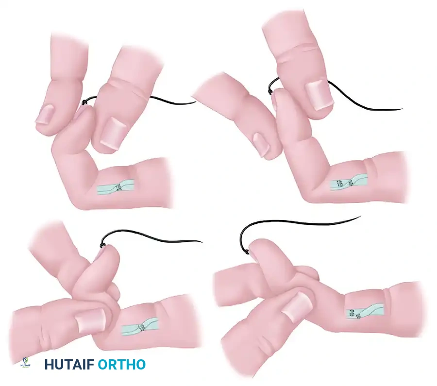
- Tensioning: Setting the correct tension is the most critical step. The tension should be set in a manner similar to a single-stage graft for the FPL. The thumb should rest in a natural cascade, typically exhibiting slightly more flexion than the index finger when the wrist is in neutral. The tenodesis effect must be confirmed intraoperatively: wrist extension should cause thumb interphalangeal joint flexion, and wrist flexion should allow full passive extension of the thumb.
6. Closure and Immobilization
- Obtain adequate hemostasis following tourniquet deflation.
- Close the wounds in a routine manner using 4-0 or 5-0 monofilament nylon sutures.
- Apply a sterile, non-adherent compressive dressing.
- Splinting: Apply a dorsal short-arm splint that immobilizes the wrist in 25 to 30 degrees of flexion. The thumb should be positioned in slight palmar abduction with the MCP and IP joints in slight flexion to remove tension from the repair.
Postoperative Care
Postoperative rehabilitation after an FDS-to-FPL transfer or a two-stage tendon graft is identical to that of a single-stage grafting technique. It requires a highly disciplined, protocol-driven approach (e.g., modified Kleinert or Duran protocols) managed by a specialized hand therapist. Early passive mobilization is initiated to prevent peritendinous adhesions, followed by graduated active motion at 4 to 6 weeks, and strengthening at 8 to 10 weeks.
FLEXOR TENOLYSIS AFTER REPAIR AND GRAFTING
Despite flawless surgical technique and rigorous postoperative therapy, tendon repair and grafting can be complicated by the adherence of the tendon to the surrounding fibro-osseous sheath. This adherence restricts tendon gliding, resulting in a profound loss of active motion. Flexor tenolysis is a highly demanding salvage procedure designed to liberate the tendon from these restrictive adhesions.
Timing and Indications
Tenolysis is never an emergency and should never be performed prematurely. The biological process of tendon healing and scar maturation must be respected.
- Timeline: Tenolysis should not be considered until it is unequivocally documented that the patient has plateaued and made no significant progress over several months despite strict compliance with an organized, progressive hand therapy program.
- Usually, a minimum of 3 months must have elapsed since the initial surgical procedure. In many complex situations, 4 to 6 months may be required to allow the inflammatory phase to completely subside and to make an accurate assessment of the patient’s true baseline.
Strict Prerequisites for Tenolysis
Before offering a tenolysis, the surgeon must ensure the following five criteria are strictly met:
- Tissue Maturation: All soft tissue and skin scars must be soft, pliable, flexible, and completely healed. Operating through indurated, erythematous, or hypertrophic scar tissue guarantees recurrent adhesion formation.
- Skeletal Stability: All associated fractures and joint injuries must be fully healed.
- Joint Mobility: The passive range of motion (PROM) in the digital joints must be maximized and as near normal as possible. Tenolysis will only restore active motion; it cannot overcome fixed passive joint contractures.
- Neurological Status: Sensibility, under ideal circumstances, should be normal. If a concomitant nerve repair was performed, there must be demonstrable clinical recovery of nerve function (e.g., advancing Tinel's sign, return of protective sensation).
- Patient Psychology and Compliance: The patient must be highly motivated, showing progression with strengthening exercises, and must realistically understand the expectations and limitations of the procedure.
💡 Clinical Pearl:
The patient must be explicitly counseled preoperatively that if the extent of scarring and adhesion is found to be excessive intraoperatively, a simple tenolysis may be impossible. In such cases, the surgeon must be prepared to excise the scarred tendon and insert a silicone Hunter rod, converting the procedure into the first stage of a two-stage flexor tendon graft.
Risks and Complications of Tenolysis
The patient must understand that tenolysis weakens the tendon by disrupting its newly formed, albeit restrictive, vascular supply and mechanically shaving away peripheral tendon fibers.
- Rupture Risk: If tenolysis is successful in restoring motion, the risk of spontaneous tendon rupture in the immediate postoperative period is 10% or greater.
- Failed Grafts: It is a fundamental rule of hand surgery that after a failed tendon graft, a fresh tendon graft should be utilized instead of attempting a tenolysis on the ischemic, scarred graft tissue.
PATHOMECHANICS OF TENDON ADHERENCE
Understanding the biomechanical consequences of tendon adherence is crucial for accurate clinical examination and surgical planning. When a tendon completely adheres to the surrounding bone or rigid fibro-osseous sheath, it ceases to function as a dynamic transmitter of muscle force and instead acts as a static, unyielding checkrein.
The Checkrein Effect
Adherence causes specific active movements of one or more joints distal to the area of adherence to be lost. Furthermore, specific passive movements are also limited because the adherent tendon tethers the distal structures.
Clinical Example: FDP Adherence to the Proximal Phalanx
Consider a scenario where the flexor digitorum profundus (FDP) tendon becomes densely adherent to the shaft of the proximal phalanx following a Zone II repair:
- Loss of Active Distal Flexion: The two distal finger joints (PIP and DIP) cannot be actively flexed by the FDP tendon because the muscle's proximal pull is halted at the proximal phalanx.
- Preservation of FDS Function: If the FDS tendon remains uninjured and non-adherent, the PIP joint can still be actively flexed by the FDS.
- Proximal Joint Function: The MCP joint can still be actively flexed by the combined forces of the FDP (pulling on the proximal phalanx), the FDS, and the intrinsic muscles (lumbricals and interossei).
- Restriction of Extension: Crucially, the adherence of the FDP to the proximal phalanx acts as a volar tether. This checks full passive and active extension of the two distal finger joints. The patient will present with a flexion contracture that cannot be passively corrected without risking tendon rupture.
Clinical Evaluation of Adherence
To differentiate between tendon adherence, joint contracture, and tendon rupture, the surgeon must employ precise blocking tests. If passive extension of the DIP joint is impossible when the PIP joint is held in extension, but becomes possible when the PIP joint is flexed, this indicates a tenodesis effect caused by tendon adherence proximal to the PIP joint, rather than a fixed capsular contracture of the DIP joint itself.
By mastering the anatomical nuances of tendon transfers, respecting the biological timelines of scar maturation, and understanding the profound biomechanical implications of tendon adherence, the orthopedic surgeon can navigate the complexities of flexor tendon reconstruction and optimize functional outcomes for the patient.
📚 Medical References
- Flexor tendon transfer for metatarsophalangeal instability of the toe, Foot Ankle 14:385, 1993.
- Thompson FM, Hamilton WG: Problems of the second metatarsophalangeal joint, Orthopedics 10:83, 1987.
- Trepman E, Yeo SJ: Nonoperative treatment of metatarsophalangeal joint synovitis, Foot Ankle Int 16:771, 1995.
- Yao L, Cracchiolo A, Farahani K, et al: Magnetic resonance imaging of plantar plate rupture, Foot Ankle Int 17:33, 1996.
- Hammer Toes Alvine FG, Garvin KL: Peg and dowel fusion of the proximal interphalangeal joint, Foot Ankle 1:90, 1980.
- Boyer ML, DeOrio JK: Metatarsal neck osteotomy with proximal interphalangeal joint resection fi xed with a single temporary pin, Foot Ankle Int 25:144, 2004.
- Cahill BR, Connor DE: A long-term follow-up on proximal phalangectomy for hammer toes, Clin Orthop Relat Res 86:191, 1972.
- Cameron H, Fedorkow D: Revision rates in forefoot surgery, Foot Ankle 3:47, 1982.
- Conklin MJ, Smith RW: Treatment of the atypical lesser toe deformity with basal hemiphalangectomy, Foot Ankle 15:585, 1994.
- Coughlin MJ: Crossover second toe deformity, Foot Ankle 8:29, 1987.
- Coughlin MJ: Lesser toe deformities, Orthopedics 10:63, 1987.
- Coughlin MJ: When to suspect crossover second toe deformity, J Musculoskeletal Med 4:39, 1987.
- Coughlin MJ, Dorris J, Polk E: Operative repair of the fi xed hammertoe deformity, Foot Ankle Int 21:94, 2000.
- Davies MS, Saxby TS: Metatarsal neck osteotomy with rigid internal fi xation for the treatment of lesser toe metatarsophalangeal joint pathology, Foot Ankle Int 20:630, 1999.
- Davis TJ, Schon LC: Branches of the tibial nerve: anatomic variation, Foot Ankle Int 16:21, 1995.
- Dhukaram V, Hossain S, Sampath J, et al: Correction of hammer toe with an extended release of the metatarsophalangeal joint, J Bone Joint Surg 84B:986, 2002.
- Duchenne GBA: Selection of the clinical works of Duchenne (translated by GV Poore), London, 1883, New Sydenham Society. Dyal CM, Davis WH, Thompson FM, et al: Clinical evaluation of the Ruiz-Mora procedure: long-term follow-up, Foot Ankle Int 18:94, 1997.
- Freiberg JA: The diagnosis and treatment of common painful conditions of the foot, Instr Course Lect 14:238, 1957.
- Giannestras NJ: Foot disorders: medical and surgical management, 2nd ed, Philadelphia, 1973, Lea & Febiger. Girdlestone GR: Physiotherapy for hand and foot, J Chartered Soc Physiother 32:167, 1947.
- Glassman F, Wolin I, Sideman S: Phalangectomy for toe deformities, Surg Clin North Am 29:275, 1949.
- Goldner JL, Ward WG: Traumatic horizontal deviation of the second toe: mechanism of deformity, diagnosis, and treatment, Bull Hosp Jt Dis 47:123, 1987.
- Johnston RB, Smith J, Daniels T: The plantar plate of the lesser toes: an anatomical study in human cadavers, Foot Ankle Int 15:276, 1994.
- Kelly A, Winson I: Use of ready-made insoles in the treatment of lesser metatarsalgia: a prospective controlled trial, Foot Ankle Int 19:217, 1998.
- Lehman DE, Smith RW: Treatment of symptomatic hammertoe with a proximal interphalangeal
You Might Also Like