Transform Your Joints: Techniques to help to reduce arthritis discomfort

Key Takeaway
This topic focuses on Transform Your Joints: Techniques to help to reduce arthritis discomfort, Managing arthritis symptoms and improving joint health involves key strategies. Regular low-impact exercise like swimming or walking, along with stretching, can help to reduce pain and stiffness. Maintaining a healthy weight, practicing good posture, and wearing proper footwear also help. Eating an anti-inflammatory diet rich in omega-3s, fruits, and vegetables can further help to reduce inflammation, as can heat or cold therapy.
Introduction and Epidemiology
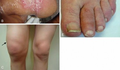
Osteoarthritis represents a progressive, degenerative arthropathy characterized by the structural deterioration of articular cartilage, subchondral bone remodeling, osteophyte formation, and synovial inflammation. As the leading cause of global disability, the epidemiological burden of degenerative joint disease is profound, predominantly affecting the weight-bearing joints, particularly the knee and hip. The prevalence of symptomatic knee osteoarthritis approaches 10 to 15 percent in adults over the age of 60, with an accelerating incidence trajectory driven by an aging demographic and the rising prevalence of metabolic syndrome and obesity.
The pathophysiology of articular degeneration is a complex interplay of mechanical, cellular, and biochemical processes. Chondrocyte senescence and altered mechanotransduction initiate a catabolic cascade. Pro-inflammatory cytokines, notably Interleukin-1 beta and Tumor Necrosis Factor alpha, upregulate the production of matrix metalloproteinases and a disintegrin and metalloproteinase with thrombospondin motifs. These enzymes systematically degrade the type II collagen network and aggrecan macromolecules within the extracellular matrix. Concurrently, systemic adiposity contributes to a state of chronic, low-grade systemic inflammation mediated by adipokines such as leptin and resistin, which further accelerate chondral degradation independent of purely mechanical joint reactive forces.
Evidence Based Non Operative Modalities
Before surgical intervention is considered, a rigorous, multimodal non-operative protocol must be exhausted. The foundational principles of conservative joint preservation aim to optimize biomechanics, attenuate systemic inflammation, and modulate nociceptive pathways.
Biomechanical Offloading and Kinesiotherapy
Targeted neuromuscular re-education and kinesiotherapy are paramount in the non-operative management of articular degeneration. Strengthening the dynamic stabilizers of the joint—specifically the quadriceps, hamstrings, and hip abductors—enhances load distribution across the articular surface. Periarticular muscle hypertrophy mitigates peak joint reactive forces during the stance phase of the gait cycle. Furthermore, correction of sagittal and coronal plane postural deviations reduces eccentric loading on the articular cartilage. Low-impact, closed-kinetic-chain exercises facilitate synovial fluid circulation, which is critical for chondrocyte nutrition via diffusion, while minimizing deleterious shear forces.
Metabolic Optimization and Nutraceuticals
Systemic metabolic optimization directly influences the local intra-articular environment. Adipose tissue is highly endocrinologically active; thus, weight reduction decreases both the mechanical load multiplier on weight-bearing joints and the circulating levels of pro-inflammatory adipokines. A reduction of even five percent of total body mass yields a clinically significant decrease in nociceptive signaling. Dietary modifications emphasizing polyunsaturated fatty acids, particularly eicosapentaenoic acid and docosahexaenoic acid, competitive inhibitors of the arachidonic acid cascade, reduce the synthesis of pro-inflammatory prostaglandins and leukotrienes.
Orthotic Interventions and Thermal Modalities
Pedorthic interventions alter the ground reaction force vector during locomotion. For instance, lateral wedge insoles can shift the mechanical axis laterally, theoretically unloading the medial compartment in patients with varus gonarthrosis, although clinical efficacy remains variable in the literature. Rigid rocker-bottom soles can limit midfoot and forefoot motion, reducing stress across the tibiotalar and metatarsophalangeal articulations. Additionally, thermal modalities serve as vital adjuncts in symptom management. Cryotherapy induces local vasoconstriction, reducing capillary permeability and acute synovial effusion, while thermotherapy decreases muscle spindle fiber sensitivity, thereby alleviating periarticular muscle spasm and capsular contracture.
Surgical Anatomy and Biomechanics
A profound understanding of osseous morphology, ligamentous restraints, and joint kinematics is requisite for successful surgical intervention when conservative measures fail. The knee joint, a modified hinge (ginglymus) joint, serves as the primary model for arthroplasty and joint reconstruction.
Osseous and Ligamentous Architecture
The tibiofemoral articulation is inherently unstable, relying heavily on its ligamentous and meniscal anatomy for congruency. The distal femur features a medial condyle that is larger and extends further distally than the lateral condyle, contributing to the physiological valgus of the mechanical axis. The proximal tibia presents a medial plateau that is concave and a lateral plateau that is convex, with a posterior slope averaging 7 to 10 degrees.
The primary static stabilizers include the anterior cruciate ligament, which prevents anterior tibial translation; the posterior cruciate ligament, which prevents posterior tibial translation; the medial collateral ligament, providing valgus restraint; and the lateral collateral ligament, providing varus restraint. The menisci are fibrocartilaginous structures that increase the contact area between the femur and tibia, effectively reducing contact stress (force per unit area) by dispersing axial loads.
Joint Kinematics and Reactive Forces
Knee kinematics involve a complex combination of rolling and gliding. During early flexion, the femoral condyles roll posteriorly on the tibial plateau. As flexion progresses, the anterior cruciate ligament dictates a transition from rolling to gliding, preventing the femur from subluxating posteriorly off the tibia. This phenomenon, known as femoral rollback, maximizes the efficiency of the extensor mechanism by increasing the patellofemoral moment arm.
The screw-home mechanism describes the obligatory external rotation of the tibia relative to the femur during the terminal degrees of extension, locking the joint in a stable, energy-efficient position. Biomechanically, joint reactive forces across the tibiofemoral joint can exceed three to four times body weight during normal ambulation and up to seven times body weight during stair descent. Any deviation from the neutral mechanical axis (a line drawn from the center of the femoral head to the center of the talar dome) exponentially increases focal contact stresses, accelerating chondral wear in the affected compartment.
Indications and Contraindications
The decision to proceed with surgical intervention, specifically Total Knee Arthroplasty, is predicated on the exhaustion of non-operative modalities, progressive radiographic deterioration, and a significant decline in the patient's functional status and quality of life.
Table of Operative versus Non Operative Indications
| Management Phase | Modality Category | Specific Indications | Clinical Thresholds |
|---|---|---|---|
| Non-Operative | Kinesiotherapy and Weight Management | Kellgren-Lawrence Grade I-III; BMI > 40; mild to moderate functional deficit. | Preserved joint space > 2mm; medically optimized for active rehabilitation. |
| Non-Operative | Pharmacologic and Intra-articular | Acute inflammatory flares; contraindications to surgery (e.g., recent myocardial infarction). | Refractory pain responsive to NSAIDs, corticosteroid, or hyaluronic acid injections. |
| Operative | Joint Preservation (Osteotomy) | Unicompartmental OA; young, active patient (typically < 60 years); malalignment. | Metaphyseal deformity; intact opposite compartment; > 90 degrees flexion arc. |
| Operative | Unicompartmental Knee Arthroplasty | Isolated medial or lateral compartment OA; intact ACL; correctable deformity. | < 15 degrees varus or < 5 degrees valgus; flexion contracture < 15 degrees. |
| Operative | Total Knee Arthroplasty | Tricompartmental OA; Kellgren-Lawrence Grade IV; severe pain at rest; functional decline. | Bone-on-bone articulation; failed 6+ months of conservative management. |
Absolute contraindications to total joint arthroplasty include active local or systemic infection, severe peripheral vascular disease, and neuropathic (Charcot) arthropathy. Relative contraindications encompass severe psychiatric disease, morbid obesity (BMI > 40 is associated with exponentially higher complication rates), and poor dentition, which must be optimized prior to elective arthroplasty to mitigate the risk of hematogenous seeding.
Pre Operative Planning and Patient Positioning
Meticulous pre-operative planning is the cornerstone of successful surgical execution, ensuring accurate implant sizing, restoration of the mechanical axis, and anticipation of intra-operative challenges.
Radiographic Evaluation and Templating
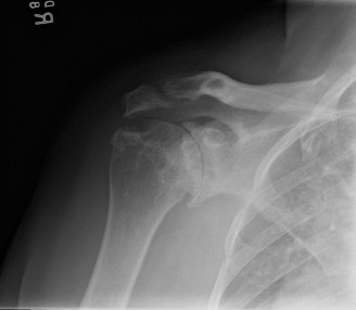
A comprehensive radiographic series is mandatory. This includes a weight-bearing anteroposterior view, a 45-degree flexion weight-bearing posteroanterior view (Rosenberg view) to assess the posterior weight-bearing cartilage, a true lateral view to evaluate patellar height and posterior tibial slope, and a Merchant or skyline view to assess patellofemoral tracking and wear. Full-length standing hip-to-ankle radiographs are critical for defining the mechanical axis and identifying extra-articular deformities that may dictate the level of intra-articular resection or necessitate concomitant osteotomy.
Digital templating involves establishing the anatomical and mechanical axes of the femur and tibia. The femoral intramedullary guide is typically set to an angle of 5 to 7 degrees of valgus relative to the anatomical axis to recreate a neutral mechanical axis. Tibial resection depth is templated to resect 8 to 10 millimeters from the intact compartment, referencing the most prominent aspect of the lateral or medial plateau depending on the deformity.
Patient Optimization and Positioning
Pre-operative medical optimization requires a multidisciplinary approach. Glycemic control is imperative, with a target Hemoglobin A1c of less than 7.0 percent to minimize surgical site infection risk. Nutritional status should be assessed via serum albumin and transferrin levels. Smoking cessation must be mandated at least four to six weeks prior to surgery to optimize microvascular perfusion and wound healing.
In the operating theater, the patient is positioned supine on a radiolucent table. A tourniquet is placed proximally on the operative thigh, though its inflation may be reserved for cementation to minimize ischemic muscle damage and post-operative pain. The operative extremity is placed in a leg holder or over a sandbag to allow a minimum of 120 degrees of flexion. A lateral post is often utilized to provide a fulcrum for valgus stress during medial compartment exposure.
Detailed Surgical Approach and Technique
The standard approach for a primary Total Knee Arthroplasty is the medial parapatellar arthrotomy, which provides excellent, extensile exposure to all three compartments of the knee.
The Medial Parapatellar Arthrotomy
A longitudinal midline skin incision is made from the superior pole of the patella to the medial aspect of the tibial tubercle. Full-thickness fasciocutaneous flaps are elevated medially and laterally. The deep dissection begins with a medial parapatellar capsulotomy. The incision courses through the quadriceps tendon, leaving a 3 to 4 millimeter cuff of tendon on the vastus medialis obliquus to facilitate robust closure. The arthrotomy extends distally around the medial border of the patella and along the medial border of the patellar tendon to the tibial tubercle.
There is no true internervous plane in the medial parapatellar approach, as the entire extensor mechanism is innervated by the femoral nerve. Care must be taken during the distal dissection to protect the infrapatellar branch of the saphenous nerve, though some degree of lateral cutaneous numbness is inevitable and should be discussed with the patient pre-operatively. The patella is then everted or laterally subluxated, and the infrapatellar fat pad is partially excised to expose the lateral tibial plateau.
Osseous Resection and Gap Balancing
The goal of osseous resection is to create rectangular, symmetric flexion and extension gaps. The distal femoral cut is performed first using an intramedullary alignment guide. The guide is set to the pre-operatively templated valgus angle (typically 5 to 7 degrees). A measured resection technique removes an amount of bone equal to the thickness of the femoral component (approximately 9 millimeters).
Attention is then directed to the proximal tibia. An extramedullary alignment guide is aligned parallel to the tibial crest in the sagittal plane and centered over the mechanical axis in the coronal plane. The resection is made perpendicular to the mechanical axis, incorporating a posterior slope of 0 to 3 degrees depending on the implant design (cruciate-retaining versus posterior-stabilized).
Femoral component rotation is critical for patellofemoral tracking and flexion gap symmetry. Rotation is determined using three anatomical landmarks: the transepicondylar axis (the most reliable), Whiteside's line (the anteroposterior axis of the trochlea), and 3 degrees of external rotation relative to the posterior condylar axis. The anterior, posterior, and chamfer cuts are then executed using a 4-in-1 cutting block.
Soft tissue balancing is performed sequentially. For a varus deformity, the deep medial collateral ligament is released from the proximal tibia. If further release is required, the superficial medial collateral ligament and posteromedial capsule are elevated. For a valgus deformity, a lateral release may include the iliotibial band, posterolateral capsule, and popliteus tendon, often performed via a pie-crusting technique to avoid catastrophic lateral collateral ligament incompetence.
Implant Fixation and Closure
Once trial components confirm appropriate tracking, stability, and gap symmetry, the definitive implants are prepared. The osseous surfaces are cleansed with pulsatile lavage and dried. High-viscosity polymethylmethacrylate bone cement is applied to the implant interfaces and the cancellous bone. The components are impacted into position, and the joint is held in terminal extension during cement polymerization to compress the implant-bone interface.
Following cement curing, the tourniquet is deflated, and meticulous hemostasis is achieved. The arthrotomy is closed in a watertight fashion using heavy, interrupted absorbable sutures, particularly ensuring secure reapproximation of the quadriceps tendon. The subcutaneous tissues and skin are closed sequentially, and a sterile compressive dressing is applied.
Complications and Management
Despite rigorous protocols, complications in joint arthroplasty can be devastating. Early recognition and aggressive management are critical for limb salvage and patient survival.
Table of Complications and Salvage Strategies
| Complication | Estimated Incidence | Pathophysiology | Salvage Strategy |
|---|---|---|---|
| Periprosthetic Joint Infection | 1.0 - 2.0 percent | Biofilm formation on implant surface; typically Staphylococcus aureus or coagulase-negative Staphylococci. | Acute (< 4 weeks): DAIR (Debridement, Antibiotics, Implant Retention). Chronic: Two-stage revision with antibiotic spacer. |
| Venous Thromboembolism | 1.0 - 3.0 percent (symptomatic) | Virchow's triad: endothelial injury from surgery, venous stasis from immobility, hypercoagulable state. | Systemic anticoagulation (DOACs, LMWH). IVC filter placement if anticoagulation is strictly contraindicated. |
| Periprosthetic Fracture | 0.5 - 2.0 percent | Altered stress shielding; osteolysis; traumatic impact on osteoporotic bone. | Supracondylar femur: Retrograde intramedullary nail or locking plate. Loose implant requires revision arthroplasty. |
| Aseptic Loosening | 2.0 - 5.0 percent (at 10 years) | Particulate wear debris (polyethylene) induces macrophage-mediated osteolysis at the bone-cement interface. | Revision arthroplasty utilizing diaphyseal engaging stems and metaphyseal cones or sleeves for bone loss. |
| Arthrofibrosis | 3.0 - 5.0 percent | Exaggerated fibroblastic response leading to dense scar tissue and capsular contracture. | Aggressive physical therapy. Manipulation under anesthesia (MUA) if < 90 degrees flexion at 6-8 weeks post-op. |
Post Operative Rehabilitation Protocols
The post-operative rehabilitation trajectory is biphasic, focusing initially on complication prevention and tissue healing, followed by functional restoration and neuromuscular integration.
Acute Inpatient Phase
Immediate post-operative mobilization is the standard of care. Patients are encouraged to bear weight as tolerated on the operative extremity within hours of surgery, utilizing an assistive device. Early mobilization mitigates the risk of venous thromboembolism, reduces the incidence of pulmonary atelectasis, and limits the formation of intra-articular adhesions.
Cryotherapy is applied continuously during the acute phase to induce vasoconstriction, thereby limiting hemarthrosis and decreasing the conduction velocity of peripheral nociceptors. The primary range of motion goal in the acute phase is achieving full terminal extension. A flexion contracture is highly detrimental to gait mechanics, increasing quadriceps fatigue and patellofemoral contact pressures. Patients are instructed to avoid placing pillows directly under the popliteal fossa, instead elevating the extremity by supporting the heel.
Outpatient Functional Restoration
Upon discharge, rehabilitation transitions to an outpatient setting. The focus shifts to achieving a functional arc of motion, ideally 0 to 120 degrees, which is required for activities of daily living such as stair climbing and rising from a seated position.
Kinesiotherapy in this phase emphasizes eccentric quadriceps control and concentric hamstring strengthening to restore dynamic joint stability. Proprioceptive training is integrated to enhance joint position sense, which is often compromised following capsular resection. Return to low-impact activities, such as stationary cycling and aquatic therapy, is encouraged at four to six weeks post-operatively, provided the surgical incision is completely healed. High-impact activities and repetitive axial loading are generally discouraged to maximize the longevity of the polyethylene bearing surface.
Summary of Key Literature and Guidelines
The management of articular degeneration and the execution of total joint arthroplasty are guided by robust, evidence-based literature. The American Academy of Orthopaedic Surgeons (AAOS) Clinical Practice Guidelines for the Management of Osteoarthritis of the Knee strongly recommend participation in self-management programs, strengthening, and low-impact aerobic exercises. The guidelines demonstrate strong evidence against the routine use of hyaluronic acid intra-articular injections for symptomatic relief, citing a lack of clinically significant efficacy compared to placebo, while supporting the short-term use of intra-articular corticosteroids.
Landmark registry data, including the Swedish Knee Arthroplasty Register and the Australian Orthopaedic Association National Joint Replacement Registry, consistently demonstrate excellent long-term survivorship of primary Total Knee Arthroplasty, with 15-year survivorship rates exceeding 93 percent. These registries highlight that aseptic loosening and periprosthetic joint infection remain the leading causes of revision burden.
Furthermore, the literature underscores the critical importance of coronal plane alignment. Historically, the goal of TKA was to achieve a neutral mechanical axis (0 degrees plus or minus 3 degrees). Recent kinematic alignment philosophies challenge this paradigm, suggesting that restoring the patient's pre-disease constitutional alignment may yield superior patient-reported outcome measures (PROMs). However, long-term survivorship data for kinematic alignment remains a subject of ongoing academic debate, and mechanical alignment remains the gold standard benchmark in contemporary orthopedic education.
You Might Also Like