Regional Anesthesia of the Foot and Ankle: Forefoot & Ankle Block Techniques
Key Takeaway
Regional anesthesia, specifically the forefoot and ankle block, is an essential skill for orthopedic surgeons performing foot and ankle procedures. These blocks provide excellent intraoperative anesthesia and prolonged postoperative analgesia while mitigating the risks associated with general anesthesia. This guide details the precise neurovascular anatomy, pharmacological principles, and step-by-step needle placement techniques required to safely and effectively execute forefoot and ankle blocks in clinical practice.
Comprehensive Introduction and Patho-Epidemiology
Regional anesthesia has fundamentally revolutionized the perioperative management and longitudinal care of foot and ankle pathology. In the contemporary era of orthopedic surgery, characterized by an emphasis on Enhanced Recovery After Surgery (ERAS) pathways and the critical necessity of opioid-sparing protocols, mastering the forefoot block and ankle block is no longer optional; it is paramount. These highly targeted techniques provide profound intraoperative anesthesia, facilitate the seamless use of a calf or ankle tourniquet for a bloodless surgical field, and deliver extended postoperative analgesia. Consequently, they drastically reduce perioperative opioid consumption, mitigate opioid-related adverse events, and facilitate early, safe mobilization.
The patho-epidemiological landscape of foot and ankle surgery frequently involves patients with a complex matrix of medical comorbidities. Diabetic foot infections, Charcot neuroarthropathy, severe peripheral vascular disease, and end-stage renal disease often present in patients who are exceptionally poor candidates for general anesthesia. When performed meticulously, regional blocks completely bypass the systemic cardiopulmonary risks associated with volatile anesthetics and systemic narcotics. This makes regional anesthesia particularly advantageous, and often life-saving, for the vasculopathic or medically fragile patient.
Furthermore, the pathophysiology of acute postoperative pain in foot and ankle surgery is uniquely intense due to the high density of nociceptive nerve endings in the periosteum and soft tissues of the pedal extremities. General anesthesia alone does nothing to blunt this localized inflammatory pain cascade once the patient emerges. Regional blockade, however, preemptively halts the afferent nociceptive signaling at the peripheral nerve level, preventing central sensitization and the phenomenon of "wind-up" pain. This comprehensive guide delineates the precise neurovascular anatomy, advanced pharmacological principles, and meticulous step-by-step surgical techniques required to execute highly successful forefoot and ankle blocks.
Detailed Surgical Anatomy and Biomechanics
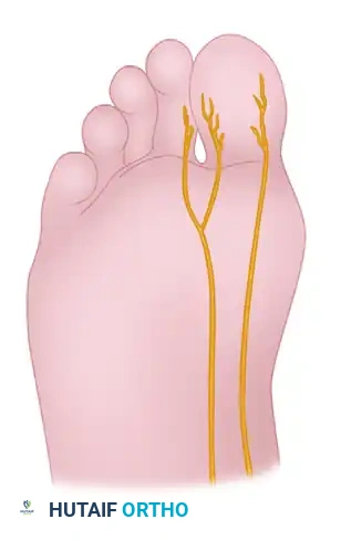
A profound, three-dimensional understanding of the pedal neurovascular arborization is the absolute foundation of successful regional anesthesia. The foot is a complex biomechanical structure innervated by five primary nerves, all of which must be conceptualized spatially depending on the planned surgical site. Failure to appreciate the depth, fascial planes, and vascular relations of these nerves results in incomplete blockade and unacceptable patient discomfort.
The Five Primary Nerves of the Foot
- Deep Peroneal Nerve (L4, L5, S1, S2): This nerve courses anterior to the ankle joint, intimately adjacent to the dorsalis pedis artery, typically positioned just lateral to the extensor hallucis longus (EHL) tendon and medial to the extensor digitorum longus (EDL). It runs deep to the extensor retinaculum. Distally, it provides critical sensory innervation to the first dorsal web space.
- Superficial Peroneal Nerve (L4, L5, S1): Originating from the common peroneal nerve, it descends through the lateral compartment of the leg. It pierces the deep crural fascia in the distal third of the leg (approximately 10-12 cm proximal to the lateral malleolus) to become subcutaneous. It then divides into the medial and intermediate dorsal cutaneous nerves, supplying the vast majority of the dorsum of the foot and the dorsal aspect of the toes (excluding the first web space and the lateral aspect of the fifth digit).
- Tibial Nerve (L4, L5, S1, S2, S3): This is the largest nerve of the ankle block. It passes through the fibro-osseous tarsal tunnel posterior to the medial malleolus, running deep to the flexor retinaculum alongside the posterior tibial artery and accompanying veins. Within or just distal to the tunnel, it bifurcates into the medial and lateral plantar nerves, supplying the entire plantar aspect of the foot, the intrinsic plantar musculature, and the plantar surfaces of all digits.
- Sural Nerve (S1, S2): Formed by contributions from the tibial and common peroneal nerves, it courses posterior to the lateral malleolus alongside the small saphenous vein. It is purely sensory at this level, supplying the lateral margin of the hindfoot, midfoot, and the lateral aspect of the fifth digit.
- Saphenous Nerve (L3, L4): The terminal sensory branch of the femoral nerve, making it the only nerve of the foot not derived from the sciatic nerve. It courses anterior to the medial malleolus, intimately associated with the great saphenous vein, supplying sensory innervation to the medial column of the ankle and midfoot.
Arterial Landmarks and Biomechanical Considerations
The dorsalis pedis artery is the critical anatomical landmark for the forefoot block. As it courses distally and reaches the base of the first intermetatarsal space, it bifurcates into the first dorsal intermetatarsal artery and the plantar penetrating branch (deep plantar artery). The plantar penetrating branch dives at a near right angle between the two heads of the first dorsal interosseous muscle to communicate with the deep plantar arch. This vascular configuration is biomechanically and anatomically analogous to the dorsal branch of the radial artery in the hand as it dives between the heads of the first dorsal interosseous muscle to form the deep palmar arch.
Surgical Warning: The proximity of these vessels to the target nerves cannot be overstated. Intravascular injection into the dorsalis pedis, posterior tibial artery, or their arborizing branches can lead to rapid, catastrophic Local Anesthetic Systemic Toxicity (LAST). Meticulous aspiration prior to every single injection aliquot is a mandatory surgical principle.
Exhaustive Indications and Contraindications
The decision to utilize a forefoot or ankle block must be tailored to the specific surgical procedure, the anticipated duration of the operation, and the patient's unique physiological profile. While regional anesthesia is overwhelmingly safe, strict adherence to indications and absolute respect for contraindications define the prudent orthopedic surgeon.
| Category | Specific Conditions / Procedures | Clinical Rationale & Considerations |
|---|---|---|
| Indications (Forefoot Block) | Hallux valgus correction, hammer toe reconstruction, neuroma excision, metatarsal osteotomies, phalangeal amputations. | Provides profound, localized anesthesia for procedures distal to the midfoot without the need to block the entire ankle. Ideal for bilateral forefoot procedures. |
| Indications (Ankle Block) | ORIF of Lisfranc/midtarsal injuries, ankle fracture closed reductions, tarsal tunnel release, Achilles tendon debridement. | Circumferential blockade allows for the use of a calf tourniquet and facilitates extensive midfoot and hindfoot reconstructions. |
| Absolute Contraindications | Patient refusal, active infection at the injection site, true allergy to amide local anesthetics. | Injecting through an infected cellulitic field can track bacteria into deep fascial planes or neurovascular sheaths. |
| Relative Contraindications | Severe coagulopathy, preexisting severe peripheral neuropathy, uncooperative patient (e.g., severe dementia). | Coagulopathy increases the risk of compressive hematoma, particularly in the non-compressible tarsal tunnel. Neuropathy requires careful documentation prior to block to avoid misattribution of postoperative deficits. |
Pre-Operative Planning, Templating, and Patient Positioning
Pharmacological Principles and Dosing
A synergistic mixture of short-acting and long-acting amide local anesthetics is highly recommended to achieve the dual goals of rapid onset for surgical efficiency and prolonged duration for postoperative analgesia.
* Short-Acting Component: 1% or 2% Lidocaine (Onset: 5-10 minutes; Duration: 1-2 hours).
* Long-Acting Component: 0.5% Bupivacaine or 0.5% Ropivacaine (Onset: 15-30 minutes; Duration: 4-8 hours, sometimes up to 12 hours depending on volume and patient metabolism).
In most adult patients, a total volume of 30 to 40 mL of a 50/50 mixture (e.g., 15 mL of 1% Lidocaine mixed with 15 mL of 0.5% Bupivacaine) is sufficient for a complete, circumferential ankle block. However, the total dose must be strictly calculated based on the patient's lean body weight to avoid exceeding maximum recommended toxic doses. For Lidocaine without epinephrine, the maximum dose is 4.5 mg/kg; for Bupivacaine, it is strictly 2.5 mg/kg.
The Critical Role of Epinephrine
Clinical Pearl: We do not routinely use epinephrine with the anesthetic solution except as a long-acting postoperative analgesic supplementing general anesthesia in complex, prolonged hindfoot procedures. When utilized, the concentration should be precisely 0.5% Bupivacaine with 1:200,000 epinephrine.
CRITICAL RULE: Epinephrine must never be used distal to the midfoot. The digital arteries of the toes are terminal end-arteries lacking sufficient collateral circulation. Epinephrine-induced vasospasm in the forefoot or digits can result in profound, irreversible ischemia leading to dry gangrene and subsequent amputation.
Patient Optimization and Positioning
The same rigorous anesthetic precautions applied to general anesthesia must be enforced for regional blocks. This includes a comprehensive history and physical examination, appropriate preoperative laboratory data, and strict adherence to NPO guidelines (abstaining from food or drink 8 to 10 hours prior to surgery).
Diabetic Exception: If minor procedures (e.g., nail surgery, single hammer toe correction, or percutaneous tenotomy) are planned for an insulin-dependent diabetic patient under local anesthesia without sedation, allowing a limited clear liquid or light meal a few hours prior to surgery is reasonable to prevent intraoperative hypoglycemia, provided the anesthesia team explicitly concurs.
For the block administration, the patient should be positioned supine. A bump may be placed under the ipsilateral hip to internally rotate the leg for easier access to the lateral structures (sural, superficial peroneal), and the leg can be externally rotated in a "frog-leg" position to access the medial structures (tibial, saphenous).
Step-by-Step Surgical Approach and Fixation Technique
While the definitive "fixation technique" depends on the specific pathology (e.g., screw fixation for a chevron osteotomy, K-wire stabilization for a hammer toe), the foundational "approach" to achieving a painless surgical field is the execution of the block itself. The forefoot block is highly efficacious for procedures involving the hallux, lesser toes, and distal metatarsals.
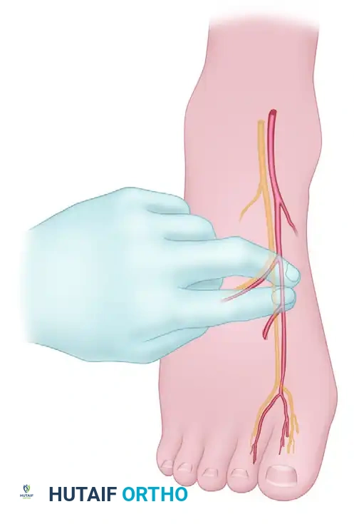
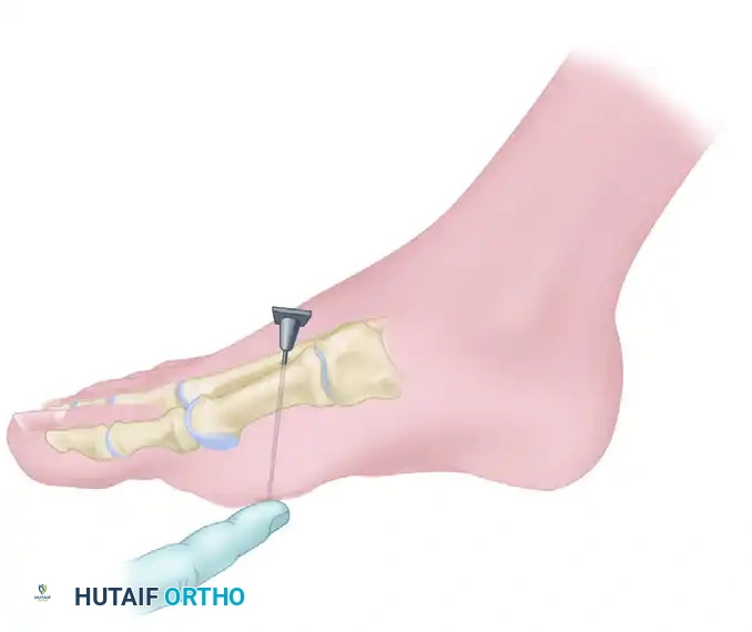
Step 1: Targeting the Deep Peroneal Nerve
Begin by meticulously palpating the dorsalis pedis artery as it courses distally over the dorsum of the foot and reaches the proximal aspect of the first intermetatarsal space. The deep peroneal nerve, which supplies the critical first web space, closely accompanies this artery, typically lying just lateral to it.
Utilizing a 1.5-inch, 25-gauge or 27-gauge needle, carefully enter the skin, avoiding direct puncture of the artery. Aspirate to ensure you are not intravascular. Inject 2 to 3 mL of the short-acting/long-acting anesthetic mixture subcutaneously and subfascially to anesthetize the deep peroneal nerve.
Step 2: Blocking the Superficial Peroneal Branches (Lateral)
If a second or third hammer toe procedure is planned, efficiency dictates that you do not withdraw the needle completely from the skin. From the same dorsal entrance point, partially withdraw and redirect the needle laterally, passing just beneath the dorsal venous arch.
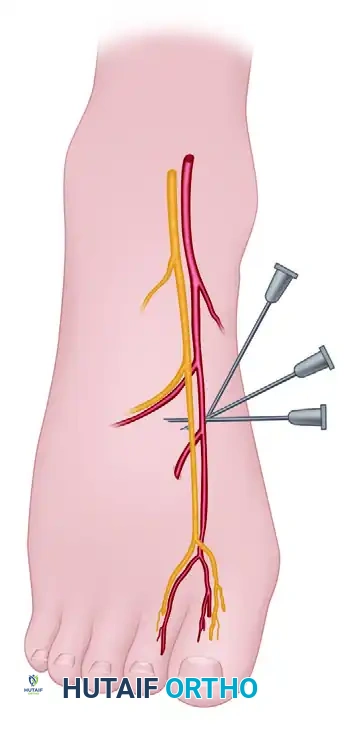
Advance the needle laterally to block the common digital branches of the superficial peroneal nerve supplying the second (and third, if necessary) intermetatarsal spaces. An injection of an additional 2 to 3 mL as the needle is slowly withdrawn is typically sufficient to create a robust transverse wheal.
Step 3: Blocking the Medial Hallucal Branch
Return the needle to the original entrance point, but now direct the trajectory medially. Maintain a path immediately beneath the dorsal veins and superficial to the extensor hallucis longus (EHL) tendon.
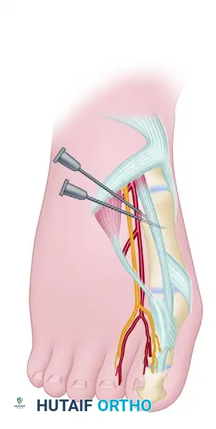
This maneuver blocks the medial hallucal branch of the dorsomedial superficial peroneal nerve. This is the specific sensory branch most frequently encountered and at risk during the standard dorsomedial approach to the medial eminence ("bunion") during hallux valgus surgery. Conclude this dorsal sensory block at the dorsomedial aspect of the forefoot, approximately 1 cm distal to the first metatarsomedial cuneiform articulation. At this stage, a total of 6 to 8 mL of anesthetic has been administered across the dorsum.
Step 4: Plantarward Progression
Entering the newly anesthetized area on the dorsomedial aspect of the forefoot (utilizing the existing numb skin), redirect the needle directly plantarward. Proceed through the subcutaneous space, remaining superficial to the abductor hallucis muscle belly, until the plantar surface of the medial side of the foot is reached.
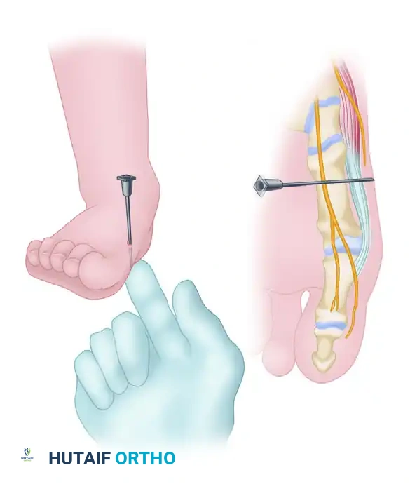
Clinical Pearl: Instilling a small amount of anesthetic agent continuously as the needle progresses plantarward (a technique known as hydrodissection) significantly lessens patient discomfort and safely pushes neurovascular structures away from the advancing needle tip.
Step 5: Anesthetizing the Proper Plantar Branch
The proper plantar branch to the medial side of the hallux becomes superficial at this specific level. It penetrates the deep fascia over the abductor hallucis and flexor hallucis brevis near the first metatarsomedial cuneiform articulation.
Palpate the tip of the needle subcutaneously on the plantar aspect with your non-dominant hand. Once felt, withdraw the needle 2 to 3 mm to ensure it is not intradermal (which would cause extreme pain and skin blanching), and instill 2 to 3 mL of the anesthetic agent.
Step 6: Completing the First Web Space Block
To complete the regional block for comprehensive hallux surgery, you must anesthetize the common digital branch of the medial plantar nerve supplying the plantar aspect of the first web space. Return to the dorsal surface at the base of the first intermetatarsal space.
Step 7: Navigating the Arterial Bifurcation
At the base of the first intermetatarsal space, as previously detailed in the anatomy section, the dorsalis pedis artery bifurcates into the first dorsal intermetatarsal artery and the plantar penetrating branch.
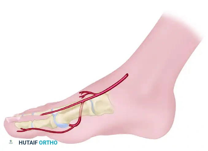
To safely avoid this critical arterial bifurcation and prevent deep hematoma or LAST:
1. Move the needle entrance point distally by 1 to 1.5 cm from the absolute base of the web space.
2. Angle the needle obliquely at 10 to 20 degrees relative to the skin.
3. Pass the 1.5-inch, 25-gauge needle plantarward between the first and second metatarsal shafts until the tip is palpable subcutaneously on the plantar surface.
4. Inject small amounts of anesthetic during advancement to minimize pain and hydrodissect.
5. Withdraw the tip 2 to 3 mm from the plantar skin and instill 4 to 5 mL of solution.
If a hammer toe procedure is planned for the lesser digits, repeat this exact technique between the second and third metatarsals. This provides robust anesthesia for the third toe. Occasionally, supplementing the block with 1 mL of anesthetic directly at the base of the third toe near the web space may be required for complete circumferential coverage.
Complications, Incidence Rates, and Salvage Management
While regional anesthesia of the foot and ankle is statistically very safe, the complications that do arise can be devastating if not immediately recognized and managed. The orthopedic surgeon must be fully prepared to handle systemic and localized adverse events.
| Complication | Estimated Incidence | Clinical Presentation | Salvage & Management Protocol |
|---|---|---|---|
| Local Anesthetic Systemic Toxicity (LAST) | < 0.1% | Perioral numbness, metallic taste, tinnitus, visual disturbances, muscle twitching, seizures, culminating in cardiovascular collapse and asystole. | IMMEDIATE: Stop injection. Call for help. Secure airway (100% O2). Administer 20% Lipid Emulsion Therapy (Intralipid): 1.5 mL/kg bolus over 1 min, followed by 0.25 mL/kg/min infusion. Avoid vasopressin, beta-blockers, and calcium channel blockers. |
| Direct Nerve Injury / Neuropathy | 0.5% - 2.0% | Patient reports a sharp, severe, electric "paresthesia" shooting down the toe during needle advancement. Postoperative persistent numbness or dysesthesia beyond 24-48 hours. | PREVENTION: Never inject against high resistance. If paresthesia occurs, withdraw needle 1-2 mm immediately before injecting. MANAGEMENT: Observation, Gabapentin/Pregabalin for neuropathic pain. Most resolve spontaneously within 3-6 months. |
| Arterial Puncture / Hematoma | 2% - 5% | Flash of pulsatile bright red blood in syringe upon aspiration. Rapid swelling and ecchymosis at the injection site. | Withdraw needle immediately. Apply firm, direct, manual pressure for a minimum of 3 to 5 minutes. Delay tourniquet inflation until hemostasis is confirmed. |
| Digital Ischemia / Gangrene | Extremely Rare | Prolonged, irreversible blanching of the digit. Cold, pulseless toe postoperatively. Usually associated with erroneous use of Epinephrine in the forefoot. | IMMEDIATE: Remove any tight dressings. Apply warm compresses. Consider topical nitroglycerin paste. Phentolamine injection (alpha-adrenergic antagonist) locally if epinephrine was mistakenly used. Consult vascular surgery if unresolved. |
Phased Post-Operative Rehabilitation Protocols
The postoperative management of a patient who has received a regional block requires specific protocols to protect the insensate limb and manage the eventual return of sensation.
Phase I: The Insensate Phase (0 to 12 Hours Post-Op)
Patients who receive a forefoot or ankle block will experience profound sensory and motor deficits lasting anywhere from 4 to 12 hours, heavily dependent on the concentration and volume of Bupivacaine or Ropivacaine utilized.
* Weight-Bearing Restrictions: Patients must be strictly counseled regarding their weight-bearing status prior to discharge. An insensate foot is at exceptionally high risk for unrecognized trauma, thermal injury, or Charcot-like acute breakdown if loaded improperly. Crutches, walkers, or a rigid controlled ankle motion (CAM) boot must be utilized strictly as dictated by the surgical procedure.
* Protection: The foot must be kept elevated above the level of the heart to minimize dependent edema, which can increase tissue pressure and mask a developing compartment syndrome in an insensate foot.
Phase II: The Transition Phase (Rebound Pain Management)
The transition from complete regional anesthesia to normal sensation is rarely gradual; it often presents as a rapid onset of severe nociception known as "rebound pain."
* Preemptive Analgesia: Patients should be explicitly instructed to take their first dose of oral postoperative analgesics (e.g., NSAIDs, Acetaminophen, or prescribed opioids) before the block completely wears off. The clinical cue for the patient is typically when they first feel a faint tingling sensation or "pins and needles" returning to the tips of the toes. Waiting until full pain returns makes it exceedingly difficult to regain analgesic control.
Summary of Landmark Literature and Clinical Guidelines
The evidence-based efficacy and reliability of the ankle and forefoot block are exceptionally well-documented in the orthopedic and anesthesiology literature.
A landmark prospective analysis by Rudkin et al. evaluated 1,000 consecutive patients undergoing foot or ankle surgery utilizing ankle block anesthesia. They reported an outstanding 95% success rate for complete surgical anesthesia. Crucially, their multivariate analysis noted that the frequency of anesthetic failure or the need for general anesthesia conversion was drastically reduced when the time between block administration and surgical incision was 20 minutes or greater. This underscores the clinical necessity of allowing adequate time for neural blockade to penetrate the nerve sheath, particularly for the large tibial nerve.
Furthermore, a pivotal study by White et al. compared intra-articular hematoma blocks to conscious sedation for the closed reduction of acute ankle fracture-dislocations in the emergency department setting. They demonstrated that regional blockade provided superior, sufficient analgesia without the systemic risks of sedation. The average time for reduction, radiographic confirmation, and splinting was significantly faster in the block group (63.8 minutes) compared to the conscious sedation group (81.5 minutes). This highlights the sheer efficiency and safety profile of regional techniques in acute trauma settings, freeing up critical nursing and monitoring resources.
Finally, the American Society of Regional Anesthesia and Pain Medicine (ASRA) guidelines strictly reinforce the absolute necessity of having a LAST rescue kit (containing 20% lipid emulsion) immediately available wherever regional blocks are performed, cementing this as a non-negotiable standard of care in modern orthopedic practice.
