Anesthesia and Surgical Preparation in Upper Extremity Surgery
Key Takeaway
The selection of anesthesia in hand and upper extremity surgery is critical for achieving a painless, motionless operative field. This guide details the indications for general versus regional anesthesia, the pharmacology of local anesthetics, ultrasound-guided brachial plexus blocks, and essential surgical preparation. Mastery of these principles minimizes complications, optimizes microsurgical precision, and significantly improves postoperative patient outcomes and rehabilitation trajectories.
Comprehensive Introduction and Patho-Epidemiology
Unsatisfactory anesthesia for hand and upper extremity operations prevents the surgeon from accomplishing their operative goals and is highly likely to compromise the final surgical result. The intricate anatomy of the upper extremity, particularly the hand and wrist, demands absolute microsurgical precision. For accurate and precise work, the operative extremity must be completely motionless, the procedure must be entirely painless, and the patient must remain hemodynamically stable and comfortable throughout the duration of the case. The physiological stress response to surgical trauma in the upper extremity can induce profound catecholamine release, leading to peripheral vasoconstriction, tachycardia, and hypertension. In the context of microvascular surgery, such as digital replantation or free tissue transfer, this sympathetically mediated vasoconstriction is disastrous, as it directly compromises anastomotic patency and flap perfusion.
The epidemiology of upper extremity surgery has undergone a massive paradigm shift over the last three decades, moving aggressively away from general anesthesia toward regional anesthetic techniques. This transition is driven by the increasing volume of complex orthopedic procedures performed in the ambulatory setting. Millions of upper extremity procedures are performed annually, ranging from routine carpal tunnel releases to complex brachial plexus reconstructions. Historically, these procedures necessitated hospital admission for pain control and monitoring. Today, the advent of sophisticated regional anesthesia protocols has facilitated a transition to outpatient management for the vast majority of these cases. This shift has not only reduced healthcare costs but has significantly decreased the incidence of nosocomial infections and deep vein thrombosis by promoting early mobilization.
All anesthetic techniques carry inherent physiological risks, and the selection of the optimal technique depends on a triad of critical factors: the specific physiological and psychological needs of the patient, the technical requirements of the surgeon, and the expertise of the anesthesiologist. Consequently, the choice of anesthesia must never be an afterthought; it must be an integral component of comprehensive preoperative planning. The ideal anesthetic plan for hand surgery provides dense sensory blockade, profound motor relaxation, and a sympathetic block that induces local vasodilation—an invaluable asset in microvascular repair.
Understanding the patho-epidemiology of perioperative complications is essential for the modern orthopedic surgeon. The incidence of postoperative nausea and vomiting (PONV), cognitive dysfunction (particularly in the elderly), and cardiopulmonary decompensation is markedly higher following general anesthesia compared to regional techniques. Conversely, regional anesthesia introduces its own epidemiological profile of complications, notably Local Anesthetic Systemic Toxicity (LAST) and iatrogenic peripheral nerve injury. Mastery of the physiological interplay between anesthetic agents, surgical tourniquet application, and the patient's baseline comorbidities is the hallmark of an expert upper extremity surgeon.
Detailed Surgical Anatomy and Biomechanics
A profound understanding of the brachial plexus anatomy and its surrounding fascial planes is the absolute foundation for successful regional anesthesia and surgical preparation. The brachial plexus is formed by the ventral rami of the lower four cervical nerves (C5-C8) and the first thoracic nerve (T1), with variable contributions from C4 (pre-fixed) or T2 (post-fixed). These roots emerge between the anterior and middle scalene muscles, carrying with them a fascial prolongation from the prevertebral fascia that forms the axillary sheath. The plexus undergoes a complex arborization: roots combine to form three trunks (superior, middle, inferior) in the posterior triangle of the neck; these divide into anterior and posterior divisions behind the clavicle; the divisions reunite to form three cords (lateral, posterior, medial) in the axilla, named for their relationship to the axillary artery; finally, these terminate into the major peripheral nerves of the upper extremity.
The sonoanatomy of the brachial plexus is equally critical, as ultrasound guidance has become the gold standard for block administration. At the interscalene level, the roots appear as a vertical array of hypoechoic nodules (often described as a "stoplight" appearance) nestled between the scalene muscles. Moving distally to the supraclavicular fossa, the trunks and divisions are tightly clustered superior and lateral to the pulsatile, anechoic subclavian artery, resting on the hyperechoic line of the first rib and pleura. At the infraclavicular and axillary levels, the cords and terminal branches are visualized in close proximity to the axillary artery and vein. The biomechanics of anesthetic spread dictate that the local anesthetic must be deposited within the fascial sheath encompassing the plexus to ensure circumferential bathing of the neural structures, a process heavily dependent on precise needle tip placement and high-pressure hydrodissection.
Beyond neural anatomy, the biomechanics of surgical instrumentation in hand surgery require specific consideration. The transition from macroscopic orthopedic surgery to the microscopic environment of the hand necessitates a shift in fundamental surgical mechanics.
Notice the design of the scalpel handle in the basic tray. An octagonal knife handle is vastly preferable to the standard flat handle (e.g., a standard #3 handle). In macroscopic orthopaedics, a scalpel is often held in a power grip, utilizing the extrinsic flexors of the forearm for forceful incisions. In hand surgery, the scalpel is manipulated using a "precision pinch" (the lumbrical plus position). The octagonal geometry allows the surgeon to roll the instrument seamlessly between the thumb, index, and long fingers, allowing for sweeping, multidirectional curvilinear incisions without repositioning the wrist. This biomechanical advantage prevents jagged skin edges and minimizes iatrogenic trauma to superficial neurovascular structures.
Furthermore, the biomechanics and physiology of the pneumatic tourniquet are paramount. A bloodless field is an absolute prerequisite for identifying microscopic neurovascular structures. Exsanguination is achieved using an Esmarch bandage prior to inflation, driving venous blood proximally. The tourniquet is then inflated to a pressure typically 100 mmHg above the patient's systolic blood pressure (or standardly 250 mmHg in a normotensive adult). The cellular tolerance to ischemia dictates surgical pacing; skeletal muscle undergoes irreversible mitochondrial damage after approximately 2.5 to 3 hours of continuous ischemia. Therefore, tourniquet time should be strictly monitored, generally not exceeding 120 minutes without a reperfusion interval of at least 15 to 20 minutes to allow for clearance of toxic metabolites and restoration of cellular ATP.
Exhaustive Indications and Contraindications
While regional anesthesia has become the gold standard for many upper extremity procedures, general anesthesia remains the preferred modality in specific clinical scenarios. The decision matrix is complex and must account for surgical duration, patient positioning, and anatomical limitations. Factors that strongly favor the administration of general anesthesia include extensive and prolonged operations anticipated to exceed the duration of long-acting regional blocks (typically >6-8 hours), such as major replantations, bilateral procedures, or complex brachial plexus reconstructions. Concurrent procedures requiring simultaneous surgical intervention on other parts of the body, such as the chest, abdomen, or the harvesting of distant tissue grafts (e.g., fibula free flaps, iliac crest bone grafts), also necessitate general endotracheal anesthesia. Furthermore, pediatric populations and highly anxious, claustrophobic patients who cannot reliably cooperate or remain immobile under regional anesthesia alone are prime candidates for general anesthesia.
Regional anesthesia offers profound advantages that fundamentally alter the perioperative experience, making it the technique of choice for the vast majority of hand and wrist surgeries. Satisfactory regional anesthesia can be safely achieved for emergency procedures performed on patients with a full stomach, significantly mitigating the risk of aspiration associated with general anesthesia. In both emergency and elective operations, a regional anesthetic effectively blocks vasoconstrictive afferent nociceptive impulses from the surgical wound. This sympathetic blockade results in maximal vasodilation, improving tissue perfusion and facilitating microvascular anastomoses. Outpatient surgery is vastly facilitated by regional anesthetic blocks, which provide extended postoperative analgesia, eliminate the need for opioid-heavy intraoperative regimens, and reduce the immediate need for intensive postoperative nursing care.
The pharmacological selection of local anesthetics is dictated by the required duration of surgical anesthesia and postoperative analgesia. Lidocaine, an amide local anesthetic, fulfills the basic requirements of rapid onset and moderate duration (1-2 hours), making it the workhorse for short procedures like carpal tunnel release or trigger finger release. Mepivacaine (Carbocaine) is similar to lidocaine but slightly longer acting. Bupivacaine (Marcaine) is highly favored by many surgeons and anesthesiologists due to its prolonged duration of action, effective for 8 hours or longer, providing excellent postoperative analgesia. Ropivacaine and Levobupivacaine are enantiomer-specific derivatives developed to provide the long-acting benefits of bupivacaine but with a significantly reduced profile for cardiac toxicity, making them ideal for high-volume blocks.
| Modality / Block Type | Primary Indications | Absolute Contraindications | Relative Contraindications |
|---|---|---|---|
| General Anesthesia | Procedures > 6-8 hours, multi-trauma, distant graft harvesting, pediatric patients, severe claustrophobia. | Patient refusal, lack of secure airway (in non-emergent settings). | Severe reactive airway disease, high malignant hyperthermia risk. |
| Interscalene Block | Shoulder arthroplasty, proximal humerus fractures, rotator cuff repair. | Contralateral phrenic nerve palsy, severe COPD. | Pre-existing cervical radiculopathy. |
| Supraclavicular Block | Mid-humerus to hand surgery, rapid onset requirement ("spinal of the arm"). | Severe pulmonary disease (risk of pneumothorax). | Coagulopathy, distorted supraclavicular anatomy. |
| Infraclavicular Block | Elbow, forearm, and hand surgery; continuous catheter placement. | Localized infection over the deltopectoral groove. | Severe coagulopathy (deep compressible site). |
| Axillary Block | Forearm, wrist, and hand surgery; high risk of pneumothorax patients. | Localized infection in the axilla, axillary lymphadenopathy, local malignancy. | Inability to abduct the arm (e.g., severe frozen shoulder). |
Pre-Operative Planning, Templating, and Patient Positioning
Preoperative planning for upper extremity surgery begins with a meticulous assessment of both the surgical pathology and the patient's physiological reserves. The surgeon and anesthesiologist must collaboratively review the patient's airway anatomy, cardiopulmonary status, and baseline neurological function. Documenting pre-existing neurological deficits—such as ulnar nerve subluxation, baseline carpal tunnel syndrome, or cervical radiculopathy—is absolutely critical prior to the administration of a regional block, as postoperative changes must be accurately differentiated from iatrogenic block complications or surgical trauma. Furthermore, the patient's weight must be accurately recorded to calculate the maximum allowable dose of local anesthetic, thereby preventing Local Anesthetic Systemic Toxicity (LAST).
Operating room setup and patient positioning are as critical as the surgical dissection itself. The patient is typically positioned supine with the operative arm extended onto a radiolucent hand table. The hand table must be rigidly secured to the main operating bed to prevent any shifting during microsurgical maneuvers. The surgeon must ensure that fluoroscopy (C-arm) can be easily brought into the field without compromising the sterile drape or forcing the surgeon into unergonomic contortions. The monitor should be positioned directly across from the surgeon to maintain a neutral cervical spine posture during fluoroscopic evaluation.
Tourniquet application requires meticulous attention to detail. A well-padded pneumatic tourniquet is applied to the proximal arm, ensuring that the padding is smooth and free of wrinkles, which can cause severe skin blistering or pressure necrosis under high inflation pressures. The tourniquet must be placed high on the brachium to avoid compression of the ulnar nerve against the medial epicondyle or the radial nerve against the spiral groove. Prior to inflation, the limb is exsanguinated using an Esmarch bandage, wrapping tightly from the distal fingertips to the proximal edge of the tourniquet. In cases of severe infection, malignancy, or acute trauma where exsanguination is contraindicated, the limb is simply elevated for 3 to 5 minutes prior to inflation to allow for gravity-assisted venous drainage.
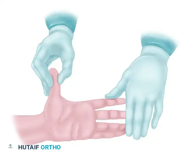
Surgical exposure in the hand frequently utilizes zigzag (Bruner) incisions to prevent postoperative flexion contractures across flexion creases. Making these incisions requires precise skin tension. As illustrated, the surgical assistant plays a vital role. The assistant must provide firm, unyielding counter-tension. By stabilizing the digits and applying longitudinal traction, the assistant flattens the palmar skin, allowing the surgeon's octagonal scalpel to glide smoothly through the dermis without skiving or creating jagged, traumatized skin edges. This collaborative tension is the foundation of atraumatic soft tissue handling, which directly correlates with reduced postoperative edema, minimized scar hypertrophy, and improved wound healing.
Step-by-Step Surgical Approach and Fixation Technique
In the context of anesthesia and surgical preparation, the "surgical approach" begins with the precise execution of the regional block and the definitive establishment of the sterile surgical field. The advent of high-frequency ultrasound has revolutionized the approach to regional anesthesia. A prospective study of the multiple-injection technique for axillary blocks demonstrated that ultrasound guidance results in significantly fewer needle passes, a shorter time to the onset of surgical anesthesia, and less procedure-related pain compared to traditional nerve stimulation techniques. The ultrasound probe is placed transversely across the anatomical landmark (e.g., the supraclavicular fossa). The needle is advanced in-plane to allow continuous visualization of the needle tip. Once the fascial sheath is breached, a small test dose of local anesthetic (hydrodissection) is injected to confirm spread around the neural structures, avoiding intraneural injection, which manifests as high resistance to injection and immediate severe pain.
The choice of approach dictates the specific block execution. The Supraclavicular Block, often termed the "spinal of the arm," provides dense, rapid-onset anesthesia of the entire upper extremity distal to the shoulder. It is performed at the level of the trunks/divisions. The Infraclavicular Block is performed at the cord level and is excellent for procedures of the elbow, forearm, and hand. It is highly suitable for continuous catheter placement due to the depth of the block and the stability of the catheter in the pectoral musculature. The Axillary Block is performed at the terminal branch level and is highly effective for forearm and hand surgery. It avoids the risk of pneumothorax entirely but requires separate, deliberate blockade of the musculocutaneous and intercostobrachial nerves to ensure complete tourniquet tolerance.
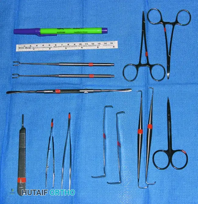
Following successful anesthesia, the transition to surgical execution requires meticulous preparation of the operative field and instrumentation. Standardization of the surgical tray enhances operative flow. The basic tray should contain instruments specifically scaled for the intricate anatomy of the hand, including small rat-tooth forceps, delicate dissecting scissors (Stevens tenotomy scissors), small hemostats, a ruler, marking pencil, double-hook Lovejoy retractors, and a fine probe. The surgical scrub nurse and the surgeon must operate in seamless synchrony to minimize wasted motion and maintain focus on the microscopic field.
Efficiency at the surgical field is maintained through the "drop technique." Using this method, the surgeon discards an instrument onto a designated sterile drape area immediately after use, and the scrub nurse returns it to its precise place on the tray. With practice, this allows the surgeon to reach for instruments without diverting their eyes from the microscopic surgical field or the surgical loupes. Instruments that are in constant rotation—such as the discarded knife, delicate tissue forceps, and dissecting scissors—are not retrieved by the nurse unless specifically requested. Special instruments, additional knife blades, and fine microsurgical sutures (e.g., 8-0 or 9-0 nylon) should be readily available on a secondary large table so they can be handed quickly to the surgeon upon request, ensuring that the definitive fixation or microvascular repair proceeds without interruption.
Complications, Incidence Rates, and Salvage Management
While highly safe when performed by experienced practitioners, complications of brachial plexus blocks and surgical preparation do occur, albeit in fewer than 1% of cases. Systemic complications are primarily related to inadvertent intravascular injection or exceeding maximum weight-based dosing, resulting in Local Anesthetic Systemic Toxicity (LAST). The pharmacodynamic profile of amide local anesthetics dictates that LAST typically presents with a bimodal distribution: initial central nervous system excitation (perioral numbness, tinnitus, metallic taste, agitation) rapidly followed by CNS depression (seizures, coma) and catastrophic cardiovascular collapse (refractory ventricular arrhythmias, asystole). Immediate availability of 20% lipid emulsion therapy is mandatory wherever regional blocks are performed; it acts as a "lipid sink," sequestering the lipophilic anesthetic molecules away from the myocardium and cerebral tissue.
Neurologic complications represent a significant medico-legal and clinical concern. Peripheral nerve injury can result from mechanical trauma (direct needle laceration), catheter-induced trauma, direct drug neurotoxicity (especially with high concentrations of local anesthetics), localized ischemia from high-pressure intraneural injection, hematoma compression, or excessive stretch during patient positioning. Fortunately, permanent neurologic sequelae occur in fewer than 1% of patients. However, patients must be counseled preoperatively that transient dysesthesias and "brachialgia" may persist for days to weeks following a brachial plexus block. This is particularly critical for patients whose occupations require fine, unimpeded manipulation of the hands, such as musicians, surgeons, and jewelers.
Pulmonary complications are anatomically linked to the proximity of the brachial plexus to the pleural dome and the phrenic nerve. Pneumothorax is the most feared anatomical complication, historically most common with the supraclavicular approach, reported as high as 6% using blind landmark techniques. The integration of ultrasound-guided techniques has drastically reduced this risk; modern prospective studies frequently report zero clinically apparent pneumothoraces in large cohorts undergoing ultrasound-guided supraclavicular blocks. Phrenic nerve palsy, resulting in ipsilateral hemidiaphragmatic paresis, is a nearly universal consequence of the interscalene block (up to 100% incidence with high volumes) and must be carefully considered in patients with severe pulmonary compromise.
| Complication | Incidence Rate | Pathophysiology/Etiology | Salvage Management / Treatment |
|---|---|---|---|
| LAST (Systemic Toxicity) | < 0.2% | Intravascular injection or overdose; blockade of cardiac sodium channels. | Airway management, ACLS (avoid vasopressin/calcium channel blockers), 20% Lipid Emulsion Therapy bolus and infusion. |
| Pneumothorax | < 0.1% (with US guidance) | Pleural puncture during supraclavicular or infraclavicular block. | Observation with 100% O2 for small (<20%) asymptomatic; Tube thoracostomy for large or symptomatic. |
| Phrenic Nerve Palsy | 100% (Interscalene), ~15% (Supraclavicular) | Local anesthetic spread to the anterior scalene fascia affecting C3-C5 roots. | Usually transient. Provide supplemental O2. Avoid block in severe COPD/contralateral pneumonectomy patients. |
| Iatrogenic Nerve Injury | < 1.0% (Transient), < 0.1% (Permanent) | Intraneural injection, needle trauma, or tourniquet neuropraxia. | Stop injection if high resistance/pain. Post-op: Gabapentinoids, EMG at 3-4 weeks if no resolution. |
| Tourniquet Palsy | Rare (< 0.1% if rules followed) | Excessive pressure or duration (>120 mins) causing mechanical compression and ischemia. | Strict adherence to time limits. Post-op observation; usually resolves spontaneously over weeks. |
Phased Post-Operative Rehabilitation Protocols
The postoperative phase begins the moment the surgical dressing is applied. The transition from the sterile field to the Post-Anesthesia Care Unit (PACU) requires meticulous attention to the physical protection of the operative site. The dressing must be non-constrictive to accommodate anticipated postoperative swelling, yet rigid enough to protect delicate tendon, nerve, or vascular repairs. This is typically achieved by incorporating a custom-molded volar plaster or fiberglass splint, heavily padded with cast padding, and secured with a loosely applied elastic bandage. Circumferential casting in the immediate postoperative period is strictly contraindicated in acute hand surgery due to the high risk of compartment syndrome as edema peaks over the first 48 to 72 hours.
A significant, and often poorly managed, consideration when utilizing long-acting regional anesthesia (such as bupivacaine or ropivacaine) is the phenomenon of "rebound pain." When the dense sensory block resolves—often abruptly between 8 to 14 hours postoperatively—the patient may experience a sudden, severe onset of nociceptive pain that can lead to emergency department visits and extreme patient dissatisfaction. To mitigate this, a multimodal analgesic protocol must be initiated before the block wears off. This preemptive strategy includes scheduled acetaminophen and NSAIDs (if not contraindicated by bone healing or bleeding risks), oral neuromodulators (e.g., gabapentin or pregabalin) for nerve-related procedures, and a short course of oral opioids reserved strictly for breakthrough pain. Thorough patient education regarding the expected timeline of block resolution is paramount.
Because the regional block masks immediate postoperative neurological function, the surgical team must rely on intraoperative anatomical certainty regarding nerve integrity. The surgeon must document that all major nerves were visually intact prior to closure. Once the block dissipates, a thorough and meticulously documented neurovascular examination must be performed. Any persistent motor or sensory deficits beyond the expected pharmacological duration of the anesthetic agent (e.g., beyond 24 hours for bupivacaine) warrant immediate clinical evaluation to rule out compressive hematoma, excessively tight dressings, or iatrogenic surgical injury.
The transition to outpatient therapy is dictated by the specific surgical procedure, but the overarching principle in hand surgery is early, controlled mobilization to prevent tendon adhesions and joint contractures. Edema control is initiated immediately through strict elevation of the extremity above the level of the heart. Depending on the repair, phased rehabilitation may begin within 3 to 5 days postoperatively under the guidance of a certified hand therapist (CHT), utilizing dynamic splinting and specific active or passive range-of-motion protocols to optimize the functional outcome while protecting the surgical reconstruction.
Summary of Landmark Literature and Clinical Guidelines
The evolution of anesthesia and surgical preparation in upper extremity surgery is heavily supported by landmark literature and established clinical guidelines. The transition from landmark-based nerve stimulation to ultrasound-guided regional anesthesia is arguably the most significant advancement in the field over the last two decades. Landmark prospective studies, such as those by Kapur et al. and Abrahams et al., definitively demonstrated that ultrasound guidance provides superior block characteristics, including faster onset times, increased success rates, and a drastic reduction in vascular puncture and pneumothorax rates. These studies form the evidence base for the current standard of care, which mandates the use of ultrasound whenever available.
The management of Local Anesthetic Systemic Toxicity (LAST) has been standardized by the American Society of Regional Anesthesia and Pain Medicine (ASRA). The ASRA practice advisories on LAST highlight the critical importance of immediate recognition of the bimodal CNS and cardiovascular symptoms. The guidelines mandate the immediate availability of 20% lipid emulsion in any facility performing regional blocks. The landmark research by Weinberg et al. on lipid resuscitation therapy fundamentally altered the algorithmic approach to local anesthetic cardiac arrest, shifting the focus away from standard ACLS protocols (which may exacerbate toxicity) toward early lipid administration to create an intravascular lipid sink.
Literature concerning tourniquet safety and ischemic preconditioning continues to guide intraoperative protocols. Studies investigating the cellular tolerance of human skeletal muscle to ischemia have validated the 120-minute threshold for safe continuous tourniquet application. Furthermore, guidelines emphasize the necessity of calculating tourniquet pressure based on the patient's specific Limb Occlusion Pressure (LOP) rather than utilizing arbitrary standard pressures, thereby minimizing the risk of tourniquet-induced neuropraxia and deep tissue injury.
Finally, the integration of Enhanced Recovery After Surgery (ERAS) protocols in upper extremity orthopedics relies heavily on the literature supporting multimodal analgesia. Clinical guidelines now strongly advocate for the minimization of perioperative opioids. The synergistic use of regional anesthesia, scheduled non-opioid analgesics, and preemptive neuromodulation has been shown in multiple randomized controlled trials to significantly improve patient satisfaction scores, decrease postoperative nausea and vomiting, and accelerate functional recovery following complex hand and upper extremity surgery. This evidence-based approach ensures that the physiological preparation of the patient perfectly complements the technical precision of the surgeon.
