Mastering Body of Navicular Fractures: Clinical Case Guide
Key Takeaway
In this comprehensive guide, we discuss everything you need to know about Mastering Body of Navicular Fractures: Clinical Case Guide. A fracture of the body of navicular bone is a serious, often high-energy intra-articular injury, typically displaced and comminuted. Management usually involves operative intervention to restore the articular surface and stabilize the fracture, aiming for a mobile, pain-free joint. Complications can include non-union, avascular necrosis, and post-traumatic osteoarthritis.
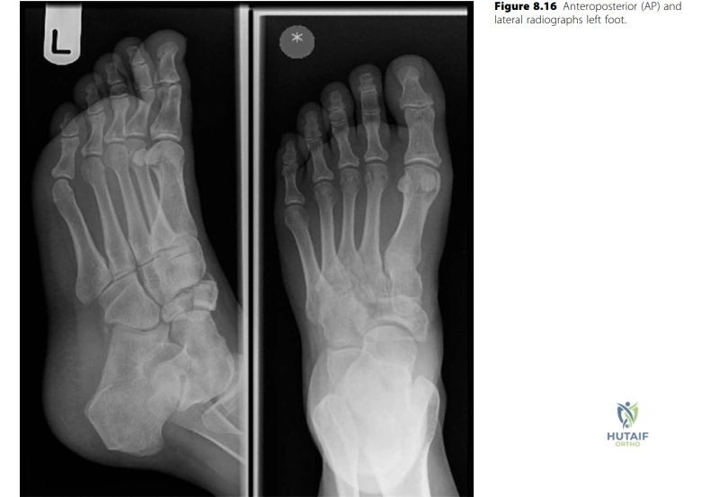
A 29-year-old female presents following a fall from a horse with an isolated, closed injury of the foot. Describe your initial assessment and interpretation of the provided radiograph.
Candidate: The radiographs show a comminuted, displaced fracture of the body of the navicular. I must assess the patient for associated injuries, particularly mid-tarsal joint involvement, and prioritize neurovascular examination and soft-tissue status to rule out compartment syndrome. My management would involve immobilization in a backslab, elevation, and a CT scan for surgical planning.
Candidates often jump straight to "I will perform an ORIF" without addressing the clinical physiology. Failing to mention the specific concern for foot compartment syndrome or the risk of occult tarsal injuries is a major oversight in a trauma viva.
A structured approach: (1) Radiographic description (body, comminution, displacement). (2) Clinical vigilance (neurovascular, skin envelope, compartment status). (3) Diagnostic step-up (CT scan is mandatory for planning articular step/gap). (4) Immediate physiological management (elevation, analgesia, backslab).
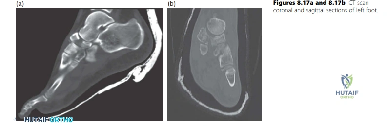
The CT scan confirms a displaced, comminuted intra-articular fracture. What are the biomechanical goals of your treatment, and in what clinical scenario would you abandon fixation for primary arthrodesis?
Candidate: The goal is to restore the talonavicular and cuneonavicular articular surfaces to prevent post-traumatic arthritis. If the comminution is too severe to reconstruct a stable construct that allows early motion, or if the bone quality is extremely poor, I would consider primary talonavicular fusion.
Failing to emphasize the importance of early motion. The navicular is critical for mid-foot kinematics; simple fusion should be a salvage procedure, not the primary choice unless reconstruction is biomechanically impossible.
Mention that the goal is "anatomical reduction of the articular surface to restore the longitudinal arch." Explicitly state: "If the fracture is unreconstructible due to severe comminution or articular impaction, primary arthrodesis provides a predictable, albeit stiff, result."
Discuss the vascular anatomy relevant to this fracture and explain why non-union and avascular necrosis are significant risks.
Candidate: The navicular has a central watershed area. The blood supply is derived from the medial plantar and dorsalis pedis arteries forming an intraosseous arcade. Damage to these vessels during trauma or excessive soft-tissue stripping during surgery leads to ischemia, necrosis, and non-union.
Vaguely stating "it has poor blood supply" without naming the specific vascular sources (dorsalis pedis/medial plantar) or the "watershed" concept. Anatomical precision is expected at the FRCS level.
Describe the "central watershed zone." Explain that the blood supply is predominantly from the dorsal and medial aspects. Emphasize that careful surgical dissection (avoiding aggressive dorsal stripping) is required to preserve this delicate blood supply.
