Fifth Metatarsal Zone II (Jones) Fractures: Advanced Diagnosis & Athlete Management
Key Takeaway
A Jones fracture is a Zone II fifth metatarsal injury at the metadiaphyseal junction, known for its precarious blood supply and high biomechanical stress. In athletes, timely surgical intervention is often crucial due to high nonunion rates with conservative care, facilitating a rapid, complete return to sport and preventing prolonged disability.
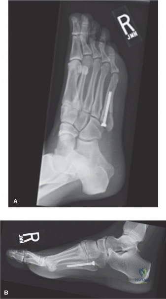
A 24-year-old professional basketball player presents to the clinic with persistent lateral foot pain following an inversion injury during a match 4 days ago. On examination, he has tenderness over the base of the fifth metatarsal. Please interpret the provided radiograph and discuss the anatomical considerations relevant to your management plan.
Candidate: The radiograph shows a transverse fracture at the metadiaphyseal junction of the fifth metatarsal, consistent with a Jones fracture (Zone II). Because this is a high-demand athlete, the blood supply in this area is precarious, and there is a high risk of nonunion. I would recommend internal fixation with an intramedullary screw.
Failing to mention the specific vascular "watershed" nature of Zone II or omitting the biomechanical rationale (cyclic bending and shear forces). Candidates often forget to mention the size of the screw required or the importance of CT imaging to size the medullary canal, which is expected at an FRCS level.
The patient has a Zone II Jones fracture. This is a watershed area with tenuous blood supply—supplied by the nutrient artery proximally and periosteal vessels distally—making it prone to delayed union. Furthermore, the area is subject to significant bending and shear forces. Given his elite athletic status, operative stabilization is the gold standard to facilitate a predictable return to sport. I would perform a pre-operative CT to assess the medullary canal diameter to ensure we can place a large-diameter (4.5mm–6.5mm) fully threaded screw, which provides superior torsional rigidity compared to smaller constructs.
You have decided to proceed with surgery. Describe your technical approach for intramedullary screw fixation and how you would mitigate the risk of injury to the surrounding neurovascular structures.
Candidate: I would make a longitudinal incision over the base of the fifth metatarsal. I would dissect down to the bone, protecting the superficial peroneal nerve branches. Under fluoroscopic guidance, I would place a guide wire into the medullary canal, ream it, and insert a fully threaded screw.
Vague description of the entry point. The entry point is the most common cause of mal-reduction or fracture propagation. Simply saying "the base" is insufficient; you must specify the starting point (tip of the tuberosity, slightly plantar) to align with the axis of the medullary canal.
I would use a lateral approach, carefully identifying and retracting the intermediate dorsal cutaneous branch of the superficial peroneal nerve. The entry point is critical: I would place the guide wire at the absolute tip of the tuberosity, slightly plantar, to ensure alignment with the long axis of the metatarsal medullary canal. I would use fluoroscopy in both AP and lateral planes to confirm the K-wire is centered. I would then perform limited reaming to optimize the screw-to-canal ratio, aiming for 70-80% fill, and insert a fully threaded cannulated screw to provide compression and rotational stability.
Post-operatively, the patient is progressing, but at 6 weeks, radiographs show no clear callus formation. How do you manage this?
Candidate: I would classify this as a delayed union. I would continue to keep the patient non-weight-bearing and potentially add a bone stimulator. If it still doesn't heal, I would consider a revision surgery with bone grafting.
Waiting too long before reassessing the construct. If the hardware is undersized or poorly placed, "continued NWB" will not achieve union. Candidates often fail to mention the need to assess for hardware failure or clinical symptoms.
I would perform a clinical and radiographic assessment. If there is no evidence of fracture progression at 6 weeks, I would scrutinize the initial radiographs for screw position and diameter. If the screw is undersized or not providing adequate compression, I would discuss early revision. Revision involves debriding the fibrous tissue at the nonunion site, re-reaming the canal, and placing a larger diameter screw, often supplemented with autologous bone graft (from the calcaneus or distal tibia) to address the biological deficit. I would also consider adjunctive therapies like PEMF bone stimulation.
