High-Energy Syndesmotic Ankle Injury: Clinical Presentation & Diagnostic Insights
Key Takeaway
Diagnosis of a high-energy syndesmotic ankle injury involves a detailed clinical exam, positive special tests like the Squeeze and External Rotation Stress tests, and critical imaging. Radiographs typically reveal a distal fibular fracture (e.g., Weber C), widened tibiofibular clear space, and reduced tibiofibular overlap, indicating syndesmotic disruption and instability.
A 32-year-old semi-professional soccer player presents with an acute rotational ankle injury. You suspect a syndesmotic injury. Before looking at the images, describe your initial clinical assessment and the "Squeeze" and "External Rotation" stress tests. Why is this patient’s history particularly concerning to you as an examiner?
Candidate: I would perform a thorough physical exam including palpation of the AITFL, PITFL, and the full length of the fibula to rule out a Maisonneuve fracture. The Squeeze test involves compressing the tibia and fibula at the mid-calf; pain distally indicates syndesmotic injury. The External Rotation test involves rotating the foot with the knee at 90 degrees. This patient is concerning because he is a high-demand athlete; any malreduction or persistent instability will lead to early post-traumatic osteoarthritis, ending his career.
Failing to mention the "Maisonneuve" search (proximal fibular palpation), missing the neurovascular exam, or failing to articulate the biomechanical consequences (42% reduction in contact area with 1mm of lateral shift) which explains why this high-demand patient has a worse prognosis.
Start with a structured approach: Physical exam (AITFL/PITFL/Maisonneuve check), neurovascular status, and diagnostic maneuvers. Explain that the Squeeze test assesses the interosseous membrane and the External rotation test stresses the AITFL/PITFL complex. Highlight the urgency: in a 32-year-old athlete, anatomical reduction is non-negotiable because the syndesmosis is the key to load sharing; a 1mm shift reduces contact area by 42%, leading to catastrophic, early arthritis.
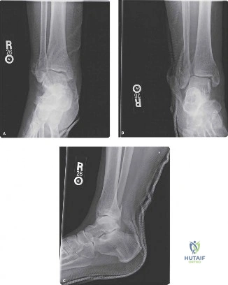
The patient's initial mortise view appears "borderline." You decide to proceed with further investigation. Look at this imaging. What are you looking for, and if this patient requires surgery, how do you verify your reduction intraoperatively?
Candidate: On the mortise view, I am assessing the tibiofibular clear space (should be <5mm) and tibiofibular overlap (should be >1mm). If these are equivocal, I would perform gravity or external rotation stress views. Intraoperatively, I would perform a Cotton (Hook) test to assess for latent instability. Most importantly, I would perform a direct open reduction of the incisura under direct vision, as fluoroscopy alone is notorious for missing malreduction.
Relying solely on "the eye" or static fluoroscopy to confirm reduction. Failing to acknowledge the high rate of malreduction (up to 52%) when not using direct visualization or postoperative CT.
Quantify the radiographic parameters (Medial Clear Space <4mm, TF Clear Space <5mm, Overlap >1mm). Emphasize that "direct visualization" is now the academic standard. Mention that the clamp must be placed 2-3cm above the joint line, parallel to the joint, and the fibula must be correctly rotated into the incisura. Mention that if the reduction is questionable, a postoperative CT is the mandatory "Gold Standard" to confirm anatomical congruence.
The patient is undergoing surgery. How do you decide between a trans-syndesmotic screw and a suture button construct? What are the implications for rehabilitation?
Candidate: Suture buttons (dynamic fixation) are generally preferred in high-demand athletes as they allow physiological micromotion and do not require routine removal, unlike static screws. With screws, we usually keep the patient non-weight-bearing until hardware removal or until syndesmotic healing is confirmed. With suture buttons, we can often initiate earlier weight-bearing and functional rehab, which is vital for this patient's return to soccer.
Failing to mention the debate regarding hardware removal. Simply saying "screws are old, suture buttons are new" without explaining the biomechanical difference (rigidity vs. dynamic motion).
Structure the answer: 1. Biomechanics (Rigid fixation vs. dynamic, physiological motion). 2. Clinical Outcomes (Equivalent scores, but suture buttons have lower malreduction rates and eliminate the need for second-look surgeries for hardware removal). 3. Rehabilitation (Suture buttons allow earlier loading). Acknowledge that while screws remain the "traditional" standard, evidence (Level I trials) supports the shift toward suture buttons in the athletic population.
