Comprehensive Guide to Rocker Bottom Sole Modifications for Midfoot Pain
Key Takeaway
A rocker bottom sole modification is a non-surgical shoe alteration designed to relieve midfoot pain by changing the foot's roll-over shape during gait. It facilitates a smoother transition, reduces motion at painful joints, offloads specific plantar pressure points, and decreases muscular effort, offering symptomatic relief for conditions like osteoarthritis, Charcot foot, and pes planus.
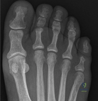
A 55-year-old female presents with chronic midfoot pain. She describes the sensation of "walking on broken glass." She has failed 6 months of conservative management including NSAIDs and standard orthotics. On examination, there is tenderness over the tarsometatarsal joints. Weight-bearing radiographs are provided below.
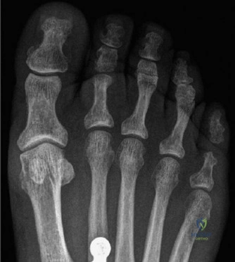
How would you describe the biomechanical rationale for using a "rocker bottom" shoe in this patient, and which configuration would you select?
Candidate: A rocker bottom shoe helps by offloading the midfoot. I would choose a heel-to-toe rocker to reduce motion across the arthritic tarsometatarsal joints, essentially bypassing the painful segment during the stance phase of gait.
Failing to mention the specific phases of gait. Candidates often just say "it takes pressure off." A high-scoring answer must link the biomechanics to the "three rockers" (heel, ankle, forefoot) and explain that the modification substitutes for the foot's lost intrinsic roll-over function.
The rocker sole alters the GRF vector and reduces the lever arm of the foot. By creating a convex profile, we effectively shorten the functional lever, thereby reducing the bending moment and joint excursion required at the midfoot. For midfoot OA, a heel-to-toe rocker is indicated to minimize motion across the entire midtarsal/TMT complex. If there was a prominent midfoot collapse (e.g., Charcot), a double rocker would be used to offload the focal pressure point by creating a concave mid-section.
The patient's pain becomes refractory to all non-operative measures. You are planning a midfoot arthrodesis. You perform a clinical assessment and order advanced imaging. What are the critical structures at risk during the dorsal approach, and how do you protect them?
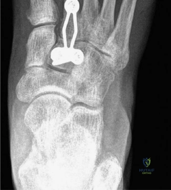
Candidate: During the dorsal approach, the main structures at risk are the dorsalis pedis artery and the deep peroneal nerve, which run between the first and second metatarsal bases. I would carefully identify and mobilize them using vessel loops and retract the EHB muscle belly laterally to protect them.
Forgetting to mention the extensor hallucis brevis (EHB). Neglecting to mention that the EHB must be mobilized or retracted is a common technical oversight. Furthermore, failing to mention the specific internervous plane (EHL/EDL) suggests a lack of anatomical familiarity.
The dorsal approach utilizes the interval between the EHL and EDL. The deep peroneal nerve and dorsalis pedis artery are the critical structures situated between the first and second metatarsal bases. Protection involves meticulous subperiosteal dissection, identification of the neurovascular bundle, and use of vessel loops. Additionally, one must identify the EHB muscle belly, which often obscures the tarsometatarsal joints; this should be reflected laterally to ensure safe access to the joints for debridement.
Six months post-operative, the patient complains of persistent pain. Radiographs show solid fusion at the site of surgery, but she now has symptoms in the adjacent joints. How do you classify this, and what is your management plan?
Candidate: This is adjacent segment disease. Because the midfoot is now rigid, stress is transferred to the adjacent joints. I would manage this conservatively with modified footwear, like a rocker bottom shoe, and if that fails, consider extending the fusion.
Jumping to surgery. Failing to mention that rocker bottom shoes are the primary solution for post-fusion adjacent segment disease suggests the candidate views surgery as the only tool in the box, rather than recognizing the biomechanical necessity of the orthotic.
This is symptomatic Adjacent Segment Disease (ASD) secondary to the altered biomechanics of a fused midfoot, which forces compensation at the Chopart or MTP joints. The management is initially non-operative: prescription of a custom orthotic with a rocker bottom modification to replace the lost sagittal motion and attenuate stress on adjacent articulations. If this fails to provide relief, then surgical expansion of the arthrodesis to include the painful segment is considered.
