Lisfranc Injury: Diagnosis, Surgical Anatomy, and Management with Focus on Second Metatarsal Base
Key Takeaway
The second metatarsal base is crucial for Lisfranc injury diagnosis as it forms the keystone of midfoot stability. Its recessed position and strong Lisfranc ligament connections mean any disruption, such as a fracture or diastasis (>2mm) at its articulation with the medial cuneiform, is pathognomonic, demanding immediate attention for accurate diagnosis and management.
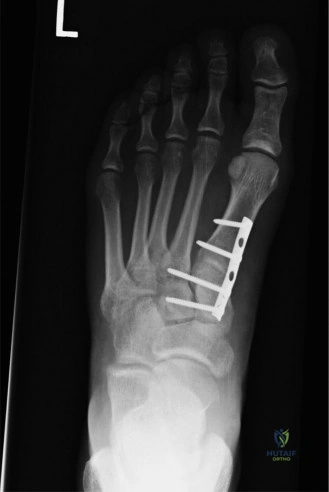
A 32-year-old male presents to the emergency department following a motor vehicle accident. He complains of severe midfoot pain and inability to weight-bear. You obtain the following radiograph. Describe the key findings and explain the significance of the "fleck sign" in this context.
Candidate: The radiograph shows a Lisfranc injury. There is widening between the base of the first and second metatarsals. The "fleck sign" is a small bony fragment near the base of the second metatarsal, which indicates that the Lisfranc ligament has pulled a piece of bone off, confirming an unstable injury.
Candidates often focus solely on the "widening" (diastasis). They fail to mention the specific anatomical attachment of the Lisfranc ligament (medial cuneiform to 2nd MT base) or neglect to state that this injury pattern mandates urgent surgical intervention because it signifies a complete rupture of the primary midfoot stabilizer.
The radiograph demonstrates a Lisfranc fracture-dislocation. The "fleck sign" represents an avulsion fracture of the base of the 2nd metatarsal at the insertion of the interosseous Lisfranc ligament. It is pathognomonic for a high-energy, unstable injury. It serves as a visual indicator of Lisfranc ligament disruption. Anatomically, the 2nd metatarsal base is the 'keystone' of the midfoot; thus, any widening >2mm or the presence of this avulsion fragment is an absolute indication for anatomical reduction and internal fixation to prevent post-traumatic arthritis.
You have taken the patient to theater for ORIF. You have achieved reduction of the first ray. Describe your technique for the "critical step" of fixing the second metatarsal to the medial cuneiform.
Candidate: I would make a dorsal incision between the first and second metatarsals. I would use a reduction clamp to pull the 2nd metatarsal base into the cuneiform, check the position with fluoroscopy, and then place a 3.5mm screw from the medial cuneiform into the base of the second metatarsal.
Failing to mention the protection of the neurovascular structures (deep peroneal nerve and dorsalis pedis artery), failing to specify the trajectory (which is prone to missing the 2nd MT base if too vertical), and forgetting to mention that the screw is often removed at 3-6 months.
Exposure is via a longitudinal dorsal incision between the EHL and EDL, carefully protecting the dorsalis pedis artery and deep peroneal nerve. After debriding the joint, I perform a 'top-down' reduction, focusing on the 2nd MT base as the keystone. I use a joy-stick K-wire or pointed reduction forceps for manipulation. The key fixation is a 3.5mm cortical screw (or headless compression screw) placed from the dorsal aspect of the medial cuneiform into the base of the 2nd metatarsal, angled to capture the lateral plantar cortex of the 2nd MT base. I confirm anatomical alignment on AP, oblique, and lateral views. The hardware is typically removed at 3–6 months post-operatively to restore physiological midfoot motion.
