Foot & Ankle Radiography: Comprehensive Guide to Interpretation, Anatomy, Biomechanics, & Pathology

Key Takeaway
Foot and ankle radiographs are foundational for assessing pain, revealing bone architecture, joint integrity, and alignment. They diagnose fractures, degenerative disease, inflammatory arthropathies, and deformities such as pes planus or hallux valgus. This guides precise orthopedic diagnosis and management strategies effectively.
You are presented with a patient suffering from chronic midfoot pain. Examine the following radiograph. What are the key radiographic features you are assessing, and how do they influence your diagnosis of a potential Lisfranc injury?
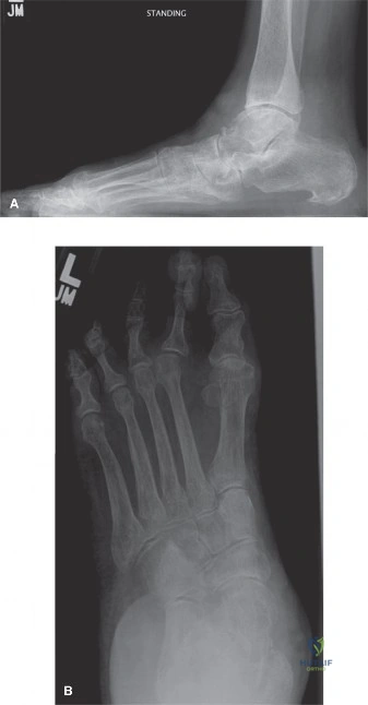
Candidate: I would look for widening between the first and second metatarsal bases. I would also check if the medial border of the second metatarsal aligns with the medial border of the middle cuneiform. I’m looking for subluxation and any small avulsion fractures at the base of the second metatarsal, which is the "fleck sign."
Candidates often focus only on the M1-M2 space. They fail to mention the critical alignment of the second metatarsal with the middle cuneiform—the "keystone" of the Lisfranc complex. They also frequently miss the importance of stating the imaging must be *weight-bearing* to reveal occult instability.
I would systematically assess the Lisfranc joint complex on weight-bearing views. My focus is three-fold: 1) Alignment: The medial border of the second metatarsal must align with the medial border of the middle cuneiform. Any step-off here is pathological. 2) Diastasis: The interval between the first and second metatarsal bases should be evaluated; widening (>2mm compared to the contralateral side) suggests ligamentous disruption. 3) Osseous involvement: I would specifically look for the "fleck sign" (avulsion of the Lisfranc ligament). If clinical suspicion remains high despite negative radiographs, I would proceed to weight-bearing stress views or CT/MRI to assess for occult ligamentous injury.
Look at this lateral hindfoot view. Describe the radiographic parameters used to assess the severity of a calcaneal fracture and why they are prognostic.
Candidate: I would measure Böhler’s angle and the angle of Gissane. Böhler’s angle should normally be 20 to 40 degrees. If it's decreased, it indicates compression of the posterior facet. The angle of Gissane is also important for the geometry of the subtalar joint.
Stating only the angle names without explaining *why* they matter. Failing to emphasize that these are 2D representations of a 3D injury, and that CT scanning is mandatory for preoperative planning to evaluate the number of articular fragments.
I focus on two key angles: 1) Böhler’s angle: Formed by the intersection of a line from the superior aspect of the anterior process to the highest point of the posterior facet, and a line from the posterior facet to the superior aspect of the tuberosity. A decrease (<20°) indicates loss of height and posterior facet depression. 2) Angle of Gissane (Crucial Angle): Assesses the lateral wall and subtalar joint geometry. Crucially, these angles provide a baseline for reduction. However, they do not delineate the degree of comminution; therefore, I would correlate these findings with a CT scan to classify the fracture (e.g., Sanders classification) to guide whether ORIF is feasible versus conservative management or primary subtalar fusion.
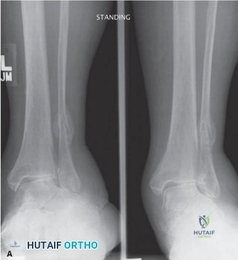
A 45-year-old patient presents with symptomatic adult-acquired flatfoot. Review this image. What radiographic parameters define the severity of this deformity, and how do they impact your surgical decision-making?
Candidate: I would measure the talonavicular coverage angle and Meary's angle. If the talonavicular coverage is increased, it shows midfoot abduction. I would also look at the calcaneal pitch. If these are severe and the joint is arthritic, I would perform a triple arthrodesis.
Jumping immediately to "fusion" (triple arthrodesis) for all cases. The candidate fails to distinguish between a *flexible* deformity (amenable to tendon transfer and corrective osteotomy) and a *fixed/arthritic* deformity (which necessitates fusion).
My assessment is stratified by flexibility: 1) Radiographic Parameters: I measure the Talar-1st Metatarsal angle (Meary's line) on the lateral view to assess sagittal collapse, and the Talonavicular coverage angle on the AP view to assess abduction. 2) Clinical-Radiographic Correlation: If the radiographs show significant deformity but the clinical examination confirms a flexible hindfoot, I would consider a joint-sparing reconstruction, such as a medializing calcaneal osteotomy combined with FDL tendon transfer. 3) Surgical Threshold: If radiographic evidence demonstrates severe joint space narrowing or fixed subluxation, I would then advocate for arthrodesis to provide a stable, plantigrade foot. The choice of procedure must be patient-centered, balancing the patient’s functional demands against the radiographic severity.
