Medial Foot & Ankle Masses: Comprehensive Diagnosis & Surgical Anatomy
Key Takeaway
Medial foot and ankle masses encompass diverse pathologies, from common accessory navicular syndrome and posterior tibial tendon dysfunction (PTTD) to ganglion cysts and rare nerve sheath tumors. Accurate diagnosis is paramount, requiring thorough investigation of the medial cord's complex bony, ligamentous, and tendinous structures to guide effective surgical or conservative management and alleviate pain.
A 15-year-old adolescent athlete presents with persistent pain over the medial aspect of the foot. On examination, there is a palpable, tender bony prominence just anterior to the medial malleolus. What is the most likely diagnosis, and how would you classify this condition to guide your management?
Candidate: The diagnosis is likely Accessory Navicular Syndrome. I would classify this using the Geist classification. Type I is a sesamoid bone within the posterior tibial tendon. Type II has a synchondrosis between the accessory ossicle and the navicular body. Type III is a fused, cornuate navicular.
Failing to mention the clinical relevance of the synchondrosis (Type II). Many candidates recite the classification but forget to state that the Type II synchondrosis is the primary source of symptoms due to micro-motion and traction from the PTT.
Identify it as Accessory Navicular Syndrome. Classify using the Geist system (Type I, II, III). Highlight that Type II—the triangular or heart-shaped ossicle with a cartilaginous synchondrosis—is the symptomatic variant due to irritation from the PTT insertion and shoe wear. Conclude that management is initially conservative (orthotics, activity modification), reserving the Kidner procedure for failures.
During the clinical assessment of a suspected accessory navicular, you order imaging. Please interpret this radiograph and explain its significance in your surgical planning.
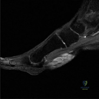
Candidate: This radiograph demonstrates an accessory navicular. I would evaluate the size and shape of the ossicle. If I were to operate, this helps me identify the synchondrosis and ensures I excise the entire fragment without leaving residual bone that could cause persistent pain.
Focusing only on the ossicle. Examiners want to see you assess the alignment of the foot (e.g., Meary’s angle or Talo-navicular coverage angle) to rule out concurrent adult-acquired flatfoot deformity, which would change the surgical plan from a simple Kidner procedure to a more complex reconstruction.
Structure the answer: 1. Confirm the presence of the accessory navicular (Type II). 2. Assess the longitudinal arch height (Meary's angle/Talo-first metatarsal angle) to rule out PTTD/flatfoot. 3. Look for signs of degenerative changes in the talonavicular joint. State that these findings dictate whether simple excision suffices or if formal tendon augmentation/calcaneal osteotomy is required.
You have decided to proceed with excision of a symptomatic soft tissue mass in the medial foot, as shown in this surgical field image. What are your specific steps to avoid iatrogenic injury to the neurovascular bundle?
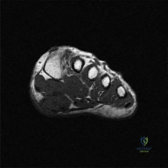
Candidate: I would use a longitudinal incision to avoid flexion creases. I would meticulously dissect the subcutaneous tissue, identifying the saphenous nerve and great saphenous vein. Deeper, I would isolate the tibial nerve and posterior tibial artery before addressing the mass, ensuring I stay in the correct plane.
Not addressing the "mass" specifically. If it is a schwannoma, simply "dissecting" can lead to nerve palsy. Candidates must distinguish between the "shelling out" technique for a schwannoma versus the necessity of formal biopsy or resection for more aggressive-looking lesions.
Emphasize a systematic approach: (1) Anatomical landmarking: identifying the saphenous nerve and vein early. (2) Deep exposure: identifying the neurovascular bundle proximal to the mass. (3) Identification of the mass origin (e.g., stalk of a ganglion vs. nerve sheath). (4) Using nerve stimulation if the mass is intimately associated with a nerve trunk. (5) Standard meticulous hemostasis and careful layered closure to prevent adhesions.
