Comprehensive Lisfranc Injury Case Study: Clinical Examination & Advanced Diagnostics
Key Takeaway
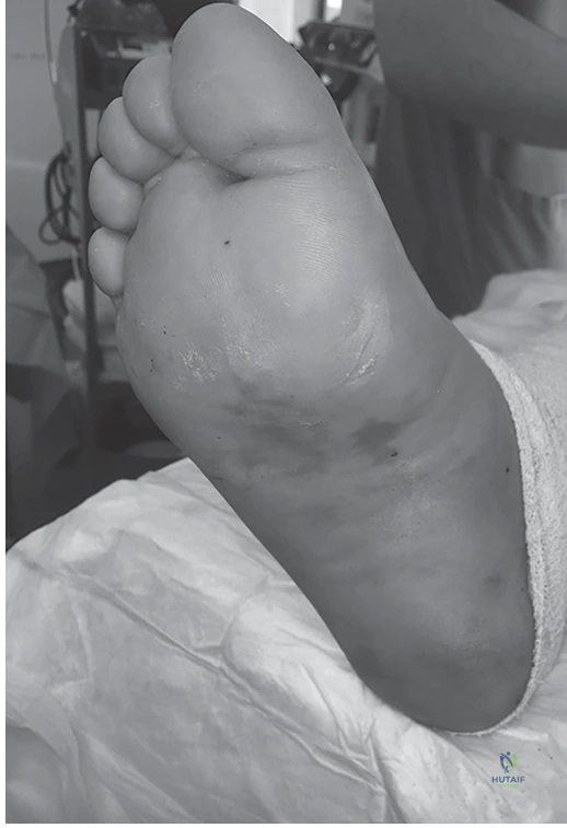
Diagnosing a Lisfranc injury requires evaluating high-energy trauma, severe midfoot pain, and swelling. Key clinical signs are plantar ecchymosis ('footprint sign') and TMT joint tenderness. Radiographs may reveal metatarsal widening or a 'fleck sign.' CT scans are crucial for confirming subtle fractures, articular incongruity, and ligament disruption, guiding treatment.
A 38-year-old male presents following a high-energy MVC. He has significant midfoot swelling and an inability to bear weight. You are presented with his initial AP foot radiograph. Describe the findings and state the significance of the "fleck sign."
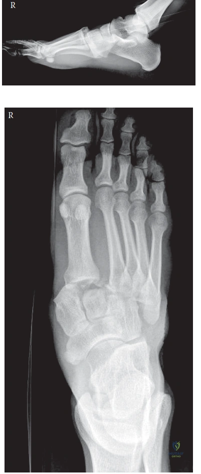
Candidate: The X-ray shows widening between the base of the first and second metatarsals. There is a small bone fragment near the base of the second metatarsal, which is the "fleck sign." This indicates a Lisfranc injury.
Candidates often stop at "Lisfranc injury." They fail to quantify the widening (>2.5mm is diagnostic) or explain the pathophysiology of the fleck sign—which is an avulsion of the Lisfranc ligament (base of the 2nd metatarsal or medial cuneiform). They also frequently fail to mention the loss of alignment of the medial border of the 2nd metatarsal with the middle cuneiform.
The radiograph demonstrates clear signs of a Lisfranc injury: 1) Widening of the 1st-2nd intermetatarsal interval (>2.5mm). 2) Loss of alignment between the medial border of the second metatarsal and the medial border of the middle cuneiform. 3) The "fleck sign," which is an avulsion fracture of the base of the second metatarsal or medial cuneiform representing the attachment of the Lisfranc ligament. This confirms complete disruption of the Lisfranc ligament complex, rendering the midfoot unstable.
Following the plain radiographs, a CT scan was obtained. Describe the role of the CT in your surgical planning for this injury, and classify the injury according to the Myerson system.
Candidate: The CT helps me see the fractures in more detail, like the comminution at the base of the second metatarsal. It helps me plan where to put my screws and if I need plates. Based on the lateral displacement of the lesser metatarsals, this is a Myerson Type B2 injury.
Failing to mention the "Keystone" concept. A candidate who doesn't mention that the CT allows assessment of the articular surface of the middle cuneiform and 2nd metatarsal base—which is vital for the reduction of the "keystone"—is missing the point of the planning phase.
The CT is essential to assess the degree of articular comminution and displacement that plain films underestimate. It dictates the need for ORIF versus bridge plating. In this case, it confirms the 'keystone' (2nd TMT joint) is involved. Following the Myerson classification, this is a Type B2 (Partial Incongruity, lateral displacement of the 2nd-5th metatarsals), which is the most common pattern and necessitates restoration of the medial and central columns.
You have decided to proceed to surgery. Outline your operative strategy regarding the order of reduction and the choice of fixation for this Lisfranc injury.
Candidate: I would use a dual incision approach. I would reduce the medial column first, then the second metatarsal. I would use a screw from the medial cuneiform to the second metatarsal to act as the Lisfranc screw and potentially plates for the other metatarsals if they are comminuted.
Ignoring the "transarticular but non-compressive" nature of the hardware. Candidates often forget to mention that they must check the hardware intraoperatively to ensure they haven't inadvertently caused an arthrodesis, or they forget to mention the rationale for hardware removal.
My strategy is: 1) Dual dorsal incisions. 2) Sequential reduction starting with the medial column (1st TMT) to restore length, followed by the "keystone" (2nd TMT to middle cuneiform) to restore the transverse arch. 3) Fixation with non-compressive cortical screws across the 1st and 2nd TMT joints to allow for biological healing of the ligamentous complex. 4) Use dorsal locking plates for the comminuted 2nd metatarsal base to provide stability while bridging the fracture. 5) Plan for elective hardware removal at 3-6 months to prevent fatigue failure and facilitate normal foot kinematics.
