FRCS Oral Examination: Abbreviated Galeazzi Case Mastery
Key Takeaway
For anyone wondering about FRCS Oral Examination: Abbreviated Galeazzi Case Mastery, An examination abbreviated Galeazzi identifies a specific fracture dislocation involving a displaced midshaft diaphyseal fracture of the radius. This injury pattern is combined with an associated injury to the distal radioulnar joint (DRUJ), commonly presenting as dorsal displacement of the distal ulna. Radiographs are crucial for confirming this characteristic orthopedic injury.
A 38-year-old carpenter presents following a fall from a height. Radiographs show a distal third radial shaft fracture with associated disruption of the distal radioulnar joint (DRUJ). How would you classify this injury, and what are the critical biomechanical principles of management?
Candidate: This is a Galeazzi fracture-dislocation. Management involves ORIF of the radius to restore length, as shortening is the primary cause of DRUJ instability. I would check the DRUJ intraoperatively and stabilize it if necessary.
Failing to mention the "anatomical restoration of radial length" as the primary goal. Candidates often forget to address the interosseous membrane (IOM) or neglect the intraoperative stability testing of the DRUJ (the "piano key sign").
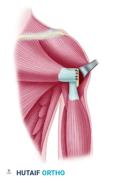
This is a Galeazzi fracture. The management is: 1. Anatomical Restoration: ORIF of the radius using a compression or locking plate to restore length, alignment, and rotation; this indirectly addresses DRUJ subluxation. 2. DRUJ Assessment: Perform the "piano key sign" test intraoperatively. If unstable after radial reduction, verify that no soft tissue (e.g., extensor carpi ulnaris) is interposed. 3. Stabilization: If persistent instability exists, use trans-syndesmotic K-wires for 4–6 weeks, typically in a position of stability (supination for dorsal, pronation for volar). 4. Rehabilitation: Protect the DRUJ during the pinning period and initiate progressive range-of-motion only post-removal.
During your volar (Henry) approach to this radius, what specific structures are at risk, and how do you protect them?
Candidate: The main risk is to the anterior interosseous nerve (AIN), which lies on the interosseous membrane between the flexor pollicis longus and flexor digitorum profundus. I would carefully dissect and identify it.
Confusing the AIN with the Posterior Interosseous Nerve (PIN). The PIN is associated with the dorsal (Thompson) approach. A candidate failing to distinguish these risks shows poor surgical anatomy knowledge.
The AIN is at risk during the volar approach, specifically when exposing the proximal-to-mid radial shaft. I protect it by identifying the interval between the FPL and FDP, staying deep to the pronator quadratus, and avoiding aggressive medial retraction or over-stripping of the interosseous membrane. If using a dorsal approach, the PIN is the primary structure at risk as it exits the supinator.
