Full Question & Answer Text (for Search Engines)
Question 1:
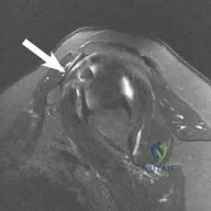
A 45-year-old male sustains a 3-part proximal humerus fracture and is scheduled for open reduction and internal fixation via a deltopectoral approach. During the superficial dissection, the cephalic vein is encountered. Which of the following best describes the internervous plane of this approach and the recommended management of the cephalic vein to preserve its major venous tributaries?
Options:
- Deltoid and Pectoralis minor; retract the vein medially
- Deltoid and Pectoralis major; retract the vein laterally
- Deltoid and Pectoralis major; retract the vein medially
- Coracobrachialis and Short head of biceps; retract the vein laterally
- Deltoid and Coracobrachialis; ligate the vein routinely
Correct Answer: Deltoid and Pectoralis major; retract the vein laterally
Explanation:
The deltopectoral approach utilizes the internervous plane between the deltoid (axillary nerve) and the pectoralis major (medial and lateral pectoral nerves). The cephalic vein is located within this interval. Standard orthopedic teaching recommends retracting the cephalic vein laterally with the deltoid muscle. This preserves the primary feeding veins which drain from the deltoid into the cephalic vein, thereby reducing postoperative deltoid swelling and venous congestion.
Question 2:
A 32-year-old female undergoes a direct anterior (Smith-Petersen) approach to the hip for a peri-acetabular osteotomy. Which of the following describes the correct superficial internervous plane and the cutaneous nerve most at risk during this portion of the dissection?
Options:
- Sartorius and Rectus Femoris; Femoral branch of the genitofemoral nerve
- Sartorius and Tensor Fasciae Latae; Lateral femoral cutaneous nerve
- Tensor Fasciae Latae and Gluteus Medius; Lateral femoral cutaneous nerve
- Tensor Fasciae Latae and Gluteus Medius; Superior gluteal nerve
- Rectus Femoris and Vastus Lateralis; Saphenous nerve
Correct Answer: Sartorius and Tensor Fasciae Latae; Lateral femoral cutaneous nerve
Explanation:
The Smith-Petersen (direct anterior) approach to the hip utilizes a superficial internervous plane between the sartorius (innervated by the femoral nerve) and the tensor fasciae latae (innervated by the superior gluteal nerve). The lateral femoral cutaneous nerve (LFCN) crosses over the sartorius approximately 2 cm distal to the ASIS and is at high risk of injury during the superficial dissection. Injury to the LFCN can result in meralgia paresthetica.
Question 3:
An orthopedic surgeon is planning an approach to the radial head to perform an open reduction and internal fixation of a displaced fracture. The surgeon elects to use the Kocher approach. Between which two muscles is the internervous plane developed?
Options:
- Extensor carpi radialis brevis and Extensor digitorum communis
- Extensor carpi radialis longus and Brachioradialis
- Anconeus and Extensor carpi ulnaris
- Brachioradialis and Pronator teres
- Flexor carpi ulnaris and Extensor carpi ulnaris
Correct Answer: Anconeus and Extensor carpi ulnaris
Explanation:
The Kocher approach to the elbow and radial head utilizes the internervous plane between the anconeus (innervated by the radial nerve) and the extensor carpi ulnaris (innervated by the posterior interosseous nerve). In contrast, the Kaplan approach is slightly more anterior and utilizes the plane between the extensor digitorum communis (PIN) and the extensor carpi radialis brevis (radial nerve proper). The Kocher approach is generally considered safer for the posterior interosseous nerve, which stays further anteriorly in the supinator.
Question 4:
During a primary total hip arthroplasty using a direct lateral (Hardinge) approach, the surgeon splits the gluteus medius and vastus lateralis. To avoid denervation of the anterior portion of the gluteus medius and tensor fasciae latae, the proximal splitting of the gluteus medius should not exceed what distance from the tip of the greater trochanter?
Options:
- 1 - 2 cm
- 3 - 5 cm
- 7 - 9 cm
- 10 - 12 cm
- 15 cm
Correct Answer: 3 - 5 cm
Explanation:
The direct lateral (Hardinge) approach to the hip does not use a true internervous plane, as it splits the gluteus medius and vastus lateralis (both innervated by the superior gluteal nerve for the former, and femoral nerve for the latter, but the split is within the substance of the gluteus medius). The superior gluteal nerve courses from posterior to anterior and supplies the gluteus medius, gluteus minimus, and TFL. Its inferior branch runs approximately 3 to 5 cm proximal to the tip of the greater trochanter. Extending the split further proximal than 5 cm risks severing this nerve, leading to a postoperative Trendelenburg gait.
Question 5:
A surgeon performs the proximal portion of the volar (Henry) approach to the forearm to expose the proximal radius. The internervous plane at this level is between which of the following muscles?
Options:
- Pronator teres and Flexor carpi radialis
- Brachioradialis and Pronator teres
- Brachioradialis and Flexor carpi radialis
- Flexor carpi ulnaris and Flexor digitorum superficialis
- Extensor carpi radialis brevis and Extensor digitorum communis
Correct Answer: Brachioradialis and Pronator teres
Explanation:
The volar (Henry) approach to the forearm provides extensile exposure to the radius. Proximally, the internervous plane is between the brachioradialis (innervated by the radial nerve) and the pronator teres (innervated by the median nerve). Distally, the plane shifts to become between the brachioradialis and the flexor carpi radialis (median nerve).
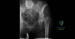
Question 6:
A 28-year-old male presents with an anterior column acetabular fracture. An ilioinguinal approach is planned. Once the external oblique aponeurosis and inguinal canal are opened, the iliopectineal fascia is identified. The 'middle window' of this approach is defined laterally by the iliopsoas/iliopectineal fascia and medially by the external iliac vessels. Which of the following structures is found within this middle window?
Options:
- External iliac artery and vein
- Spermatic cord and genitofemoral nerve
- Iliopsoas muscle and femoral nerve
- Obturator nerve and artery
- Sciatic nerve and inferior gluteal artery
Correct Answer: Iliopsoas muscle and femoral nerve
Explanation:
The ilioinguinal approach creates three surgical windows. The lateral window is lateral to the iliopsoas (contains the iliacus and lateral femoral cutaneous nerve). The middle window is between the iliopectineal fascia (lateral) and the external iliac vessels (medial); it contains the iliopsoas muscle and the femoral nerve. The medial window is medial to the external iliac vessels and provides access to the superior pubic ramus, the quadrilateral surface, and the retropubic space (Space of Retzius).
Question 7:
During a modified Stoppa approach for a pelvic ring fracture, the surgeon elevates the peritoneum from the superior pubic ramus and quadrilateral plate. A significant vascular structure traversing vertically over the superior pubic ramus is encountered and must be ligated to prevent catastrophic hemorrhage. This structure is an anastomosis between which two vascular systems?
Options:
- Internal pudendal and External pudendal
- External iliac and Obturator
- Superior gluteal and Inferior gluteal
- Femoral and Deep femoral
- Internal iliac and Median sacral
Correct Answer: External iliac and Obturator
Explanation:
The structure is the corona mortis (crown of death), which is an aberrant anastomosis between the external iliac system (specifically the inferior epigastric vessels) and the obturator system. It is present in approximately 30-40% of hemi-pelves and crosses over the superior pubic ramus at an average of 5-6 cm from the pubic symphysis. Ligation is critical during the modified Stoppa or ilioinguinal approach to prevent severe intrapelvic bleeding.
Question 8:
A 40-year-old male sustains a midshaft humerus fracture requiring plate fixation. A posterior approach to the humerus is chosen. To safely identify and protect the radial nerve, the surgeon must know its reliable anatomical landmarks. The radial nerve typically crosses the posterior aspect of the humerus at approximately what distance proximal to the lateral and medial epicondyles respectively?
Options:
- 10 cm proximal to the lateral epicondyle; 15 cm proximal to the medial epicondyle
- 14 cm proximal to the lateral epicondyle; 20 cm proximal to the medial epicondyle
- 20 cm proximal to the lateral epicondyle; 14 cm proximal to the medial epicondyle
- 5 cm proximal to the lateral epicondyle; 10 cm proximal to the medial epicondyle
- 18 cm proximal to the lateral epicondyle; 18 cm proximal to the medial epicondyle
Correct Answer: 14 cm proximal to the lateral epicondyle; 20 cm proximal to the medial epicondyle
Explanation:
The posterior approach to the humerus involves identifying the radial nerve as it passes through the spiral groove. A reliable anatomical landmark is that the radial nerve lies directly on the posterior aspect of the humerus approximately 14 cm proximal to the lateral epicondyle, and it crosses the medial intermuscular septum roughly 20 cm proximal to the medial epicondyle.
Question 9:
Which of the following surgical approaches to the hip is accurately matched with its proper internervous plane?
Options:
- Watson-Jones : Tensor Fasciae Latae and Gluteus Medius
- Smith-Petersen : Sartorius and Rectus Femoris
- Kocher-Langenbeck : Gluteus Maximus and Gluteus Medius
- Hardinge : Gluteus Medius and Vastus Lateralis
- Smith-Petersen : Sartorius and Tensor Fasciae Latae
Correct Answer: Smith-Petersen : Sartorius and Tensor Fasciae Latae
Explanation:
The Smith-Petersen approach uses a true internervous plane between the Sartorius (femoral n.) and the Tensor Fasciae Latae (superior gluteal n.). The Watson-Jones (anterolateral) approach utilizes the interval between the TFL and Gluteus Medius, which is not a true internervous plane since both are innervated by the superior gluteal nerve. The Hardinge approach involves a direct split of the Gluteus Medius and Vastus Lateralis, offering no internervous plane. The Kocher-Langenbeck splits the Gluteus Maximus.
Question 10:
During a Kocher-Langenbeck approach for a posterior wall acetabular fracture, the short external rotators of the hip are tagged and tenotomized near their femoral insertion. Which muscle should be preserved or have its femoral insertion left intact to protect the main blood supply to the femoral head?
Options:
- Piriformis
- Superior gemellus
- Obturator internus
- Quadratus femoris
- Inferior gemellus
Correct Answer: Quadratus femoris
Explanation:
The deep branch of the medial femoral circumflex artery (MFCA) provides the primary blood supply to the adult femoral head. It runs anterior to the quadratus femoris and posterior to the obturator externus. To protect the MFCA during a posterior approach, the quadratus femoris (or at least its inferior half) and the obturator externus should be preserved. Tenotomizing the piriformis and the triceps coxae (superior gemellus, obturator internus, inferior gemellus) is standard and safe.
Question 11:
The anterior (Smith-Robinson) approach to the cervical spine allows access from C3 to T1. The internervous plane utilized in this approach lies between muscles innervated by which two nerves?
Options:
- Spinal accessory nerve and Ansa cervicalis
- Vagus nerve and Hypoglossal nerve
- Recurrent laryngeal nerve and Phrenic nerve
- Cervical plexus branches and Facial nerve
- Ansa cervicalis and Recurrent laryngeal nerve
Correct Answer: Spinal accessory nerve and Ansa cervicalis
Explanation:
The anterior approach to the cervical spine goes between the sternocleidomastoid muscle laterally (innervated by the spinal accessory nerve, CN XI) and the strap muscles medially (sternohyoid, sternothyroid, omohyoid), which are innervated by the ansa cervicalis. The recurrent laryngeal nerve is a critical structure at risk, particularly on the right side due to its more variable and oblique course.
Question 12:
A surgeon is performing an open reduction and internal fixation of a distal tibia (pilon) fracture via a standard anterolateral approach. Which internervous plane is developed, and what nerve is directly at risk during the distal extent of this exposure?
Options:
- Tibialis anterior and Extensor hallucis longus; Deep peroneal nerve
- Extensor digitorum longus and Peroneus tertius; Superficial peroneal nerve
- Peroneus brevis and Peroneus tertius; Sural nerve
- Between the Tibia and Fibula (no true muscle plane); Superficial peroneal nerve
- Extensor hallucis longus and Extensor digitorum longus; Deep peroneal nerve
Correct Answer: Between the Tibia and Fibula (no true muscle plane); Superficial peroneal nerve
Explanation:
The anterolateral approach to the distal tibia and ankle joint is truly an approach between the tibia and the fibula, without a strict internervous plane since both the extensor digitorum longus and the peroneus tertius are supplied by the deep peroneal nerve. During the superficial dissection, the superficial peroneal nerve (specifically its intermediate dorsal cutaneous branch) crosses the operative field from medial to lateral and is at high risk of injury.
Question 13:
During a posterolateral approach to the tibia to bone graft an ununited fracture, an internervous plane is developed. Which of the following accurately describes the muscle interval and nerve supply for this approach?
Options:
- Flexor hallucis longus (Tibial n.) and Tibialis posterior (Tibial n.)
- Gastrocnemius/Soleus (Tibial n.) and Flexor digitorum longus (Tibial n.)
- Lateral head of Gastrocnemius (Tibial n.) and Peroneus longus/brevis (Superficial peroneal n.)
- Tibialis anterior (Deep peroneal n.) and Peroneus longus (Superficial peroneal n.)
- Soleus (Tibial n.) and Extensor digitorum longus (Deep peroneal n.)
Correct Answer: Lateral head of Gastrocnemius (Tibial n.) and Peroneus longus/brevis (Superficial peroneal n.)
Explanation:
The posterolateral approach to the tibia utilizes the true internervous plane between the posterior compartment muscles (lateral head of gastrocnemius, soleus, and FHL), which are all innervated by the tibial nerve, and the lateral compartment muscles (peroneus longus and brevis), which are innervated by the superficial peroneal nerve.
Question 14:
The classic posterior approach to the shoulder joint requires developing an internervous plane between the infraspinatus and teres minor. What are the respective nerve supplies to these muscles?
Options:
- Suprascapular nerve and Axillary nerve
- Axillary nerve and Suprascapular nerve
- Spinal accessory nerve and Dorsal scapular nerve
- Axillary nerve and Radial nerve
- Suprascapular nerve and Lower subscapular nerve
Correct Answer: Suprascapular nerve and Axillary nerve
Explanation:
The posterior approach to the shoulder develops an internervous plane between the infraspinatus (innervated by the suprascapular nerve) and the teres minor (innervated by the axillary nerve). Care must be taken not to injure the axillary nerve, which exits the quadrangular space just inferior to the teres minor.
Question 15:
A distal humerus fracture is treated using a posterior approach with an olecranon osteotomy. During the approach, the ulnar nerve is identified and mobilized. The ulnar nerve enters the forearm between the two heads of which muscle?
Options:
- Flexor digitorum superficialis
- Pronator teres
- Flexor carpi ulnaris
- Flexor carpi radialis
- Supinator
Correct Answer: Flexor carpi ulnaris
Explanation:
The ulnar nerve runs through the cubital tunnel posterior to the medial epicondyle. As it transitions into the proximal forearm, it passes between the humeral and ulnar heads of the flexor carpi ulnaris (FCU) muscle.
Question 16:
A surgeon uses the posteromedial approach to the ankle for fixation of a posterior malleolus fracture. The dissection takes place posterior to the medial malleolus. To safely access the posterior tibia, the surgeon must retract the neurovascular bundle. Which of the following represents the correct order of structures passing behind the medial malleolus, from anterior/medial to posterior/lateral?
Options:
- Tibialis posterior, Flexor digitorum longus, Posterior tibial artery, Posterior tibial vein, Tibial nerve, Flexor hallucis longus
- Flexor digitorum longus, Tibialis posterior, Tibial nerve, Posterior tibial artery, Posterior tibial vein, Flexor hallucis longus
- Tibialis posterior, Flexor hallucis longus, Posterior tibial artery, Posterior tibial vein, Tibial nerve, Flexor digitorum longus
- Flexor hallucis longus, Posterior tibial artery, Posterior tibial vein, Tibial nerve, Tibialis posterior, Flexor digitorum longus
- Tibialis posterior, Flexor digitorum longus, Tibial nerve, Posterior tibial artery, Posterior tibial vein, Flexor hallucis longus
Correct Answer: Tibialis posterior, Flexor digitorum longus, Posterior tibial artery, Posterior tibial vein, Tibial nerve, Flexor hallucis longus
Explanation:
The correct sequence of structures passing behind the medial malleolus is remembered by the mnemonic 'Tom, Dick, And Very Nervous Harry': Tibialis posterior, flexor Digitorum longus, posterior tibial Artery, posterior tibial Vein, tibial Nerve, and flexor Hallucis longus.
Question 17:
During a lateral extensile approach to the calcaneus for open reduction and internal fixation of a displaced intra-articular fracture, a full-thickness subperiosteal flap is elevated. Which nerve is most at risk in the proximal and posterior portion of the vertical limb of this incision?
Options:
- Superficial peroneal nerve
- Deep peroneal nerve
- Saphenous nerve
- Sural nerve
- Medial plantar nerve
Correct Answer: Sural nerve
Explanation:
The lateral extensile approach involves an L-shaped incision. The vertical limb is placed halfway between the fibula and the Achilles tendon. The sural nerve runs in close proximity to the small saphenous vein in this region and crosses the posterior aspect of the lateral malleolus. It is at direct risk during the incision and elevation of the corner of the full-thickness flap.
Question 18:
When exposing the entire length of the radius via the volar (Henry) approach, the supinator must be detached to expose the proximal third of the radius. To safely detach the supinator without injuring the posterior interosseous nerve, how should the forearm be positioned during the detachment?
Options:
- Full pronation
- Full supination
- Neutral rotation
- 90 degrees of flexion and full pronation
- Forearm position has no effect on the nerve position
Correct Answer: Full supination
Explanation:
During the proximal Henry approach, the supinator is elevated to expose the proximal radius. The posterior interosseous nerve (PIN) runs within the substance of the supinator. By fully supinating the forearm, the insertion of the supinator moves laterally and anteriorly, which safely rotates the PIN away from the surgical field. The muscle can then be safely elevated subperiosteally from medial to lateral.
Question 19:
An anterolateral approach to the femur is performed for the treatment of a proximal third shaft fracture. The surgeon develops the plane between the rectus femoris and the vastus lateralis. Which structure crosses this surgical interval proximally and must be protected or ligated?
Options:
- Ascending branch of the lateral femoral circumflex artery
- Descending branch of the lateral femoral circumflex artery
- Transverse branch of the medial femoral circumflex artery
- Deep femoral artery
- Profunda femoris vein
Correct Answer: Descending branch of the lateral femoral circumflex artery
Explanation:
The anterolateral approach to the femur utilizes the interval between the rectus femoris and the vastus lateralis (both innervated by the femoral nerve, so no true internervous plane). Proximally, the descending branch of the lateral femoral circumflex artery and vein cross this interval obliquely. They must be identified, isolated, and ligated to allow adequate retraction and to prevent significant bleeding.
Question 20:
A surgeon is utilizing an extensile posterior approach to the knee to manage a complex popliteal artery injury and posterior tibial plateau fracture. As dissection proceeds through the popliteal fossa, the surgeon must be aware of the relationship of the neurovascular structures. From superficial/lateral to deep/medial, what is the anatomical arrangement in the central popliteal fossa?
Options:
- Popliteal Artery, Popliteal Vein, Tibial Nerve
- Popliteal Vein, Tibial Nerve, Popliteal Artery
- Tibial Nerve, Popliteal Vein, Popliteal Artery
- Tibial Nerve, Popliteal Artery, Popliteal Vein
- Popliteal Artery, Tibial Nerve, Popliteal Vein
Correct Answer: Tibial Nerve, Popliteal Vein, Popliteal Artery
Explanation:
In the popliteal fossa, the structures are arranged such that the Tibial Nerve is the most superficial and lateral. Deep and slightly medial to the nerve is the Popliteal Vein. The deepest and most medial structure against the joint capsule is the Popliteal Artery. Mnemonic: N-V-A from superficial/lateral to deep/medial.
Question 21:
A surgeon is performing an anterolateral approach (Watson-Jones) to the hip for a femoral neck fracture. What is the internervous plane for the superficial dissection of this approach?
Options:
- Tensor fasciae latae and Gluteus medius
- Sartorius and Tensor fasciae latae
- Gluteus medius and Gluteus minimus
- Tensor fasciae latae and Rectus femoris
- Gluteus maximus and Gluteus medius
Correct Answer: Tensor fasciae latae and Gluteus medius
Explanation:
The Watson-Jones approach utilizes the plane between the Tensor fasciae latae (TFL) and Gluteus medius. Note that this is not a true internervous plane, as both muscles are innervated by the superior gluteal nerve.
Question 22:
During a posterior approach to the shoulder, the internervous plane is developed between the infraspinatus and teres minor. Which of the following correctly pairs these muscles with their respective innervations?
Options:
- Infraspinatus (Suprascapular nerve) and Teres minor (Axillary nerve)
- Infraspinatus (Axillary nerve) and Teres minor (Suprascapular nerve)
- Infraspinatus (Suprascapular nerve) and Teres minor (Musculocutaneous nerve)
- Infraspinatus (Spinal accessory nerve) and Teres minor (Axillary nerve)
- Infraspinatus (Subscapular nerve) and Teres minor (Suprascapular nerve)
Correct Answer: Infraspinatus (Suprascapular nerve) and Teres minor (Axillary nerve)
Explanation:
The posterior approach to the shoulder uses the true internervous plane between the infraspinatus (supplied by the suprascapular nerve) and teres minor (supplied by the axillary nerve).
Question 23:
A direct lateral (Hardinge) approach is used for a total hip arthroplasty. The gluteus medius is split during the approach. To avoid denervating the anterior portion of the gluteus medius and tensor fasciae latae, the proximal split should not extend beyond what distance from the tip of the greater trochanter?
Options:
- 1 cm
- 3 cm
- 5 cm
- 7 cm
- 10 cm
Correct Answer: 5 cm
Explanation:
The superior gluteal nerve innervates the gluteus medius, minimus, and TFL. Its branches traverse the deep surface of the gluteus medius approximately 3 to 5 cm proximal to the tip of the greater trochanter. Splitting the muscle beyond 5 cm places the nerve at significant risk.
Question 24:
A surgeon utilizes the volar (Henry) approach to the forearm to plate a middle-third radial shaft fracture. During the deep dissection in the proximal third of the forearm, the supinator muscle must be elevated from the radius. To minimize risk to the posterior interosseous nerve (PIN), how should the supinator be managed?
Options:
- It should be divided at its musculotendinous junction.
- It should be incised at its ulnar border and reflected radially.
- It should be detached from its radial insertion and reflected ulnarly.
- It should be split longitudinally in the midline.
- It should be detached from the lateral epicondyle.
Correct Answer: It should be detached from its radial insertion and reflected ulnarly.
Explanation:
In the proximal third of the volar Henry approach, the posterior interosseous nerve (PIN) runs within the supinator. The muscle should be detached from its insertion on the radius and reflected ulnarly, carrying the PIN with it to protect it. Supination of the forearm moves the PIN further laterally, away from the surgical field.
Question 25:
When performing an ilioinguinal approach for an acetabular fracture, the middle window is defined by which of the following boundaries?
Options:
- Medial to the external iliac vessels and lateral to the rectus abdominis
- Lateral to the iliopsoas and medial to the anterior superior iliac spine
- Between the iliopsoas/femoral nerve laterally and the external iliac vessels medially
- Between the external iliac vessels laterally and the corona mortis medially
- Medial to the symphysis pubis and lateral to the external iliac vein
Correct Answer: Between the iliopsoas/femoral nerve laterally and the external iliac vessels medially
Explanation:
The ilioinguinal approach creates three windows. The lateral window is lateral to the iliopsoas. The middle window is between the iliopsoas/femoral nerve laterally and the external iliac vessels medially (separated by the iliopectineal fascia). The medial window is medial to the external iliac vessels.
Question 26:
In the modified Stoppa approach for anterior intrapelvic access, which of the following structures must often be identified and ligated on the superior pubic ramus to prevent catastrophic bleeding?
Options:
- Internal pudendal artery
- Superior gluteal artery
- Corona mortis
- Obturator artery main trunk
- Inferior epigastric artery
Correct Answer: Corona mortis
Explanation:
The corona mortis is a venous and/or arterial anastomosis between the external iliac (or inferior epigastric) and obturator vessels. It is located on the posterior aspect of the superior pubic ramus and is at significant risk during the Stoppa approach, necessitating careful identification and ligation.
Question 27:
During a deltopectoral approach to the shoulder, the conjoined tendon is identified attaching to the coracoid process. Which of the following structures makes up the conjoined tendon, and what nerve is most at risk if retractors are placed aggressively medial to it?
Options:
- Short head of biceps and coracobrachialis; Musculocutaneous nerve
- Long head of biceps and coracobrachialis; Axillary nerve
- Pectoralis minor and short head of biceps; Median nerve
- Short head of biceps and coracobrachialis; Median nerve
- Pectoralis minor and coracobrachialis; Musculocutaneous nerve
Correct Answer: Short head of biceps and coracobrachialis; Musculocutaneous nerve
Explanation:
The conjoined tendon consists of the short head of the biceps brachii and the coracobrachialis. The musculocutaneous nerve enters the coracobrachialis approximately 5-8 cm distal to the coracoid process and is highly susceptible to traction injury with vigorous medial retraction.
Question 28:
A posterolateral (Kocher) approach is performed on the elbow for radial head replacement. Which ligament is most at risk of iatrogenic injury if the origin of the extensor mass on the lateral epicondyle is aggressively elevated anteriorly?
Options:
- Annular ligament
- Radial collateral ligament
- Lateral ulnar collateral ligament (LUCL)
- Medial ulnar collateral ligament
- Quadrate ligament
Correct Answer: Lateral ulnar collateral ligament (LUCL)
Explanation:
The lateral ulnar collateral ligament (LUCL) originates on the lateral epicondyle and inserts on the supinator crest of the ulna. It acts as the primary restraint to posterolateral rotatory instability (PLRI) of the elbow. If the dissection is taken too far anteriorly or distally off the lateral epicondyle, the LUCL can be severed, leading to iatrogenic PLRI.
Question 29:
The posterolateral approach to the distal tibia (often used for posterior malleolus fractures) exploits the internervous plane between which two muscle groups?
Options:
- Tibialis posterior and Flexor digitorum longus
- Flexor hallucis longus and Peroneus brevis
- Soleus and Flexor hallucis longus
- Peroneus longus and Peroneus brevis
- Extensor digitorum longus and Peroneus brevis
Correct Answer: Flexor hallucis longus and Peroneus brevis
Explanation:
The posterolateral approach to the ankle utilizes the internervous plane between the peroneal muscles (supplied by the superficial peroneal nerve) and the flexor hallucis longus (supplied by the tibial nerve). Retraction of the FHL medially also protects the posterior tibial neurovascular bundle.
Question 30:
A surgeon uses the medial approach to the knee to perform an opening wedge high tibial osteotomy. The superficial medial collateral ligament (sMCL) is located deep to which of the following structures?
Options:
- Pes anserinus tendons
- Medial patellofemoral ligament
- Semimembranosus insertion
- Posterior oblique ligament
- Iliotibial band
Correct Answer: Pes anserinus tendons
Explanation:
The superficial MCL lies deep to the pes anserinus tendons (sartorius, gracilis, semitendinosus) at its distal tibial insertion. The pes must often be retracted or elevated to fully expose the distal aspect of the sMCL and medial tibia.
Question 31:
During a Smith-Petersen (anterior) approach to the hip, the deep dissection involves working between the rectus femoris and the gluteus medius/minimus. Which blood vessels typically cross this field and require ligation to mobilize the rectus femoris safely?
Options:
- Medial femoral circumflex artery branches
- Ascending branches of the lateral femoral circumflex artery
- Descending branch of the lateral femoral circumflex artery
- Inferior gluteal artery branches
- First perforating branch of the profunda femoris
Correct Answer: Ascending branches of the lateral femoral circumflex artery
Explanation:
In the deep interval of the Smith-Petersen approach (between the rectus femoris and gluteus medius/minimus), the ascending branch of the lateral femoral circumflex artery (often termed the 'vascular leash of Henry') crosses the field transversely and must be ligated to gain full access to the hip joint capsule.
Question 32:
A posterior approach to the humerus is selected for open reduction and internal fixation of a distal third humerus fracture. The radial nerve is identified in the spiral groove. Approximately how far proximal to the lateral epicondyle does the radial nerve piece the lateral intermuscular septum to pass from the posterior to the anterior compartment?
Options:
- 5 cm
- 10 cm
- 14 cm
- 20 cm
- 25 cm
Correct Answer: 10 cm
Explanation:
The radial nerve pierces the lateral intermuscular septum approximately 10 cm proximal to the lateral epicondyle. It crosses the posterior aspect of the humerus approximately 20 cm proximal to the medial epicondyle and 14 cm proximal to the lateral epicondyle.
Question 33:
An anterior approach to the cervical spine (Smith-Robinson) is performed at the C5-C6 level. The dissection passes medial to the carotid sheath and lateral to the visceral axis (trachea/esophagus). Which of the following fascial layers must be divided to enter the retropharyngeal space and access the longus colli muscles?
Options:
- Superficial cervical fascia
- Investing fascia
- Pretracheal fascia
- Alar fascia
- Prevertebral fascia
Correct Answer: Prevertebral fascia
Explanation:
After splitting the platysma (superficial fascia) and passing the investing fascia, the dissection retracts the pretracheal fascia/visceral structures medially and the carotid sheath laterally. The prevertebral fascia lies immediately anterior to the longus colli muscles and the cervical spine and must be incised to expose the vertebral bodies.
Question 34:
A direct posterior approach is chosen to access the posterior malleolus. The patient is prone. Which of the following nerves is at greatest risk during the superficial surgical dissection of the posterolateral approach to the distal tibia?
Options:
- Deep peroneal nerve
- Superficial peroneal nerve
- Sural nerve
- Saphenous nerve
- Tibial nerve
Correct Answer: Sural nerve
Explanation:
The sural nerve runs parallel and lateral to the Achilles tendon in the posterolateral aspect of the ankle. It is superficial and highly vulnerable during the superficial dissection of the posterolateral approach to the distal tibia/ankle.
Question 35:
In the Kaplan approach (anterolateral) to the proximal radius, the internervous plane is developed between the extensor digitorum communis (EDC) and the extensor carpi radialis brevis (ECRB). Why is this approach considered to have a higher risk of neurologic injury compared to the Kocher approach?
Options:
- It directly exposes the ulnar nerve.
- The posterior interosseous nerve (PIN) lies within the EDC muscle belly.
- The PIN crosses the surgical field closer to the joint line compared to the Kocher interval.
- It risks the superficial radial nerve in the proximal forearm.
- The median nerve is highly susceptible to traction injury during this approach.
Correct Answer: The PIN crosses the surgical field closer to the joint line compared to the Kocher interval.
Explanation:
The Kaplan approach (between ECRB and EDC) places the posterior interosseous nerve (PIN) at higher risk because the PIN lies closer to the Kaplan interval (more anterior) as it enters the supinator. The Kocher approach (between Anconeus and ECU) is more posterior, making it safer for the PIN, although the LUCL is at higher risk in the Kocher approach.
Question 36:
When performing an extensile lateral approach to the calcaneus for a displaced intra-articular fracture, a full-thickness subperiosteal flap is created. Which of the following structures is explicitly preserved and elevated within this full-thickness flap?
Options:
- Sural nerve, peroneal tendons, and calcaneofibular ligament
- Superficial peroneal nerve and extensor digitorum brevis
- Sural nerve, tibialis posterior tendon, and spring ligament
- Saphenous nerve and great saphenous vein
- Tibial nerve and posterior tibial artery
Correct Answer: Sural nerve, peroneal tendons, and calcaneofibular ligament
Explanation:
The extensile lateral approach to the calcaneus requires a 'no-touch' full-thickness flap to avoid skin necrosis. The flap includes the skin, subcutaneous tissue, sural nerve, peroneal tendons, and the calcaneofibular ligament (CFL), all of which are elevated anteriorly and superiorly together as a single tissue block off the lateral wall of the calcaneus.
Question 37:
An anterolateral (Watson-Jones) approach is utilized. The deep dissection requires identification of the capsule. During this deep dissection, which muscle must frequently be detached from the anterior aspect of the greater trochanter to optimize exposure of the femoral neck?
Options:
- Gluteus maximus
- Tensor fasciae latae
- Gluteus minimus
- Piriformis
- Iliopsoas
Correct Answer: Gluteus minimus
Explanation:
In the Watson-Jones approach (between TFL and Gluteus medius), access to the superior capsule and femoral neck is often obstructed by the gluteus minimus. Its anterior fibers are typically detached from the greater trochanter and retracted superiorly to fully expose the hip capsule.
Question 38:
A volar approach to the wrist is performed for carpal tunnel release. The palmar cutaneous branch of the median nerve must be protected. What is its typical anatomical course in relation to the main median nerve and surrounding structures at the wrist crease?
Options:
- It branches 5 cm proximal to the crease and runs ulnar to the palmaris longus.
- It branches 5 cm proximal to the crease and runs between the palmaris longus and flexor carpi radialis.
- It branches at the level of the crease and runs deep to the transverse carpal ligament.
- It branches from the ulnar nerve and crosses the wrist radially.
- It runs deep to the flexor digitorum superficialis tendons.
Correct Answer: It branches 5 cm proximal to the crease and runs between the palmaris longus and flexor carpi radialis.
Explanation:
The palmar cutaneous branch of the median nerve typically originates about 5 cm proximal to the wrist crease, on the radial side of the median nerve. It runs distally in the interval between the palmaris longus (PL) and the flexor carpi radialis (FCR), passing superficial to the transverse carpal ligament. Carpal tunnel incisions are placed ulnar to the PL to avoid it.
Question 39:
During a medial approach to the tibia to access the entire shaft, what is the structure at risk that runs parallel to the medial border of the tibia, particularly in the distal half of the leg?
Options:
- Deep peroneal nerve
- Sural nerve
- Great saphenous vein and saphenous nerve
- Posterior tibial artery
- Tibial nerve
Correct Answer: Great saphenous vein and saphenous nerve
Explanation:
The saphenous nerve and great saphenous vein run superficially along the medial aspect of the leg, closely following the medial border of the tibia. They are the primary superficial structures at risk during a medial approach to the tibial shaft.
Question 40:
A surgeon performs the Ludloff (medial) approach to the hip for an open reduction of a pediatric developmental dysplasia of the hip. What is the deep internervous plane utilized in this approach?
Options:
- Adductor longus and Gracilis
- Adductor brevis and Pectineus
- Iliopsoas and Pectineus
- Adductor magnus and Adductor brevis
- Sartorius and Rectus femoris
Correct Answer: Adductor brevis and Pectineus
Explanation:
The Ludloff medial approach to the hip exploits the superficial plane between the adductor longus (obturator nerve) and pectineus (femoral nerve). The deep plane continues between the adductor brevis (obturator nerve) and the pectineus (femoral nerve) to access the lesser trochanter and inferior hip capsule.