Surgical Masterclass: Management of Distal Upper Extremity Bone Tumors
Key Takeaway
Join this masterclass on treating enchondromas, unicameral bone cysts, and giant cell tumors of the distal upper extremity. We'll meticulously cover preoperative planning, precise surgical techniques including curettage, cryosurgery, and en bloc resection, alongside critical anatomical considerations and postoperative protocols. This comprehensive guide ensures optimal patient outcomes for these challenging bone lesions.
Comprehensive Introduction and Patho-Epidemiology
The surgical management of primary bone tumors in the distal upper extremity requires an intricate understanding of musculoskeletal oncology, advanced reconstructive techniques, and the highly unforgiving anatomy of the hand, wrist, and forearm. While the majority of osseous neoplasms encountered in this anatomic region are benign, their clinical behavior ranges from completely latent to locally aggressive and highly destructive. The fundamental objective of the orthopedic oncologist or hand surgeon is to achieve complete local eradication of the neoplastic tissue while preserving the maximal functional capacity of the limb. This delicate balance demands a rigorous, systematic approach to diagnosis, staging, and surgical execution, particularly when addressing the three most prevalent entities in this region: enchondromas, unicameral bone cysts (UBCs), and giant cell tumors of bone (GCTs).
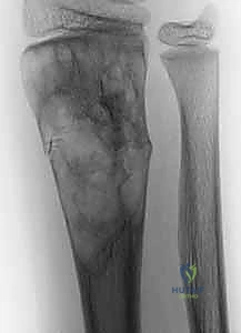
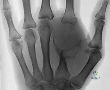
Enchondromas represent the most frequently encountered primary bone tumors in the hand, accounting for up to ninety percent of all bone tumors in the phalanges and metacarpals. These benign, medullary cartilaginous neoplasms arise from ectopic rests of hyaline cartilage that fail to undergo normal endochondral ossification during skeletal development. Recent molecular studies have identified somatic mutations in the isocitrate dehydrogenase genes (IDH1 and IDH2) in a significant proportion of solitary enchondromas, linking their pathogenesis to altered cellular metabolism and epigenetic dysregulation. While typically asymptomatic and discovered incidentally, they commonly present as pathologic fractures due to progressive endosteal scalloping and cortical thinning. The rare but catastrophic risk of malignant transformation into secondary chondrosarcoma necessitates careful clinical and radiographic surveillance, particularly in patients with syndromic presentations such as Ollier disease or Maffucci syndrome, where the risk of malignant degeneration is exponentially magnified.
Unicameral bone cysts (UBCs), or simple bone cysts, are benign, serous fluid-filled cavities lined by a thin fibrous membrane. Although they exhibit a strong predilection for the proximal humerus and proximal femur in the skeletally immature population, they occasionally manifest in the distal radius and, exceedingly rarely, within the tubular bones of the hand. The prevailing etiologic theory suggests a localized venous outflow obstruction within the metaphyseal cancellous bone, leading to increased intraosseous pressure, focal bone resorption, and the accumulation of reactive fluid rich in prostaglandins, interleukins, and proteolytic enzymes. These lesions typically abut the physis and migrate diaphyseally as the child grows. In the distal upper extremity, UBCs frequently present following a minor traumatic event resulting in a pathologic fracture through the mechanically compromised metaphyseal cortex.
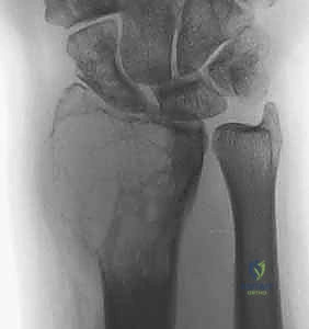
Giant cell tumors of bone (GCTs) are locally aggressive neoplasms characterized by a highly vascularized stroma populated by neoplastic mononuclear cells and abundant, reactive osteoclast-like multinucleated giant cells. The neoplastic mononuclear cells express high levels of Receptor Activator of Nuclear Factor Kappa-B Ligand (RANKL), which drives the recruitment and activation of the destructive giant cells. GCTs typically arise in the epiphyses of long bones in skeletally mature individuals, with the distal radius serving as the third most common site systemically. Lesions in the hand and carpus are relatively rare but are notorious for exhibiting a more aggressive clinical course, higher local recurrence rates, and a paradoxically higher incidence of benign pulmonary metastasis (ranging from two to ten percent). The Campanacci grading system remains the gold standard for radiographic classification, categorizing lesions from Grade 1 (latent, intramedullary) to Grade 3 (aggressive, exhibiting cortical destruction and soft tissue extension), directly guiding the aggressiveness of the surgical intervention.
Detailed Surgical Anatomy and Biomechanics
Phalanges and Metacarpals
The surgical anatomy of the tubular bones of the hand is characterized by extreme spatial constraints, demanding meticulous dissection to avoid devastating functional morbidity. When addressing enchondromas of the phalanges, surgeons must navigate the delicate balance between adequate tumor exposure and the preservation of the extensor mechanism, flexor tendon sheaths, and digital neurovascular bundles. The digital arteries and nerves course along the mid-axial line, protected by Cleland’s and Grayson’s ligaments. A mid-axial approach, either ulnar or radial depending on the lesion's eccentricity, is generally preferred as it allows for safe mobilization of the neurovascular bundle volarly and the extensor apparatus dorsally. The periosteum in this region is thin but mechanically vital; it must be carefully incised and elevated as a continuous sleeve to facilitate post-resection closure and contain any grafted material.
Metacarpal lesions are predominantly approached via longitudinal dorsal incisions. The complex dorsal venous network and the superficial sensory branches of the radial and ulnar nerves lie immediately subcutaneous and are highly susceptible to iatrogenic injury or traction neuropraxia. Deep to the superficial fascia, the extensor tendons (extensor digitorum communis, extensor indicis proprius, and extensor digiti minimi) must be identified within their respective sagittal bands and retracted. The interosseous muscles, originating from the metacarpal shafts, form the lateral borders of the surgical corridor. Biomechanically, the metacarpals endure significant axial and bending loads during grip and pinch activities; therefore, cortical windows created for tumor extirpation must be carefully designed to minimize stress risers, typically employing an oblong or elliptical geometry rather than sharp, angular cuts.
Distal Radius and Carpus
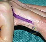
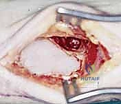
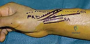
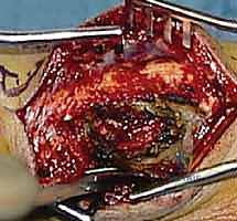
The distal radius is a frequent harbor for GCTs and presents a formidable anatomical challenge due to its proximity to the radiocarpal and distal radioulnar joints (DRUJ), as well as dense neurovascular structures. The palmar (volar) approach, utilizing the classic Henry interval between the flexor carpi radialis (FCR) and the radial artery, provides excellent access to the volar cortex. Retraction of the FCR ulnarly protects the median nerve, while the radial artery is mobilized radially. The pronator quadratus muscle serves as a critical anatomical landmark and a natural biological barrier to volar tumor extension; it must be meticulously elevated from its radial insertion to expose the underlying bone. In cases of massive Grade 3 GCTs, the tumor may breach the volar cortex, necessitating an en bloc resection that may sacrifice the pronator quadratus and require complex soft tissue reconstruction.
Dorsal approaches to the distal radius exploit the intervals between the six extensor compartments. The third compartment, containing the extensor pollicis longus (EPL) tendon wrapping around Lister's tubercle, is a common entry point. The terminal branches of the posterior interosseous nerve (PIN) lie on the floor of the fourth compartment and provide deep nociceptive innervation to the dorsal wrist capsule; while often sacrificed during extensive capsular resections, its identification is anatomically relevant. Biomechanically, the distal radius articular surface bears approximately eighty percent of the axial load transmitted across the wrist. Consequently, subchondral bone preservation during intralesional curettage is paramount. If the subchondral plate is breached or structurally compromised by the tumor, the joint is at high risk for catastrophic collapse, necessitating structural allografting, cement augmentation, or, in severe cases, conversion to an arthrodesis or osteoarticular allograft reconstruction.
Exhaustive Indications and Contraindications
The decision to proceed with surgical intervention for distal upper extremity bone tumors hinges on a complex matrix of patient symptomatology, radiographic findings, biologic tumor behavior, and the structural integrity of the host bone. Prophylactic surgical intervention is heavily weighed against the natural history of the specific lesion.
Indications for Surgical Intervention
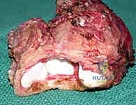
For benign, latent lesions such as asymptomatic, incidentally discovered enchondromas or small UBCs, nonoperative observation with serial radiography is the standard of care. However, surgical intervention becomes unequivocally indicated when these lesions become symptomatic, typically manifesting as localized pain, swelling, or an impending/actual pathologic fracture. In the hand, an enchondroma that occupies greater than fifty percent of the cortical diameter in both orthogonal planes is considered at high risk for mechanical failure and warrants prophylactic intralesional curettage and grafting. Furthermore, any rapid expansion, onset of rest pain, or cortical breakthrough in a previously stable cartilaginous lesion raises the high clinical suspicion of malignant transformation to chondrosarcoma, mandating immediate biopsy and subsequent wide en bloc resection.
Conversely, the diagnosis of a Giant Cell Tumor (GCT) is a near-absolute indication for surgical intervention due to its relentless local aggressiveness and potential for joint destruction. Intralesional extended curettage with high-speed burring and the use of local chemical or thermal adjuvants is the workhorse procedure for Campanacci Grade 1 and 2 lesions. For massive Grade 3 GCTs with extensive cortical destruction, intra-articular extension, or in cases of multiple local recurrences, wide en bloc resection with complex osteoarticular reconstruction or wrist arthrodesis becomes the indicated surgical pathway. The advent of neoadjuvant denosumab therapy has provided an indication for medical downstaging of massive, initially unresectable GCTs, allowing for subsequent, less morbid joint-sparing curettage, though the timing and duration of therapy remain subjects of intense academic debate.
Contraindications
Absolute contraindications to limb-sparing tumor resection in the upper extremity include the presence of active, uncontrolled local or systemic infection, which precludes the use of structural allografts or polymethylmethacrylate (PMMA) cement due to the catastrophic risk of deep periprosthetic infection. Severe medical comorbidities rendering the patient unfit for prolonged general or regional anesthesia also serve as absolute contraindications, occasionally necessitating primary amputation if the tumor represents a life-threatening or severely painful burden. Relative contraindications include massive soft tissue contamination from a poorly planned prior biopsy; if a biopsy tract was placed in a manner that violates multiple anatomic compartments or crucial neurovascular bundles, a joint-sparing intralesional approach may be contraindicated, forcing the surgeon into a much more morbid wide resection to achieve negative oncologic margins.
| Tumor Type | Primary Indications for Surgery | Primary Contraindications | Preferred Surgical Procedure |
|---|---|---|---|
| Enchondroma | Pathologic fracture, intractable pain, impending fracture (>50% cortical diameter), suspected malignant transformation. | Asymptomatic incidental finding, active local osteomyelitis. | Intralesional curettage + bone grafting. Wide resection if malignant. |
| Unicameral Bone Cyst | Recurrent pathologic fractures, progressive enlargement threatening the physis, failure of conservative management. | Small, asymptomatic lesions, lesions resolving post-fracture. | Curettage + grafting, occasionally percutaneous steroid/bone marrow injection. |
| Giant Cell Tumor | Almost all cases upon diagnosis due to local aggressiveness, joint threat, or recurrence. | Poorly placed biopsy tract preventing joint salvage, systemic instability. | Extended intralesional curettage + high-speed burring + adjuvants + PMMA/graft. |
Pre-Operative Planning, Templating, and Patient Positioning
Advanced Imaging and Biopsy Principles
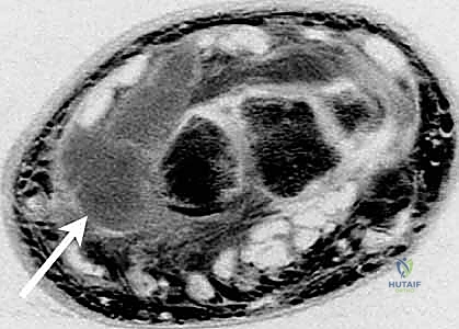
The foundation of successful musculoskeletal oncology surgery is exhaustive preoperative planning, beginning with a comprehensive radiographic assessment. Standard orthogonal plain radiographs provide initial data regarding the lesion's anatomic location, matrix calcification (e.g., "rings and arcs" pathognomonic for enchondromas), and the degree of cortical expansion or lysis. However, advanced cross-sectional imaging is mandatory for surgical templating. Magnetic Resonance Imaging (MRI), utilizing T1, T2, STIR, and gadolinium-enhanced sequences, is the gold standard for delineating the intraosseous extent of the tumor, identifying skip metastases, and crucially, mapping any extraosseous soft tissue extension and its relationship to the neurovascular bundles. For GCTs, a staging computed tomography (CT) scan of the chest is an absolute requirement to rule out pulmonary metastases prior to any surgical intervention.
If the diagnosis remains equivocal following advanced imaging, a tissue biopsy is required. The cardinal rule of orthopedic oncology dictates that the biopsy must be meticulously planned by the treating surgeon who will perform the definitive resection. The biopsy tract must be oriented longitudinally, avoiding transverse incisions that contaminate adjacent fascial compartments. Meticulous hemostasis is critical to prevent hematoma formation, which acts as a vehicle for tumor cell dissemination. The entire biopsy tract must be considered contaminated and must be excised en bloc during the definitive surgical procedure. In the hand and distal radius, a core needle biopsy under fluoroscopic or ultrasound guidance is often preferred over an open incisional biopsy to minimize soft tissue contamination and preserve future reconstructive options.
Templating and Defect Reconstruction Strategy
Preoperative templating involves calculating the anticipated volume of the post-resection osseous defect and formulating a precise reconstructive strategy. For small, contained defects in the phalanges following enchondroma curettage, autologous cancellous bone graft (frequently harvested from the distal radius or olecranon) or synthetic osteoconductive bone void fillers (e.g., calcium phosphate or tricalcium phosphate) are typically sufficient. However, for massive defects in the distal radius following GCT resection, the surgeon must decide between structural allograft reconstruction and PMMA cementation. PMMA offers immediate structural stability, allows for early aggressive rehabilitation, and facilitates the early radiographic detection of local recurrence (which appears as a new radiolucent zone at the cement-bone interface). Conversely, structural allografts provide biological restoration of the bone stock but carry risks of nonunion, fracture, and hardware failure.
Patient Positioning and Anesthesia
Optimal patient positioning is critical for surgical access and intraoperative fluoroscopy. The patient is typically positioned supine with the operative extremity extended on a radiolucent hand table. A pneumatic tourniquet is applied to the proximal arm to ensure a bloodless surgical field, which is absolutely essential for distinguishing between normal cancellous bone and residual neoplastic tissue during intralesional curettage. Regional anesthesia, such as a supraclavicular or axillary brachial plexus block, is highly advantageous, providing excellent intraoperative muscle relaxation and superior postoperative analgesia. However, general anesthesia may be required for prolonged procedures involving complex microvascular reconstructions or concomitant distant bone graft harvesting (e.g., iliac crest). Prior to inflation of the tourniquet, the limb is exsanguinated via elevation rather than an Esmarch bandage to prevent the theoretical risk of systemic tumor cell embolization.
Step-by-Step Surgical Approach and Fixation Technique
Exposure and Cortical Window Creation
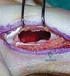
The surgical approach is executed precisely as templated, utilizing the appropriate internervous and intermuscular planes. Once the periosteum overlying the lesion is exposed, it is carefully incised longitudinally and elevated. The creation of the cortical window is a critical step; it must be large enough to allow complete visualization of the entire tumor cavity, including all hidden recesses, yet designed to minimize structural weakening of the bone. For tubular bones, an oblong or elliptical window is fashioned using a high-speed burr or fine osteotomes, avoiding sharp corners that act as stress risers. The cortical lid can occasionally be preserved in saline and replaced at the conclusion of the procedure, acting as a biological barrier to contain the graft or cement.
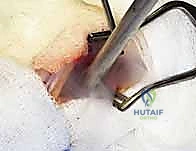
Extended Intralesional Curettage
The core of the procedure for benign and locally aggressive tumors is extended intralesional curettage. Gross tumor is initially removed using a series of progressively sized curettes. It is imperative to systematically scrape the endosteal surface in multiple directions to eradicate all visible neoplastic tissue. However, simple curettage alone leaves microscopic disease within the cancellous bone interstices, leading to unacceptable recurrence rates (historically up to 40-50% for GCTs). Therefore, the procedure must be "extended" using a high-speed spherical burr. The burr is utilized to aggressively abrade the cavity walls, expanding the margin by 1 to 2 millimeters into normal-appearing, bleeding cancellous bone. Continuous pulsatile lavage is employed during burring to clear debris and prevent thermal necrosis of the adjacent articular cartilage.
Application of Adjuvants
Following mechanical burring, chemical or thermal adjuvants are applied to further sterilize the cavity and induce necrosis of any remaining microscopic tumor cells. Phenol (typically 85% concentration) is a potent protein coagulant frequently used; it is carefully swabbed onto the cavity walls for several cycles, followed by immediate neutralization with absolute alcohol and copious saline irrigation to prevent systemic toxicity or damage to adjacent soft tissues. Alternatively, liquid nitrogen (cryotherapy) can be utilized to achieve cellular destruction through intracellular ice crystal formation and osmotic shock. While highly effective at reducing local recurrence, cryotherapy carries a significant risk of collateral damage, including adjacent nerve palsy and delayed pathologic fracture due to extensive bone necrosis. Argon beam coagulation is another excellent modality, providing precise thermal necrosis of the cavity surface with minimal depth of penetration.
Reconstruction and Prophylactic Fixation
Once the cavity is oncologically sterilized, the defect must be reconstructed. For enchondromas and UBCs, packing the void tightly with cancellous autograft or allograft chips is the standard approach, promoting osteoconduction and eventual remodeling. For GCTs, packing the cavity with PMMA bone cement is widely favored. The exothermic reaction of the curing PMMA provides an additional layer of thermal adjuvant therapy to the cavity walls. Furthermore, PMMA provides immediate compressive strength, allowing for early weight-bearing and range of motion. If the subchondral bone is exceedingly thin (less than 2-3mm), a thin layer of cancellous bone graft or a gelatin sponge may be placed prior to cementation to protect the articular cartilage from thermal injury.
Prophylactic internal fixation is judiciously applied based on the mechanical integrity of the remaining host bone. If the cortical window exceeds fifty percent of the bone's diameter, or if the subchondral plate is compromised, internal fixation is mandatory to prevent postoperative catastrophic fracture. In the hand, this may involve the application of low-profile titanium mini-plates or crossed Kirschner wires. In the distal radius, modern volar locking plate technology is frequently utilized. The plate can be applied to bridge the cemented or grafted defect, providing rigid angular stability and allowing for immediate, aggressive postoperative rehabilitation.
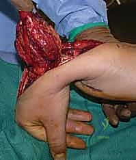
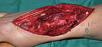
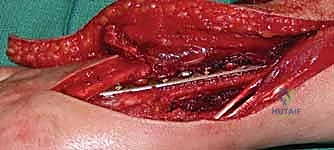
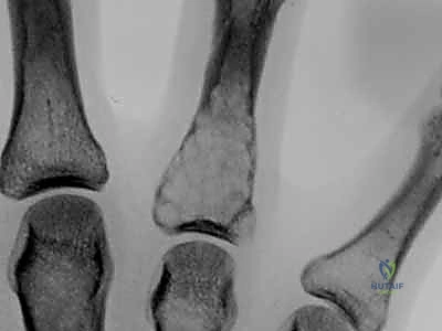
Clinical & Radiographic Imaging Archive