Comprehensive Introduction and Patho-Epidemiology
Cerebral palsy (CP) represents a highly heterogeneous group of non-progressive, permanent disorders of the development of movement and posture, causing profound activity limitation. These disorders are attributed to non-progressive disturbances, such as hypoxic-ischemic encephalopathy, periventricular leukomalacia, or intraventricular hemorrhage, that occurred in the developing fetal or infant brain. While the inciting neurologic lesion is definitively static, the resulting musculoskeletal pathology is relentlessly progressive. The orthopaedic surgeon’s fundamental role is not to cure the neurologic deficit but to aggressively manage the secondary musculoskeletal manifestations—namely spasticity, muscle contractures, altered bone growth, and joint instability—to optimize function, alleviate pain, and facilitate comprehensive nursing care.
The foundation of any surgical decision-making paradigm in cerebral palsy relies heavily on the Gross Motor Function Classification System (GMFCS). Developed by Palisano and colleagues, the GMFCS stratifies patients into five distinct levels based on self-initiated movement, providing a highly reliable prognostic indicator for ambulation and a rigid framework for establishing realistic, patient-specific surgical goals. In ambulatory patients (GMFCS Levels I-III), the primary orthopaedic goal is the preservation or enhancement of ambulation, gait efficiency, and energy conservation. Conversely, in non-ambulatory patients (GMFCS Levels IV-V), the surgical focus shifts entirely to the prevention of pain, the facilitation of perineal hygiene, the optimization of sitting balance, and the prevention of severe decubitus ulcers.
Hip displacement is the second most common orthopaedic manifestation in cerebral palsy, superseded only by equinus deformity of the foot and ankle. The incidence of hip displacement is directly and linearly proportional to the GMFCS level. While children classified as GMFCS Level I have a negligible risk of hip displacement (approaching 0%), the risk escalates exponentially, reaching up to 90% in patients classified as GMFCS Level V. This displacement is rarely present at birth; rather, it develops progressively during the first decade of life secondary to the persistent, abnormal forces exerted by spastic musculature across the growing skeleton. Without proactive surveillance and timely intervention, this progressive subluxation inevitably culminates in a painful, rigid, and chronically dislocated hip, which represents a catastrophic failure of orthopaedic management and severely diminishes the patient's quality of life.
The natural history of the spastic hip dictates that early identification through standardized radiographic surveillance is paramount. The concept of the "hip at risk" was popularized to describe the hip that has begun to subluxate but has not yet developed irreversible bony deformation. In this early phase, the pathology is primarily driven by dynamic muscle imbalance, and soft-tissue interventions can effectively alter the trajectory of the disease. However, once the subluxation progresses and the proximal femur and acetabulum become structurally deformed, soft-tissue releases alone are universally doomed to fail, necessitating massive, complex bony reconstructive procedures to restore joint congruity and stability.
Detailed Surgical Anatomy and Biomechanics
The normal development of the pediatric hip joint is exquisitely dependent on the Hueter-Volkmann principle, which dictates that normal bone growth requires balanced mechanical forces and concentric seating of the femoral head within the acetabulum. In the neurotypically developing child, the physiologic process of walking and weight-bearing, combined with balanced muscle pull, stimulates the proximal femur to remodel, gradually decreasing femoral anteversion from approximately 40 degrees at birth to 15 degrees at maturity, and decreasing the neck-shaft angle from 150 degrees to 120-130 degrees. Simultaneously, the spherical femoral head acts as a template, stimulating the acetabulum to develop a deep, hemispherical concavity.
In the child with cerebral palsy, this delicate biomechanical symphony is entirely disrupted. A profound and persistent muscle imbalance exists around the hip joint. The primary deforming forces are the overactive, spastic flexors (iliopsoas) and adductors (adductor longus, adductor brevis, gracilis, and the adductor portion of the magnus). These powerful muscles relentlessly overpower the relatively weak and frequently under-recruited abductors (gluteus medius and minimus) and extensors (gluteus maximus). This constant, unbalanced vector pulls the proximal femur proximally, laterally, and posteriorly.
Because the spastic child often lacks normal weight-bearing capabilities and the requisite balanced muscle forces, the normal physiologic remodeling of the proximal femur fails to occur. The patient develops a severe coxa valga (neck-shaft angle frequently exceeding 150 degrees) and persistent, excessive femoral anteversion (often measuring 50 to 70 degrees). This abnormal proximal femoral geometry directs the femoral head anterolaterally. However, the strong pull of the spastic adductors and flexors creates a resultant posterosuperior subluxation vector. As the femoral head is driven against the posterosuperior and lateral rim of the acetabulum, it causes progressive mechanical erosion and inhibits normal acetabular development.
Over time, this pathologic biomechanical environment results in severe acetabular dysplasia. The acetabulum becomes shallow, capacious, and markedly deficient anterolaterally—often described as a "teardrop" or "saucer-shaped" acetabulum. The capsular structures become attenuated laterally and contracted medially, further tethering the hip in an abnormal position. Understanding this complex, multi-planar deformity is critical for the orthopaedic surgeon, as successful reconstruction requires simultaneous correction of the proximal femoral geometry (varus and derotation) and the acetabular volume and coverage (pelvic osteotomy). Failure to address all components of this pathomechanical triad inevitably leads to recurrent subluxation and surgical failure.
Exhaustive Indications and Contraindications
The decision-making process for surgical intervention in the spastic hip requires a meticulous analysis of the patient's age, GMFCS level, clinical examination (degree of contracture), and precise radiographic parameters. Surgical timing is critical; intervening too early with bony surgery may lead to recurrence as the child grows, while intervening too late results in irreversible joint destruction, rendering reconstructive efforts futile and necessitating salvage procedures.
The gold standard for quantifying hip displacement is Reimers' Migration Percentage (MP), which measures the percentage of the femoral head ossific nucleus that lies lateral to Perkin’s line on a standardized anteroposterior pelvis radiograph. An MP greater than 30% defines a "hip at risk," while an MP exceeding 40% to 50% generally mandates comprehensive bony reconstruction. The Acetabular Index (AI) must also be scrutinized to quantify the degree of acetabular dysplasia.
Below is a comprehensive table detailing the indications and contraindications for the various tiers of surgical intervention in the cerebral palsy hip:
| Surgical Procedure | Primary Indications | Absolute Contraindications | Relative Contraindications | Target GMFCS Levels |
|---|---|---|---|---|
| Soft-Tissue Release (Adductor/Psoas) | Age 2-6 years; MP 30-40%; Hip abduction < 30° in extension; Dynamic contracture without fixed bony deformity. | MP > 50%; Severe acetabular dysplasia (AI > 25° in older child); Fixed bony coxa valga. | Previous failed soft-tissue release; Athetosis/Dystonia without spasticity. | I - V (Primarily preventative in IV-V) |
| Varus Derotation Osteotomy (VDRO) | MP > 40-50%; Coxa valga (NSA > 140°); Excessive anteversion (> 40°); Failed soft-tissue release. | Severe, end-stage degenerative joint disease; Chronically dislocated, stiff, painful hip in older patient. | Severe malnutrition/osteopenia; Uncontrolled seizure disorder preventing rehab. | II - V |
| Pelvic Osteotomy (Dega/San Diego) | Concomitant with VDRO when AI > 25°; Intraoperative failure to achieve anterolateral coverage after femoral repositioning. | Open triradiate cartilage is required for Dega (Contraindicated if closed); Severe pelvic obliquity driven by spine. | Medically fragile patient unable to tolerate prolonged surgical time/blood loss. | II - V |
| Salvage: Resection Arthroplasty (Castle) | Chronically dislocated, painful hip; Severe DJD; Non-ambulatory patient; Failed reconstruction. | Ambulatory patient; Pain-free dislocated hip (prophylactic resection is not indicated). | Poor soft-tissue envelope for interposition; Active pelvic osteomyelitis. | IV - V ONLY |
| Total Hip Arthroplasty (THA) | Mature patient; Painful osteoarthritis; High-functioning ambulatory or sitting patient. | Active infection; Non-ambulatory patient with severe spasticity/contractures (high dislocation risk). | Severe cognitive impairment precluding post-op precautions; Inadequate bone stock. | I - III (Rarely IV) |
Pre-Operative Planning, Templating, and Patient Positioning
Meticulous preoperative planning is the cornerstone of successful hip reconstruction in cerebral palsy. The clinical examination must be exhaustive, focusing on differentiating dynamic spasticity from fixed myostatic contractures using the Tardieu scale. The Thomas test is utilized to quantify fixed flexion contractures, while the Phelps test assesses gracilis contracture. The surgeon must also evaluate the spine for scoliosis and pelvic obliquity, as a "windblown" pelvis will drastically alter the apparent coverage of the hip and dictate whether spinal fusion should precede or follow hip reconstruction.
Radiographic evaluation begins with a high-quality, standardized anteroposterior (AP) pelvis radiograph. The patient must be positioned supine with the patellae facing strictly anteriorly to accurately assess the neck-shaft angle and Reimers' Migration Percentage. A frog-leg lateral radiograph is obtained to assess the sphericity of the femoral head and rule out concurrent pathologies such as avascular necrosis or slipped capital femoral epiphysis. In cases of severe, complex deformities, or when revision surgery is contemplated, a 3D Computed Tomography (CT) scan is absolutely essential. The 3D CT allows for precise quantification of femoral anteversion and provides a topographical map of the acetabular deficiency, guiding the exact placement and volume of the pelvic osteotomy graft.
Templating for the Varus Derotation Osteotomy (VDRO) requires calculating the exact wedge of bone to be resected or the angle of the implant to be utilized. The goal is to achieve a postoperative neck-shaft angle of 110 to 120 degrees and a residual femoral anteversion of 10 to 15 degrees. The surgeon must select the appropriate fixation construct preoperatively. For younger children with smaller anatomy, a pediatric fixed-angle blade plate (e.g., 90-degree or 100-degree) is often utilized. For older children or those with osteopenic bone, pediatric locking proximal femoral plates (LCP) provide superior pull-out strength and allow for more versatile fixation.
Patient positioning in the operating room is critical for intraoperative assessment. The patient is placed supine on a fully radiolucent Jackson or OSI table. A Foley catheter is placed to monitor urine output during these potentially blood-loss-heavy procedures. The entire lower extremity, from the iliac crest to the toes, is prepped and draped free. This "free draping" technique is non-negotiable, as the surgeon must be able to visually assess the rotational profile of the limb, perform intraoperative range of motion testing to ensure impingement-free concentric reduction, and manipulate the limb for precise fluoroscopic imaging. A sterile bump is kept on the field to elevate the ipsilateral pelvis during the pelvic osteotomy phase.
Step-by-Step Surgical Approach and Fixation Technique
Comprehensive hip reconstruction in cerebral palsy typically involves a staged, single-anesthetic approach: soft-tissue releases, followed by proximal femoral osteotomy, and culminating in pelvic osteotomy. Blood conservation strategies, including the use of intravenous tranexamic acid (TXA) and cell salvage systems, are initiated prior to incision.
Soft-Tissue Releases
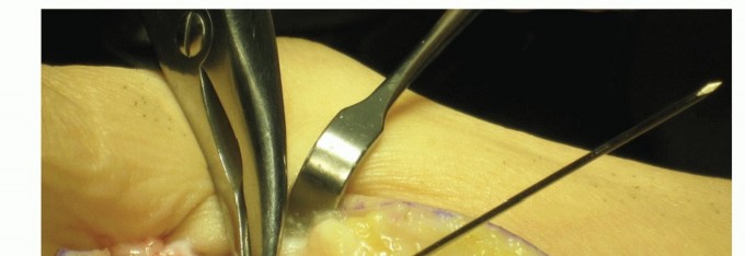
The procedure begins with the adductor release. With the hip abducted, a 3-4 cm longitudinal incision is made centered over the adductor longus origin at the pubic symphysis. The deep fascia is incised, and the adductor longus is isolated bluntly. It is tenotomized sharply near its origin. The gracilis, located immediately posterior and medial, is identified and similarly released. The adductor brevis is inspected; if significant tension remains, a fractional lengthening of the anterior epimysium is performed. The psoas recession is then performed via a separate anterior approach or extended medial approach. The iliopsoas tendon is identified as it crosses the pelvic brim. The tendinous portion is sharply transected, allowing the muscle belly to stretch, thereby preserving active flexion for swing-phase clearance in ambulatory patients.
Proximal Femoral Varus Derotation Osteotomy (VDRO)
A direct lateral approach to the proximal femur is utilized. A longitudinal incision is made extending from the tip of the greater trochanter distally for 8-10 cm. The fascia lata is incised in line with the skin. The vastus lateralis is identified, elevated off the lateral intermuscular septum, and reflected anteriorly, exposing the proximal femoral shaft and the vastus ridge. Using fluoroscopic guidance, a guidewire is advanced through the lateral cortex, up the femoral neck, and into the exact center of the femoral head epiphysis. The angle of this wire relative to the femoral shaft determines the final varus correction.
If a blade plate is used, a seating chisel is driven over the wire to create the track. If a locking plate is used, the proximal locking screws are placed. A transverse osteotomy is then performed using an oscillating saw at the intertrochanteric level, just proximal to the lesser trochanter. To decompress the joint and reduce the risk of avascular necrosis (AVN) and sciatic nerve palsy in chronically dislocated hips, a 1-2 cm cylindrical segment of the distal femur is routinely resected. The distal femoral fragment is then externally rotated to correct the excessive anteversion. The plate is secured to the distal fragment using cortical screws. Rigid, absolute stability must be achieved to permit early mobilization.
The Dega Pelvic Osteotomy
Following the VDRO, intraoperative fluoroscopy is used to assess acetabular coverage. If the Acetabular Index remains > 25 degrees or if anterolateral uncoverage persists, a Dega osteotomy is performed. A bikini incision is made just below the iliac crest. The cartilaginous iliac apophysis is split, and the iliacus and gluteal muscles are elevated subperiosteally to expose the inner and outer tables of the ilium down to the greater sciatic notch.
Under fluoroscopy, an incomplete transiliac osteotomy is performed using a curved osteotome. The osteotomy begins just above the Anterior Inferior Iliac Spine (AIIS) and curves posteriorly toward the sciatic notch. Crucially, the osteotomy cuts through the outer table and the middle cancellous bone but leaves the inner table of the pelvis and the triradiate cartilage intact. These intact medial structures act as a plastic hinge. Lamina spreaders are inserted into the osteotomy site, and the acetabular roof is levered downwards, hinging on the triradiate cartilage, to dramatically improve anterolateral coverage. The triangular bone wedge harvested from the femoral shortening during the VDRO is meticulously decorticated and impacted into the open pelvic osteotomy site. The periosteum and iliac apophysis are meticulously repaired over the graft to ensure stability.
Complications, Incidence Rates, and Salvage Management
Surgical intervention for the spastic hip is fraught with potential complications, driven by the patient's underlying osteopenia, poor nutritional status, relentless muscle spasticity, and the sheer magnitude of the surgical insult. The orthopaedic surgeon must be hyper-vigilant in both the intraoperative execution and postoperative management to mitigate these risks.
Avascular necrosis (AVN) of the femoral head is one of the most devastating complications. It is primarily caused by excessive pressure on the femoral head following reduction of a chronically dislocated hip, which occludes the delicate retinacular vessels derived from the medial circumflex femoral artery. The absolute best prevention for AVN is adequate femoral shortening (resecting 1-2 cm of the femur) during the VDRO to fully decompress the joint. Recurrent dislocation is another major failure mode, almost universally resulting from the surgeon's failure to recognize and adequately treat concurrent acetabular dysplasia with a pelvic osteotomy at the time of the index VDRO.
Below is a detailed table of specific complications, their approximate incidence rates, etiologies, and salvage management strategies:
| Complication | Incidence Rate | Primary Etiology | Salvage / Management Strategy |
|---|---|---|---|
| Avascular Necrosis (AVN) | 5% - 15% | Failure to shorten femur; excessive joint pressure; vascular injury during approach. | Observation if asymptomatic; Core decompression (rare in CP); Resection arthroplasty if severe collapse/pain. |
| Recurrent Subluxation/Dislocation | 10% - 20% | Failure to perform pelvic osteotomy; inadequate varus/derotation; progressive spasticity. | Revision VDRO and/or Pelvic Osteotomy; Optimization of spasticity management (ITB pump). |
| Implant Failure / Loss of Fixation | 3% - 8% | Severe osteopenia; uncontrolled postoperative muscle spasms; inadequate initial fixation. | Revision open reduction and internal fixation (ORIF) with longer locking plates; Spica casting. |
| Delayed Union / Nonunion | 2% - 5% | Poor nutrition (low Vitamin D/Albumin); excessive periosteal stripping; thermal necrosis. | Optimization of nutritional status; Revision with autologous bone grafting and stable fixation. |
| Deep Surgical Site Infection | 2% - 6% | Poor perineal hygiene; immunocompromise; prolonged operative time. | Aggressive surgical debridement (I&D); Intravenous antibiotics; Hardware retention if stable, removal if loose. |
| Sciatic Nerve Palsy | 1% - 3% | Excessive traction during reduction of a high dislocation without adequate femoral shortening. | Immediate removal of cast/splint; Hip flexion to relieve tension; Surgical revision to shorten femur if severe. |
When reconstructive efforts fail, or when a patient presents late with a chronically dislocated, arthritic, and agonizingly painful hip, salvage procedures are indicated. The Proximal Femoral Resection-Interposition Arthroplasty (Castle Procedure) is the workhorse salvage operation for the non-ambulatory (GMFCS IV-V) patient. By resecting the proximal femur at the subtrochanteric level and interposing the vastus lateralis muscle over the stump, the painful bone-on-bone articulation is eliminated. While this procedure sacrifices joint stability, it reliably eradicates pain, restores perineal access for nursing care, and allows the patient to sit comfortably in a customized wheelchair. Total Hip Arthroplasty (THA) is reserved strictly for mature, highly functional patients (GMFCS I-III) due to the unacceptably high rates of dislocation and aseptic loosening in the severely spastic, non-ambulatory population.
Phased Post-Operative Rehabilitation Protocols
The technical success of the intraoperative reconstruction is entirely dependent upon the rigor and meticulousness of the postoperative rehabilitation protocol. Cerebral palsy patients are uniquely vulnerable in the postoperative period due to their propensity for violent muscle spasms, altered pain perception, and baseline neurologic deficits.
Phase I: Immediate Postoperative Period (Days 0-5)
The immediate focus is on immobilization and aggressive pain management. Depending on the surgeon's preference and the rigidity of the fixation, the patient is placed in either a bilateral long-leg spica cast or a custom-molded rigid abduction orthosis (A-frame) in the operating room. Pain management is the most critical aspect of this phase. Muscle spasms are a primary source of excruciating postoperative pain and can be violent enough to cause implant failure. A multimodal, aggressive neuropharmacologic regimen is mandatory. This typically includes continuous epidural analgesia for the first 48-72 hours, scheduled intravenous diazepam or enteral baclofen to suppress spasticity, gabapentin for neuropathic pain modulation, and scheduled NSAIDs/acetaminophen to reduce opioid requirements.
Phase II: Immobilization and Healing (Weeks 1-6)
During this phase, the patient remains strictly non-weight-bearing in their spica cast or orthosis. The focus shifts to nursing care, specifically the prevention of decubitus ulcers and the maintenance of perineal hygiene. Nutritional support is paramount; many GMFCS IV-V patients require temporary or permanent gastrostomy tube feeding to ensure adequate caloric and protein intake for bone healing. Caregivers are extensively educated on safe transfer techniques to avoid placing torque on the osteotomy sites.
Phase III: Mobilization and Early Therapy (Weeks 6-12)
At the 6-week mark, AP pelvis radiographs are obtained to confirm early callus formation and hardware stability. Once clinical and radiographic healing is confirmed, the spica cast or orthosis is discontinued. Intensive physical therapy commences immediately. The initial focus is strictly on restoring passive range of motion, particularly hip extension and abduction, to prevent the soft tissues from scarring in a contracted position. Aquatic therapy is highly beneficial in this phase, as the buoyancy of the water unloads the joints and the warmth helps relax spastic musculature. Active-assisted strengthening of the abductors and extensors is gradually introduced.
Phase IV: Functional Restoration and Long-Term Surveillance (Months 3-12+)
For ambulatory patients (GMFCS I-III), intensive gait training with appropriate assistive devices (e.g., reverse walkers) is initiated. The goal is to return the patient to their preoperative baseline of ambulation by 6 months, and ideally surpass it by 12 months as the biomechanical advantage of the reconstructed hip is realized. For non-ambulatory patients, the focus is on wheelchair seating and positioning, ensuring the pelvis is level and the spine is balanced.
Because cerebral palsy is a lifelong, dynamic condition, the orthopaedic surgeon's job is never truly finished. Patients require annual clinical and radiographic follow-up until skeletal maturity. The surgeon must monitor for hardware complications, late recurrent subluxation, and the development of compensatory deformities in adjacent joints, such as knee flexion contractures or severe planovalgus foot deformities, which frequently require subsequent staged interventions.
Summary of Landmark Literature and Clinical Guidelines
The modern paradigm of hip surveillance and reconstruction in cerebral palsy is built upon decades of rigorous academic research and international consensus. The orthopaedic surgeon must be intimately familiar with the landmark literature that dictates current standards of care.
The foundational concept of radiographic surveillance was established by Reimers in 1980 with his seminal paper detailing the Migration Percentage. Reimers definitively proved that clinical examination alone is insufficient to detect early hip displacement, mandating the use of standardized AP pelvis radiographs. This metric remains the universal language of CP hip pathology today.
The paradigm shifted significantly with the implementation of national hip surveillance programs, most notably the CPUP (Cerebral Palsy Follow-Up Program) initiated in Sweden by Hagglund and colleagues in 1994. The CPUP demonstrated unequivocally that systematic, population-based radiographic screening, coupled with early soft-tissue or bony intervention, could virtually eradicate the incidence of the chronically dislocated, painful hip in the CP population. The success of the Swedish model has led to the widespread adoption of similar surveillance protocols globally.
In North America, the American Academy for Cerebral Palsy and Developmental Medicine (AACPDM) and the Pediatric Orthopaedic Society of North America (POSNA) have published exhaustive consensus guidelines based on the GMFCS. These guidelines dictate the frequency of radiographic screening: for example, a GMFCS Level V patient requires an AP pelvis radiograph at 12-18 months of age, and every 6 months thereafter until age 7, followed by annual radiographs until skeletal maturity.
Furthermore, landmark studies by Shore et al. and Flynn et al. have validated the efficacy of the comprehensive single-stage reconstruction (VDRO combined with pelvic osteotomy) over isolated femoral or pelvic procedures in patients with an MP > 50%. These studies highlighted that failure to address the acetabular dysplasia at the time of femoral reconstruction is the single greatest predictor of surgical failure and recurrent dislocation. Mastery of this literature ensures that the orthopaedic surgeon practices evidence-based medicine, providing the highest standard of care to this vulnerable and complex patient population.
This academic synthesis is based on established protocols from Hutaifortho's Operative Orthopaedics and has been medically reviewed by Prof. Dr. Mohammed Hutaif, Consultant Orthopedic & Spine Surgeon. It is designed to assist orthopedic residents, fellows, and practicing surgeons in surgical preparation and board reviews (AAOS, FRCS, Arab Board).
