Achieving Success with Uncemented Hip Arthroplasty Metal Socket
Key Takeaway
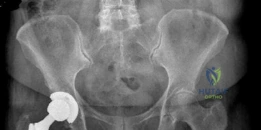
Looking for accurate information on Achieving Success with Uncemented Hip Arthroplasty Metal Socket? Total hip arthroplasty addresses severe hip osteoarthritis, as demonstrated in a 69-year-old female's case. The procedure replaces the hip joint with prosthetic components, often including an uncemented acetabular shell and stem, and a ceramic femoral head with a polyethylene liner. Consideration of hip arthroplasty metal components, like the porous-coated shell, is vital for fixation and long-term success in restoring mobility and reducing pain.
A 58-year-old patient presents with a 2-year history of debilitating right hip pain, refractory to conservative management. Clinical assessment reveals an antalgic gait and restricted internal rotation. You are planning a primary total hip arthroplasty using an uncemented acetabular component. Below is the patient's current AP pelvic radiograph.
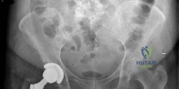
Question: Define the "Safe Zone" for acetabular component orientation and discuss why static radiographic measurements might be misleading when considering functional outcomes.
Candidate: The Lewinnek Safe Zone is defined as 40 ± 10 degrees of inclination and 15 ± 10 degrees of anteversion. However, this is static; it doesn't account for pelvic tilt. If a patient has a stiff spine or is standing versus sitting, their functional anteversion changes, which can lead to impingement or dislocation even if the cup looks perfect on an AP X-ray.
Focusing solely on the Lewinnek numbers. A weak answer fails to mention "Spinopelvic Mechanics." Examiners are looking for candidates who understand that sagittal plane pelvic tilt changes the functional orientation of the acetabulum during sitting and standing.
Define the Lewinnek Safe Zone (40°±10° Inclination; 15°±10° Anteversion). Immediately pivot to the concept of functional acetabular orientation. Discuss the relationship between the lumbar spine and pelvis: identify "stiff" vs. "flexible" spines. Explain that in patients with limited pelvic compensation (e.g., fusion or stenosis), the cup may be "safe" on the AP radiograph but functionally malpositioned during functional arcs of motion, leading to anterior impingement (in extension) or posterior dislocation (in flexion).
During the preparation of the acetabulum for the uncemented shell, you ream to the appropriate size. However, upon placing your trial shell, you notice it lacks the desired "press-fit" stability. The radiograph below shows the appearance of the acetabulum after initial preparation.
Question: What are the structural and biological requirements for a successful uncemented acetabular fixation, and how would you manage the lack of primary stability in this scenario?
Candidate: Successful fixation requires primary mechanical stability (achieved by press-fit/interference fit) and secondary biological fixation (osseointegration). If I lack primary stability, I must check if the reaming is too deep or if bone quality is poor. I would consider using supplementary screw fixation in the safe posterior-superior quadrant, or perhaps downsizing the reamer and using a multi-holed shell.
Suggesting a larger cup size as the primary solution. This leads to acetabular fracture or "bottoming out." A poor answer also fails to mention the specific anatomical quadrants for screw placement, which is a critical safety component of the FRCS exam.
Structure the answer by distinguishing Primary Stability (mechanical interference, usually 1-2mm under-reaming) and Secondary Stability (osseointegration into porous surfaces like Titanium or Tantalum). When primary stability is absent: 1. Assess for technical errors (inadequate seating, debris). 2. Evaluate bone stock (Paprosky classification). 3. Options: Use of screw-in acetabular shells, supplemental screws in the posterior-superior or anterior-superior quadrants (avoiding the "Death Zone" of the inferior/anterior-inferior quadrants containing neurovascular structures). If stability remains inadequate, consider a larger, more porous shell or, in extreme bone loss, transition to a cemented construct or revision cage.
