Pathologic Subtrochanteric Femur Fracture in Paget's Disease: Clinical & Diagnostic Insights
Key Takeaway
A subtrochanteric femoral fracture in Paget's disease is diagnosed with clinical assessment, X-rays showing Pagetic changes and the fracture, and CT scans. CT is vital for detailing fracture morphology, medullary canal narrowing, and bone quality. These alterations significantly challenge surgical planning due to compromised bone structure.
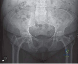
A 72-year-old male presents following a low-energy fall with a deformed left thigh. Initial radiographs are shown below. Describe the radiological features and provide your immediate differential diagnosis for this fracture.
Candidate: The radiograph shows a displaced, comminuted subtrochanteric femur fracture. There is evidence of underlying Paget's disease, specifically cortical thickening, coarsened trabeculae, and bone enlargement. My differential includes a pathological fracture through Pagetic bone, metastatic disease, or potentially a Paget's sarcoma given the age and the severity of the lesion.
Focusing only on the fracture mechanics and ignoring the systemic disease. Candidates often miss the "pathological" nature of the fracture and fail to systematically describe the Pagetic radiographic hallmarks, which are vital for preoperative planning.
Structure your answer: 1. Describe the fracture (AO/OTA 32-A3.3). 2. Systematically describe Pagetic signs (cortical thickening, trabecular coarsening, bone enlargement, and bowing). 3. State the differential diagnosis in order of likelihood (Pagetic fracture, metastatic disease, Paget's sarcoma). 4. Immediately mention that while Paget's is the working diagnosis, one must rule out malignant transformation (Paget's Sarcoma) during surgery.
You have decided to proceed with surgical fixation. Given the patient has active Paget's disease and the CT scan confirms a sclerotic, narrowed medullary canal, what are your major intraoperative concerns and how do you mitigate them?
Candidate: My main concerns are iatrogenic fracture during reaming, excessive bleeding due to hypervascularity, and poor implant purchase. To mitigate these, I would ream slowly and sequentially, have blood products ready for transfusion, and have a backup plan for a long locking plate if the canal is too narrow for an IM nail.
Failing to mention the need for a backup plan (plating) or the specific need for bone grafting in the comminuted medial zone. Also, failing to mention the multidisciplinary approach (involvement of endocrinology for post-operative bisphosphonates).
Categorize into: 1. Canal Management: Slow, sequential reaming to prevent iatrogenic fracture; consider smaller diameter nails. 2. Fixation: IM nail as the primary construct, but with a backup plan of long plate osteosynthesis + cement augmentation for screws. 3. Vascularity: Proactive cross-matching of blood due to Pagetic hypervascularity. 4. Biology: Harvesting local bone graft to address the comminution and promote union in metabolic bone.
The surgery was successful. How do you manage the patient's Paget's disease post-operatively, and what is the long-term risk profile for this patient?
Candidate: I would start intravenous bisphosphonates (like Zoledronic acid) once the patient is stable, after consultation with endocrinology. Long-term, the main risks are non-union of the fracture, secondary OA of the hip, and a small but significant risk of sarcomatous transformation (Paget's sarcoma), so I will monitor him with serial radiographs and ALP levels.
Ignoring the "systemic" nature of the disease. Failing to mention monitoring of ALP as a marker of treatment efficacy or ignoring the necessity of long-term sarcoma surveillance.
A high-scoring answer connects the systemic metabolic treatment (bisphosphonates to suppress turnover) with the biomechanical requirements of the fracture (protected weight-bearing). It must explicitly mention the 1% lifetime risk of sarcomatous transformation, necessitating lifelong surveillance. It concludes by defining the endpoint of monitoring: normalization of serum ALP and radiographic evidence of union.
