Patient Presentation & History
We present the case of a 72-year-old male, a retired civil engineer, with a significant past medical history including T2N0M0 adenocarcinoma of the prostate, treated five years prior with radical prostatectomy and adjuvant radiation therapy, currently in remission with stable PSA levels. He also has well-controlled hypertension and Type 2 Diabetes Mellitus.
The patient presented to the orthopedic trauma clinic with a 3-month history of insidious onset left distal thigh pain. Initially, the pain was intermittent, dull, and primarily nocturnal, rated 3/10 on the Visual Analog Scale (VAS). Over the past four weeks, the pain had become more constant, exacerbated by weight-bearing and activity, and now rated 6-7/10 VAS. He reported no specific mechanism of injury, fall, or direct trauma. He denied any constitutional symptoms such as fever, chills, night sweats, or unexplained weight loss. He also denied any numbness, tingling, or weakness in the limb. His primary care physician had initially attributed the pain to osteoarthritis or muscular strain and prescribed NSAIDs, which provided minimal relief. Given his oncologic history and worsening, unremitting pain, further investigation was warranted due to high suspicion for metastatic disease.
Clinical Examination
Upon examination, the patient was ambulatory with a noticeable antalgic gait, favoring the left lower extremity.
*
Inspection:
No obvious swelling, erythema, skin changes, or deformity noted around the left distal thigh or knee. The limb alignment appeared normal. There was no evidence of muscle atrophy.
*
Palpation:
Localized tenderness was elicited over the lateral aspect of the left distal femoral metadiaphysis, approximately 10 cm proximal to the lateral femoral epicondyle. No palpable mass was identified, and the skin temperature was normal.
*
Range of Motion (ROM):
* Left hip ROM was full and pain-free.
* Left knee ROM: Flexion to 120 degrees, extension to 0 degrees, with mild pain at the extremes of flexion. There was no mechanical block or crepitus. Pain was elicited with axial loading and rotational stress across the knee joint, transmitting forces to the distal femur.
*
Neurological Assessment:
Sensation was intact to light touch and pinprick in L2-S1 dermatomes. Motor strength was 5/5 in all major muscle groups (hip flexion, extension, abduction, adduction; knee flexion, extension; ankle dorsiflexion, plantarflexion) bilaterally. Deep tendon reflexes (patellar, Achilles) were 2+ and symmetrical.
*
Vascular Assessment:
Distal pulses (femoral, popliteal, dorsalis pedis, posterior tibial) were palpable, symmetrical, and strong bilaterally. Capillary refill was brisk (<2 seconds).
Given the localized pain, antalgic gait, and history of malignancy, the clinical picture mandated urgent radiographic investigation for a potential osseous lesion.
Imaging & Diagnostics
Initial imaging focused on the symptomatic area, the left distal femur.
*
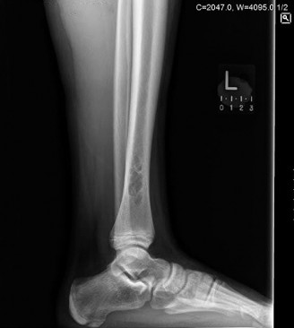
Plain Radiographs (AP and Lateral views of the left femur):
These revealed a well-defined, eccentric, lytic lesion in the lateral cortex of the left distal femoral metadiaphysis. The lesion measured approximately 3.5 x 2.0 cm, exhibiting a characteristic multiloculated or "bubbly" appearance with a distinct sclerotic rim. There was no overt cortical breach or periosteal reaction identified. The overall bone architecture otherwise appeared intact. The appearance was somewhat atypical for a rapidly aggressive metastatic lesion, which often presents with less defined margins, more diffuse lytic destruction, or a mixed lytic-blastic pattern. However, the presence of any lytic lesion in an oncology patient automatically raises concerns for metastasis.
-
Magnetic Resonance Imaging (MRI) of the left femur with and without contrast: Performed to further characterize the lesion, assess soft tissue extension, and rule out impending pathological fracture.
- T1-weighted images demonstrated a well-circumscribed lesion with low signal intensity, primarily involving the cortical and medullary bone of the distal femoral metaphysis.
- T2-weighted and STIR sequences showed heterogeneous high signal intensity within the lesion, consistent with a fibrous component and fluid content, but notably lacking the diffuse marrow edema typically associated with acute metastasis or aggressive primary bone tumors.
- Post-contrast T1-weighted images revealed peripheral, rim-like enhancement, with central non-enhancing areas. Crucially, there was no significant surrounding marrow edema extending beyond the lesion's immediate margins, which would be highly suggestive of malignancy. The lack of extensive soft tissue invasion further supported a less aggressive process.
-
Computed Tomography (CT) of the left femur: Utilized for high-resolution assessment of cortical integrity and lesion morphology. The CT confirmed the eccentric cortical location of the lesion, with clear demonstration of its well-defined sclerotic margins and internal trabeculations, contributing to the multiloculated appearance. Cortical thinning was noted over the lesion, but without complete breach or frank fracture line. The CT was instrumental in evaluating the integrity of the cortex, which is critical for assessing fracture risk.
-
Technetium-99m Bone Scintigraphy (Bone Scan): Performed as part of the metastasis workup given the patient's history. The bone scan demonstrated mildly increased uptake in the left distal femur corresponding to the lesion. This uptake was less intense and more focal than what is typically seen in active, widespread metastatic disease, which often shows multifocal areas of significantly increased radiotracer accumulation. While not completely "cold" (which is common for NOF), the limited uptake was still somewhat reassuring in the context of a potential benign lesion.
-
Fluorodeoxyglucose Positron Emission Tomography-Computed Tomography (FDG PET/CT): Given the equivocal bone scan and the need to definitively rule out metastatic progression, an FDG PET/CT was performed. This demonstrated low-grade FDG uptake (SUVmax ~2.0) within the left distal femoral lesion. This level of uptake is typically considered non-malignant or only mildly active, and significantly lower than the high SUVmax values (often >5-10) associated with most metastatic carcinomas or aggressive primary bone tumors. Furthermore, the PET/CT confirmed no other areas of suspicious FDG avidity throughout the rest of the skeleton or body, providing confidence that the known prostate cancer was still in remission.
-
Biopsy Decision: Despite the reassuring imaging features pointing away from aggressive malignancy, the patient's oncologic history and the symptomatic nature of the lesion necessitated histological confirmation. A CT-guided core needle biopsy of the lesion was performed. Histopathological analysis revealed spindle cell proliferation arranged in storiform patterns, interspersed with multinucleated giant cells and hemosiderin deposition. Immunohistochemistry was negative for cytokeratins (ruling out carcinoma) and S100 protein (ruling out melanoma/neural tumors), and positive for CD68 in giant cells. These findings were classic for a Non-Ossifying Fibroma (NOF), also known as a Fibrous Cortical Defect when smaller.
Differential Diagnosis
The presentation of a lytic bone lesion in an elderly patient with a history of malignancy necessitates a broad differential diagnosis. The primary concern is always metastatic disease, but other benign and malignant processes must be considered.
| Feature | Non-Ossifying Fibroma (NOF) | Metastatic Carcinoma (e.g., from prostate) | Fibrous Dysplasia |
|---|---|---|---|
| Typical Age | Adolescents/Young Adults (often incidental, can persist) | Typically >40 years, aligns with primary cancer demographics | Children, adolescents, young adults (can be diagnosed later) |
| Common Location | Metaphyseal (distal femur, proximal/distal tibia) | Any bone, axial skeleton, proximal long bones (femur, humerus) | Long bones, skull, ribs, pelvis (monostotic or polyostotic) |
| X-ray Findings | Eccentric, lytic, well-defined sclerotic rim, multiloculated/bubbly, non-expansile, often cortical. | Lytic, blastic, or mixed. Poorly defined margins, cortical destruction, periosteal reaction, pathological fracture common. | "Ground-glass" matrix, ill-defined margins, fusiform expansion, cortical thinning. Shepherd's crook deformity in proximal femur. |
| MRI Findings | T1 low, T2 high (heterogeneous), peripheral/rim enhancement post-contrast. No extensive marrow edema. | T1 low, T2 high (often diffuse marrow edema), strong heterogeneous enhancement. Cortical breach, soft tissue extension. | T1 low/intermediate, T2 high (less homogeneous than NOF). Heterogeneous enhancement. |
| Bone Scintigraphy | Often "cold" or mildly increased uptake (focal) | "Hot" (significantly increased uptake), often multifocal. | Variable uptake, usually mildly to moderately increased. |
| PET/CT (FDG) | Low to no FDG uptake (SUVmax typically <2-3) | High FDG uptake (SUVmax typically >5-10) | Variable, usually low to moderate uptake. |
| Clinical Presentation | Often asymptomatic, incidental. Can cause pain if large, or lead to pathological fracture. | Pain (often severe, nocturnal), pathological fracture, constitutional symptoms. | Pain, deformity, pathological fracture. Often asymptomatic until fracture. |
| Biopsy Findings | Spindle cell fibroblasts in storiform pattern, giant cells, hemosiderin, no mitotic activity, no malignancy. | Malignant cells consistent with primary tumor. | Immature woven bone trabeculae in a fibrous stroma, no osteoblastic rimming. |
| Prognosis | Excellent, benign, usually regresses or heals spontaneously. | Depends on primary cancer stage, extent of metastases, and treatment response. | Good, benign but can cause deformity and recurrent fractures. |
Surgical Decision Making & Classification
The primary considerations for surgical intervention in this patient were:
1.
Symptom control:
The patient's pain significantly impacted his quality of life and mobility.
2.
Risk of pathological fracture:
Despite being benign, large NOFs can weaken the bone, especially in high-stress areas like the distal femur, leading to an impending or actual pathological fracture.
3.
Definitive diagnosis confirmation:
While imaging was highly suggestive, histological confirmation was prudent given the oncologic history. The core biopsy provided this confirmation.
Upon diagnosis of a symptomatic NOF and exclusion of metastatic disease, the treatment strategy shifted. Conservative management with activity modification and serial imaging is often appropriate for asymptomatic or small NOF lesions, as many regress spontaneously. However, in this case, the lesion was symptomatic and relatively large (3.5 cm), with cortical thinning, making the risk of pathological fracture a significant concern. While the Mirels' score is primarily used for metastatic lesions, the principles apply to any lytic lesion causing cortical compromise. A lesion size >2.5 cm and cortical involvement >50% would generally put a patient at a higher risk of fracture, warranting prophylactic intervention. Given the patient's age and desire for continued active lifestyle, prophylactic stabilization and lesion management were deemed appropriate.
Classification (Relevant to surgical decision-making):
*
Lesion Location:
Distal femoral metadiaphysis. This is a common location for NOF and also a high-stress area, particularly prone to bending forces.
*
Risk of Fracture:
Although no acute fracture was present, the significant cortical involvement and patient symptoms suggested an impending fracture. A "Mirels-like" assessment for a benign lesion would lean towards prophylactic intervention. While NOF does not have a specific fracture classification, its size and cortical involvement are crucial.
*
Pathology:
Benign fibrous lesion. This allows for local intralesional treatment rather than wide resection.
The decision was made for surgical intervention consisting of intralesional curettage of the NOF, followed by bone grafting, and prophylactic internal fixation to prevent a future pathological fracture and provide immediate pain relief. This approach aimed to completely remove the benign lesion, reconstruct the cortical defect, and provide biomechanical stability.
Surgical Technique / Intervention
The surgical procedure involved careful planning to address both the lesion and provide prophylactic fixation.
Patient Positioning: The patient was positioned supine on a radiolucent operating table. A high thigh tourniquet was applied. The entire left lower extremity was prepped and draped in a sterile fashion to allow full manipulation of the limb and access for fluoroscopy.
Approach: A standard lateral approach to the distal femur was utilized. An incision was made over the lateral aspect of the distal femur, centered over the lesion, extending proximally and distally as needed. The vastus lateralis muscle was identified and incised longitudinally along its anterior border, then reflected anteriorly and medially to expose the lateral femoral cortex. Care was taken to protect the neurovascular structures, particularly the peroneal nerve and popliteal vessels, during deep dissection.
Lesion Exposure and Curettage: Fluoroscopy was used to precisely localize the NOF. A cortical window, approximately 3 cm x 2 cm, was carefully created over the lesion using an oscillating saw and osteotomes, ensuring removal of the thinned lateral cortex directly overlying the NOF. The lesion itself was identified as a yellowish-brown, gritty, and fibrous mass. Thorough intralesional curettage was performed using curettes of various sizes, ensuring complete removal of all visible fibrous tissue, including curettage of the sclerotic margins. High-speed burrs were used to meticulously debride the inner aspects of the cavity and ensure no residual lesion tissue was left behind. The cavity was irrigated copiously.
Bone Grafting: Following complete curettage, the resultant cortical defect and intramedullary cavity required reconstruction. An allograft cancellous chip bone graft was prepared and packed tightly into the cavity to fill the defect and promote healing. This provided structural support and osteoconductive properties. In some cases, autograft (e.g., from the iliac crest) might be preferred, but allograft was chosen to minimize donor site morbidity.
Fixation Construct: Given the size of the defect, the cortical thinning, and the patient's age and activity level, prophylactic internal fixation was deemed essential to prevent a pathological fracture through the reconstructed site. A locking compression plate (LCP) system, specifically a Distal Femoral LCP, was chosen due to its ability to provide stable fixation in metaphyseal bone and its load-sharing characteristics.

(Representative intraoperative fluoroscopic image showing placement of a locking compression plate across the reconstructed distal femoral metaphysis, with screws engaging both proximal and distal fragments, bypassing the bone graft site.)
The LCP was contoured to match the lateral femoral cortex. It was positioned strategically to span the entire length of the curetted and grafted defect, with sufficient screws placed proximally and distally in healthy bone. Bicortical locking screws were utilized where appropriate to maximize stability. The plate provided an immediate stable construct, allowing for protected early weight-bearing and mitigating the risk of fracture during the bone graft incorporation phase.
Closure: The wound was copiously irrigated. The vastus lateralis muscle was repaired. Subcutaneous tissues and skin were closed in layers. A sterile dressing was applied.
Post-Operative Protocol & Rehabilitation
Immediate Post-Operative Period (Day 0-7):
*
Pain Management:
Multimodal analgesia regimen including opioids, NSAIDs (if not contraindicated), and acetaminophen. Nerve blocks (e.g., adductor canal block) were considered for immediate post-op pain control.
*
Weight-bearing:
Touch-down weight-bearing (TDWB) or 20 lbs flat-foot weight-bearing was initiated immediately using crutches or a walker. This protected the bone graft and fixation while allowing early mobilization.
*
Range of Motion:
Gentle active-assisted and passive range of motion exercises for the knee and hip were started immediately, within pain limits, to prevent stiffness.
*
DVT Prophylaxis:
Standard chemical DVT prophylaxis (e.g., LMWH) was administered.
*
Wound Care:
Daily wound checks for signs of infection.
Early Rehabilitation (Weeks 1-6):
*
Physical Therapy:
Aggressive physical therapy program initiated.
*
Weight-bearing progression:
Gradually increased weight-bearing as tolerated, typically progressing to partial weight-bearing (PWB) at 3-4 weeks, depending on pain and radiographic evidence of healing.
*
Strengthening:
Isometric quadriceps and hamstring strengthening exercises, progressing to isotonic exercises. Hip abductor and adductor strengthening.
*
ROM:
Continue with knee and hip ROM exercises to achieve full flexion and extension.
*
Gait Training:
Progression from walker/crutches to a single cane as strength and balance improved.
Intermediate Rehabilitation (Weeks 6-12):
*
Radiographic Assessment:
Follow-up radiographs (AP and lateral views of the femur) at 6 weeks and 12 weeks post-operatively to assess bone graft incorporation and hardware integrity. Signs of callus formation and diminishing lucency around the graft indicated healing.
*
Weight-bearing:
Progression to full weight-bearing (FWB) once radiographic evidence of adequate healing and pain tolerance allowed, typically around 8-12 weeks.
*
Advanced Strengthening:
Introduction of closed-chain exercises, functional training, proprioceptive exercises, and balance activities. Stationary cycling and swimming were encouraged.
Late Rehabilitation (Months 3-6+):
*
Activity Progression:
Gradual return to pre-injury activities, avoiding high-impact sports initially.
*
Monitoring:
Continued clinical and radiographic follow-up to ensure complete healing, bone remodeling, and absence of recurrence. Hardware removal is generally not necessary unless symptomatic.
*
Oncologic Surveillance:
Continued routine surveillance for the underlying prostate cancer as per oncology guidelines.
The patient tolerated the procedure well and followed the rehabilitation protocol diligently. At 6 months post-operatively, he had excellent pain relief, full knee and hip range of motion, and was ambulating without an assistive device. Radiographs showed excellent bone graft incorporation and remodeling of the distal femur.
Pearls & Pitfalls (Crucial for FRCS/Board Exams)
Pearls
- Don't Assume Metastasis: Even in patients with a known history of malignancy, not every new lytic lesion is a metastasis. Always maintain a broad differential. Misdiagnosing a benign lesion as malignant can lead to inappropriate and potentially harmful treatments (e.g., unnecessary systemic chemotherapy, radiation, or aggressive resections).
- Radiographic Clues for NOF: A well-defined, eccentric, cortical, lytic lesion with a sclerotic rim, often multiloculated or "bubbly" appearance, is highly suggestive of NOF. Common locations are the metaphyses of long bones (distal femur, proximal/distal tibia).
-
Advanced Imaging Differentiation:
- MRI: NOF typically shows low T1, high T2, and peripheral/rim enhancement with minimal surrounding marrow edema. Metastases often show more diffuse marrow edema and heterogeneous, aggressive enhancement patterns.
- Bone Scan/PET/CT: NOFs are typically "cold" or show very low uptake on bone scans and minimal to low FDG avidity on PET/CT (SUVmax generally <2-3). Metastatic lesions are usually "hot" on bone scans and show significantly higher FDG uptake on PET/CT. This distinction is paramount in the oncology patient.
- Biopsy Threshold: If any doubt remains after comprehensive imaging, a biopsy (CT-guided core or open incisional) is mandatory for definitive histological diagnosis, especially in oncology patients. Adequacy of the sample is critical to avoid sampling error.
- Mirels' Criteria (Adapted for Benign Lesions): While primarily for metastases, the principles of Mirels' scoring for impending pathological fracture should be considered for large, symptomatic, or structurally compromising benign lesions. Lesions greater than 50% cortical involvement or >2.5 cm in size in weight-bearing bones warrant strong consideration for prophylactic fixation.
- Treatment of Symptomatic NOF: For symptomatic NOFs, large lesions, or those with impending fracture risk, intralesional curettage and bone grafting (allograft or autograft) is the standard of care.
- Prophylactic Fixation: In high-stress areas or where cortical integrity is significantly compromised by curettage and grafting, prophylactic internal fixation (e.g., IM nail or locking plate) is crucial to prevent pathological fracture and allow early mobilization. Intramedullary nailing is often preferred for load-sharing in diaphyseal or peritrochanteric regions, while locking plates are excellent for metaphyseal defects.
Pitfalls
- Diagnostic Oversimplification: The most common pitfall is to automatically attribute any new lytic lesion in an oncology patient to metastasis without a thorough diagnostic workup. This can lead to overtreatment of a benign condition.
- Inadequate Imaging Protocol: Failing to obtain specific sequences on MRI (e.g., STIR, post-contrast) or specialized imaging (bone scan, PET/CT) can lead to missed diagnostic clues.
- Sampling Error in Biopsy: An improperly performed biopsy (e.g., inadequate tissue, targeting necrotic areas) can result in a false-negative diagnosis, delaying appropriate management. Ensure the biopsy targets the viable, enhancing portions of the lesion.
- Underestimating Fracture Risk: Over-reliance on the "benign" label can lead to underestimation of the mechanical weakness posed by a large NOF, resulting in a preventable pathological fracture.
- Incomplete Curettage: Leaving residual NOF tissue can lead to recurrence, although NOF often resolves spontaneously. Thorough intralesional curettage, often augmented with high-speed burrs, is essential.
- Insufficient Fixation: If fixation is chosen, using an inadequate construct or not bypassing the lesion/grafted area sufficiently can lead to construct failure and subsequent fracture. Long implants are often preferred in pathological fracture prophylaxis.
- Ignoring Patient's Overall Condition: While focused on the orthopedic issue, it's crucial to remember the patient's overall oncologic status and prognosis when planning treatment, especially in frail or heavily treated oncology patients. Treatment must align with patient goals and quality of life.
