Ewing Sarcoma: Comprehensive Orthopedic Insights into Epidemiology, Surgical Anatomy & Biomechanics
Key Takeaway
Ewing Sarcoma is an aggressive pediatric bone tumor. Orthopedic management encompasses understanding its epidemiology, precise surgical anatomy, and critical biomechanics, especially for locations like long bones and pelvis. Treatment involves wide-margin resection and complex reconstruction. Biomechanical planning ensures functional recovery and skeletal integrity, vital for optimizing patient outcomes.
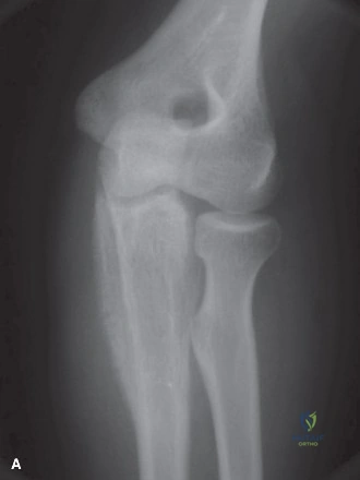
A 14-year-old male presents with a 3-month history of insidious, progressive pain in the left mid-thigh. He reports associated night pain and recent low-grade fevers. On examination, there is a palpable, tender, firm mass in the diaphysis of the femur. Radiographs show a permeative diaphyseal lesion with a "moth-eaten" appearance and an aggressive "onion-skin" periosteal reaction. Look at the imaging provided.
Candidate: Based on the age, location in the diaphysis, and the aggressive periosteal reaction, my top differential is Ewing Sarcoma. I would also consider Osteomyelitis, which can mimic this presentation, and potentially Lymphoma or Osteosarcoma.
Failure to immediately prioritize malignancy over infection. Candidates often jump to imaging orders without explicitly stating the need for biopsy, or they ignore the "systemic symptoms" (fever) which are red herrings meant to trap the candidate into thinking only of Osteomyelitis.
"Given the clinical presentation of a symptomatic, permeative, diaphyseal bone lesion with lamellated periosteal reaction in an adolescent, the primary diagnosis is Ewing Sarcoma. The differential includes Osteomyelitis, Eosinophilic Granuloma, and Lymphoma. My management would include an immediate multidisciplinary workup: staging with MRI of the femur, CT of the chest to rule out pulmonary metastases, and a core needle biopsy performed by the surgeon who will eventually perform the definitive resection to ensure the tract is excisable."
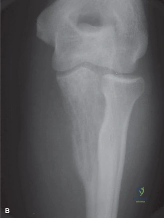
The biopsy confirms Ewing Sarcoma. You are now discussing surgical planning for a pelvic lesion in a different patient. Why is the pelvic location specifically considered a poor prognostic indicator compared to an appendicular site?
Candidate: Pelvic Ewing sarcomas are harder to treat because they are located deep in the pelvis. Achieving wide surgical margins is difficult because the tumor is often close to large blood vessels and nerves, which makes it more likely that the resection will be marginal or intralesional.
Providing a vague answer. Failing to use the specific Enneking oncologic terminology (e.g., "wide margins," "anatomical compartments," "local recurrence risk").
"The pelvic location carries a worse prognosis primarily due to the anatomical complexity that hinders the achievement of wide surgical margins. Unlike the appendicular skeleton, the pelvis lacks distinct anatomical compartments, allowing for earlier soft tissue invasion. Furthermore, the proximity to the lumbosacral plexus, major pelvic vessels, and viscera frequently necessitates a compromise in margin status to avoid unacceptable morbidity, directly increasing the risk of local recurrence."
You have decided on a limb-salvage procedure for an appendicular Ewing Sarcoma. What are the key biomechanical and oncological considerations when choosing between an endoprosthesis and an intercalary allograft?
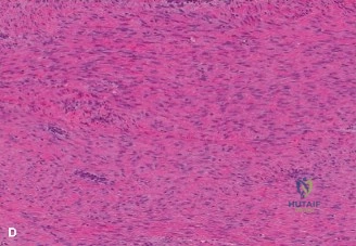
Candidate: Endoprostheses are better for early weight-bearing and return to function. Allografts are better for young patients because they replace bone with bone, but they have high risks of non-union, fracture, and infection.
Forgetting to address the impact of neoadjuvant chemotherapy/radiation on biological healing, which significantly influences the choice of allograft.
"The choice depends on the patient's age and activity demands. The endoprosthesis provides immediate structural stability and rapid rehabilitation, but carries risks of aseptic loosening and mechanical failure over time. The intercalary allograft offers biological restoration of bone stock, which is preferable in a younger patient; however, it is biologically inferior in patients undergoing adjuvant chemotherapy/radiation due to high rates of non-union, structural fatigue, and deep infection. In short: the prosthesis is for immediate function; the allograft is for biological longevity at the expense of a higher complication profile."
