Orthopedic Ob Trauma B Review | Dr Hutaif Trauma & Frac -...
Key Takeaway
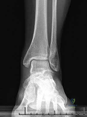
Looking for accurate information on ORTHOPEDIC MCQS ONLINE OB 20 TRAUMA 2B? A posterior knee dislocation, the injury shown in figure, poses a significant risk of popliteal artery injury (18-45%). If the limb remains ischemic after reduction, emergent vascular exploration and reconstruction are imperative. Delaying revascularization beyond 6-8 hours substantially increases amputation rates, potentially reaching 85%. Preoperative angiography should be avoided to prevent treatment delays.
Orthopedic Ob Trauma B Review | Dr Hutaif Trauma & Frac -...
Comprehensive 100-Question Exam
00:00
Start Quiz
Question 1
A 24-year-old male is brought to the trauma bay following a high-speed motorcycle collision. He has bilateral closed femoral shaft fractures and a severe closed head injury. His initial vitals are: HR 130, BP 85/50 mmHg. Arterial blood gas reveals pH 7.15, base excess -10, and lactate 6.0 mmol/L. Resuscitation is initiated. According to the principles of Damage Control Orthopedics (DCO), what is the most appropriate initial management of his bilateral femur fractures?
Explanation
Question 2
A 38-year-old female pedestrian is struck by a vehicle. She arrives hypotensive (BP 75/40 mmHg) with a mechanically unstable pelvis. An anterior-posterior compression (APC) type III injury is suspected. A pelvic binder is to be applied. What is the optimal anatomic landmark to center the pelvic binder to maximize reduction of the pelvic volume?
Explanation
Question 3
A 42-year-old male falls from a height of 20 feet. Pelvic radiographs and CT scan are obtained. Which of the following radiographic findings is pathognomonic for an associated both-column acetabular fracture?
Explanation
Question 4
A 28-year-old male sustains a closed, isolated Pauwels type III (vertical shear) femoral neck fracture. Which of the following fixation constructs is most biomechanically advantageous for mitigating the high shear forces across this fracture pattern?
Explanation
Question 5
A 35-year-old female presents with a highly comminuted intra-articular distal femur fracture (OTA/AO 33-C3) after a motor vehicle collision. A coronal plane fracture of the lateral femoral condyle (Hoffa fragment) is identified on CT scan. What is the optimal surgical approach and initial fixation strategy for this specific fragment?
Explanation
Question 6
A 29-year-old male sustains a closed comminuted tibial shaft fracture. On the morning following intramedullary nailing, he complains of severe leg pain out of proportion to the injury. His blood pressure is 110/60 mmHg. Intracompartmental pressure measurements are obtained. Which of the following intracompartmental pressures is an absolute indication for emergent four-compartment fasciotomy?
Explanation
Question 7
A 40-year-old male sustains a Gustilo-Anderson IIIB open midshaft tibia fracture. Following initial debridement and external fixation, a free tissue transfer is required for soft tissue coverage. Current literature suggests that to minimize the risk of deep infection and flap failure, soft tissue coverage should ideally be performed within what time frame from the time of injury?
Explanation
Question 8
A 45-year-old male presents with a high-energy closed severe pilon fracture. There is significant soft tissue swelling, fracture blisters, and ecchymosis around the ankle. What is the most appropriate management plan regarding the timing and method of definitive internal fixation?
Explanation
Question 9
A 31-year-old male sustains a Hawkins type II talar neck fracture following an MVA. He undergoes open reduction and internal fixation. At the 8-week follow-up, an AP radiograph of the ankle demonstrates a subchondral radiolucent band in the talar dome. What does this finding indicate?
Explanation
Question 10
A 50-year-old male undergoes ORIF of a displaced intra-articular calcaneus fracture using an extensile lateral approach. He is a 1-pack-per-day smoker. Which of the following is the most common postoperative complication associated with this specific surgical approach?
Explanation
Question 11
A 22-year-old collegiate football player sustains an axial load to a plantarflexed foot. Weight-bearing radiographs reveal widening of the interval between the medial and middle cuneiforms, and between the first and second metatarsal bases, with no obvious fractures (purely ligamentous Lisfranc injury). What is the optimal surgical treatment associated with the best functional outcome for this specific injury pattern?
Explanation
Question 12
A 65-year-old female sustains a 4-part proximal humerus fracture. Recent anatomical studies by Hertel et al. have redefined the understanding of the blood supply to the proximal humerus. According to these studies, preservation of which of the following is the most critical predictor of humeral head viability?
Explanation
Question 13
A 55-year-old female undergoes volar locking plate fixation for a displaced distal radius fracture. At 6 months postoperatively, she presents with an inability to actively flex the interphalangeal joint of her thumb. What is the most likely cause of this complication?
Explanation
Question 14
A 32-year-old male sustains a Galeazzi fracture-dislocation. Intraoperatively, after achieving anatomic open reduction and rigid internal fixation of the radial shaft with a compression plate, the distal radioulnar joint (DRUJ) remains unstable in both supination and pronation. What is the most appropriate next step in management?
Explanation
Question 15
A 44-year-old female undergoes open reduction and internal fixation of an intercondylar distal humerus fracture (13-C2) using an olecranon osteotomy approach. Regarding the management of the ulnar nerve during this procedure, current evidence suggests:
Explanation
Question 16
An 82-year-old female with a history of a cemented right total hip arthroplasty performed 12 years ago presents after a ground-level fall. Radiographs demonstrate a fracture around the tip of the femoral stem. The stem is radiographically loose, but there is adequate remaining femoral bone stock. According to the Vancouver classification, how should this fracture be managed?
Explanation
Question 17
A 68-year-old female who has been taking alendronate for 8 years presents with a 3-month history of insidious onset left thigh pain. Radiographs reveal focal lateral cortical thickening and a transverse radiolucent line extending partially through the lateral cortex of the left subtrochanteric femur. What is the most appropriate management?
Explanation
Question 18
A 25-year-old male sustains a low-velocity, civilian handgun wound to the mid-thigh. Radiographs show a midshaft femur fracture with a non-comminuted, short oblique pattern. The bullet has exited the limb. The patient is neurovascularly intact. What is the most appropriate infection prophylaxis protocol for this injury?
Explanation
Question 19
A 33-year-old male motorcyclist is struck by a truck and suffers an open pelvic ring injury with a massive perineal laceration extending to the rectum. He is hemodynamically unstable but responds transiently to blood products. Upon arrival to the OR, after initial pelvic stabilization with an external fixator and preperitoneal packing, what is the mandatory next step regarding the perineal wound?
Explanation
Question 20
A 42-year-old male sustains a high-energy OTA/AO 41-C3 bicondylar tibial plateau fracture. CT scanning reveals a significant posteromedial shear fragment that is displaced distally. What is the optimal surgical approach and positioning to address this specific fracture component?
Explanation
Question 21
When performing a posteromedial approach to the tibial plateau for a coronal shear fracture (Moore Type I), the standard surgical interval to expose the posterior aspect of the medial condyle is between the:
Explanation
Question 22
A 42-year-old male presents with a high-energy closed pilon fracture. Initial management included a spanning external fixator. Definitive internal fixation is planned. What is the most reliable clinical indicator that the soft tissues are ready for definitive surgical management?
Explanation
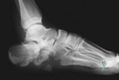
Question 23
A 35-year-old roofer falls from a height and sustains a displaced intra-articular calcaneus fracture. He undergoes open reduction and internal fixation via an extensile lateral approach. Which of the following arteries provides the primary blood supply to the lateral soft-tissue flap elevated in this approach?
Explanation
Question 24
A 28-year-old snowboarder sustains a Hawkins Type II talar neck fracture. At 8 weeks postoperatively, a plain AP radiograph of the ankle demonstrates a subchondral radiolucent band in the dome of the talus. What does this finding indicate?
Explanation
Question 25
The Lisfranc ligament is a crucial stabilizing structure of the midfoot. Between which two osseous structures does the true Lisfranc ligament travel?
Explanation
Question 26
During the ilioinguinal approach to the acetabulum, the surgeon must be careful to identify and ligate the 'corona mortis' to prevent life-threatening hemorrhage. This structure typically represents an anastomosis between which two vascular systems?
Explanation
Question 27
A 40-year-old male is involved in a high-speed motor vehicle collision. Radiographs of the pelvis demonstrate an acetabular fracture. Which of the following radiographic findings on an obturator oblique view is pathognomonic for a both-column fracture?
Explanation
Question 28
A 25-year-old female sustains a vertically oriented, displaced femoral neck fracture (Pauwels Type III) after a fall from a horse. Which of the following fixation constructs offers the highest biomechanical stability against shear forces for this fracture pattern?
Explanation
Question 29
In the treatment of intertrochanteric femur fractures, the integrity of the lateral trochanteric wall is a critical determinant of construct stability. According to orthopedic literature, a lateral wall thickness less than what threshold is considered an absolute indication for a cephalomedullary nail rather than a sliding hip screw?
Explanation
Question 30
A 30-year-old polytrauma patient presents with bilateral femoral shaft fractures, a pulmonary contusion, and a grade III spleen laceration. Which of the following physiological parameters is an absolute indication for temporary external fixation (Damage Control Orthopedics) rather than early total care with intramedullary nailing?
Explanation
Question 31
A 34-year-old motorcyclist sustains a coronal shear fracture of the lateral femoral condyle. What is the standard eponym for this fracture, and what is the standard direction of screw fixation to secure the fragment?
Explanation
Question 32
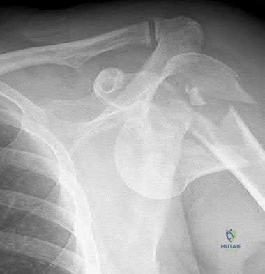
A 28-year-old male sustains a traumatic knee dislocation during a football game. Upon arrival at the ED, the knee is reduced. His pedal pulses are palpable, but his Ankle-Brachial Index (ABI) is measured at 0.82. What is the most appropriate next step in management?
Explanation
Question 33
A 22-year-old male is admitted with a closed midshaft tibia fracture. Twelve hours later, he complains of severe leg pain out of proportion to the injury, unrelieved by opioids. His blood pressure is 120/70 mmHg. Intracompartmental pressure in the anterior compartment is 45 mmHg. What is the patient's delta pressure, and is fasciotomy indicated?
Explanation
Question 34
A 45-year-old male farm worker caught his leg in an auger, sustaining a highly contaminated open diaphyseal tibia fracture. There is a 12 cm soft tissue laceration with extensive periosteal stripping. On examination, the foot is pulseless, and vascular surgery determines that an arterial repair is required to salvage the limb. What is the Gustilo-Anderson classification?
Explanation
Question 35
A 19-year-old male presents with a low-velocity gunshot wound to the right knee. Radiographs confirm a retained intact bullet freely mobile within the joint space, with no associated fracture. What is the most appropriate definitive management for the retained intra-articular bullet?
Explanation
Question 36
A 42-year-old female sustains a "terrible triad" injury of the elbow. She is taken to the operating room for definitive fixation. To optimize stability and clinical outcomes, what is the generally recommended sequence of surgical reconstruction?
Explanation
Question 37
A 38-year-old male sustains a subtrochanteric fracture of the right femur. During open reduction and intramedullary nailing, the surgeon notes classic multi-planar displacement of the proximal fragment. Which set of deforming forces accurately describes the displacement of the proximal fragment?
Explanation
Question 38
A 65-year-old female presents with a volar shear fracture of the distal radius (volar Barton fracture). Which of the following is the most appropriate surgical approach and internal fixation method?
Explanation
Question 39
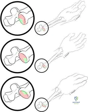
A 28-year-old male gymnast complains of chronic central wrist pain following a fall on an extended wrist 3 months ago. Radiographs show a widened scapholunate interval (Terry Thomas sign). The scapholunate interosseous ligament (SLIL) has three distinct regions. Which region is the primary mechanical stabilizer of the scapholunate joint?
Explanation
Question 40
A 50-year-old female falls onto an outstretched hand and sustains an isolated coronal shear fracture of the capitellum. Radiographs reveal a large osseous fragment consisting of the capitellum and the lateral half of the trochlea without posterior comminution. According to the Dubberley classification, what type of fracture is this?
Explanation
Question 41
During the resuscitation of a polytrauma patient with severe orthopedic injuries, monitoring lactate clearance is critical. A delay in normalizing serum lactate levels beyond what timeframe is most highly predictive of increased mortality, ARDS, and multi-organ failure?
Explanation
Question 42
A 35-year-old male sustains an anterior-posterior compression type III (APC III) pelvic ring injury. During surgical fixation via an anterior ilioinguinal approach, massive hemorrhage occurs near the superior pubic ramus while developing the medial window. Which of the following vascular structures is most likely injured?
Explanation
Question 43
A 45-year-old male is brought to the ED after a severe crush injury to the pelvis. He is hemodynamically unstable (BP 60/40 mmHg). A pelvic binder is applied, and FAST exam is negative. Despite massive transfusion, he remains in profound hemorrhagic shock. If angiography is delayed, what is the most appropriate next step for temporary hemorrhage control?
Explanation
Question 44
A 28-year-old female presents with a closed pelvic ring fracture after being run over by a truck. Examination reveals a large, fluctuant swelling over the greater trochanter with overlying skin bruising and reduced sensation. Which of the following best describes the pathophysiology of this soft tissue injury?
Explanation
Question 45
A 25-year-old male is diagnosed with a posterior wall acetabular fracture after a dashboard injury.
On CT scan, the fracture involves 30% of the posterior wall. What is the most accurate method to determine if this hip requires operative fixation due to instability?
Explanation
Question 46
A polytrauma patient undergoes damage control orthopedics (DCO) for bilateral femur fractures with temporary external fixation. Which of the following laboratory parameters best indicates that the patient is adequately resuscitated and cleared for Early Total Care (ETC) conversion to intramedullary nailing?
Explanation
Question 47
A 40-year-old male falls from a roof, sustaining a highly comminuted transforaminal sacral fracture extending centrally. According to the Denis classification, this involves Zone III. What is the most likely associated neurological complication?
Explanation
Question 48
A 22-year-old male with a diaphyseal femur fracture develops confusion, tachypnea (RR 30), and hypoxia on post-injury day 2. A petechial rash is noted over his axilla and conjunctiva. According to Gurd's criteria, what is the most critical physiological driver of his respiratory insufficiency?
Explanation
Question 49
During the ilioinguinal approach for an associated both-column acetabular fracture
, the surgeon develops the 'middle window'. Which structures define the borders of this window, and what critical structure lies within it?
Explanation
Question 50
A 32-year-old male sustains a high-energy tibial plateau fracture. The orthopedic surgeon suspects acute compartment syndrome. The patient's blood pressure is 110/60 mmHg. Intra-compartmental pressures are: Anterior 40 mmHg, Lateral 35 mmHg, Deep Posterior 45 mmHg, Superficial Posterior 30 mmHg. What is the absolute indication for four-compartment fasciotomy in this patient?
Explanation
Question 51
A 38-year-old pedestrian is struck by a vehicle, sustaining an anterior-posterior compression (APC) type II pelvic ring injury. Which of the following best describes the ligamentous disruption pattern in this specific injury?
Explanation
Question 52
A 26-year-old male sustains a low-velocity civilian gunshot wound to the mid-thigh. Radiographs show a highly comminuted midshaft femur fracture. The patient has normal distal pulses and intact sensation. The entry and exit wounds are 1 cm each with no gross contamination. What is the most appropriate management?
Explanation
Question 53
According to the Pape/Hannover criteria for polytrauma patients, which of the following findings would classify a patient as 'borderline' and thus potentially contraindicate Early Total Care (ETC) of major long bone fractures?
Explanation
Question 54
A 29-year-old male motorcyclist sustains an ipsilateral midshaft femur fracture and midshaft tibia fracture (floating knee). He is hemodynamically stable. What is the most widely recommended surgical sequence and its primary rationale?
Explanation
Question 55
A 30-year-old male sustains a high-energy diaphyseal femur fracture. Due to the high risk of a concomitant, missed ipsilateral femoral neck fracture, what is the 'gold standard' imaging protocol?
Explanation
Question 56
A 45-year-old farmer sustains a Gustilo-Anderson IIIB open tibia fracture from a tractor rollover. The wound is heavily contaminated with soil and manure. In addition to a first-generation cephalosporin and an aminoglycoside, which antibiotic must be added to the initial regimen?
Explanation
Question 57
A pelvic binder is applied to a hypotensive patient with an open-book pelvic fracture in the trauma bay. What is the correct anatomical landmark for centering the binder, and what is the most significant complication of leaving it on for >24 hours?
Explanation
Question 58
A 45-year-old male presents with a hemodynamically unstable pelvic crush injury. Angiography shows active extravasation from the 'corona mortis'. Which of the following describes the most common arterial vessels communicating at this anatomic structure?
Explanation
Question 59
A 30-year-old female sustains a Denis Zone 3 sacral fracture following a fall from height. Which of the following is the most likely neurologic complication associated with this specific injury pattern?
Explanation
Question 60
A 35-year-old male undergoes open reduction and internal fixation of a posterior wall acetabular fracture via the Kocher-Langenbeck approach. Postoperatively, he exhibits a foot drop. Which specific neural structure was most likely injured or overly retracted during the procedure?
Explanation
Question 61
A 28-year-old male is admitted with a severe closed tibia fracture. His current blood pressure is 110/70 mmHg. Intracompartmental pressure testing of the anterior compartment yields a value of 45 mmHg. What is the most appropriate next step in management?
Explanation
None
