Full Question & Answer Text (for Search Engines)
Question 1:
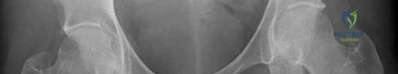
A 45-year-old male is brought to the ED after a high-speed MVC. He is hypotensive (BP 80/50). Pelvic radiograph shows an APC-III injury. Where is the most appropriate anatomical landmark to center a pelvic binder?
Options:
- Greater trochanters
- Iliac crests
- Symphysis pubis and ASIS
- Umbilicus
- Proximal thirds of the femurs
Correct Answer: Greater trochanters
Explanation:
A pelvic binder should be centered over the greater trochanters to effectively close the pelvic ring, particularly in anteroposterior compression (APC) injuries. Placing it over the iliac crests is a common error and is less effective mechanically, as it does not adequately reduce the symphysis.
Question 2:
A 25-year-old male sustained severe polytrauma. According to current guidelines for Damage Control Orthopedics (DCO), which of the following laboratory values indicates that the patient is in extremis and should undergo temporizing external fixation for his bilateral femur fractures rather than definitive intramedullary nailing?
Options:
- Lactate 2.0 mmol/L
- Base excess -2 mEq/L
- Platelet count 150,000/microL
- Arterial pH 7.20
- Core temperature 36.5°C
Correct Answer: Arterial pH 7.20
Explanation:
Markers of a patient in extremis or borderline who would benefit from DCO include arterial pH < 7.24, core temperature < 35°C, massive transfusion requirement (>10 units), and clinical coagulopathy. A pH of 7.20 reflects severe acidosis, making definitive long-bone nailing unacceptably risky due to the 'second hit' phenomenon.
Question 3:
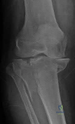
A 30-year-old male sustains a low-velocity gunshot wound to the thigh, resulting in a minimally displaced diaphyseal femur fracture. The bullet passed cleanly through the thigh without injuring major vessels or nerves. What is the most appropriate initial management of the fracture and wounds?
Options:
- Immediate surgical debridement of the entire bullet track and IM nailing
- Local wound care with superficial debridement and IM nailing
- Intravenous antibiotics for 7 days and skeletal traction
- Immediate external fixation and delayed definitive fixation
- Formal debridement of the entry and exit wounds with primary closure
Correct Answer: Local wound care with superficial debridement and IM nailing
Explanation:
Low-velocity gunshot wounds resulting in femur fractures generally behave like closed fractures or low-grade open fractures. They do not require formal operative debridement of the entire bullet tract. Standard local wound care, superficial debridement of the skin edges, and standard intramedullary nailing are appropriate.
Question 4:
A 40-year-old patient has a Gustilo-Anderson IIIB open tibia fracture from a motorcycle crash on a highway. What is the currently recommended antibiotic prophylaxis upon initial presentation?
Options:
- First-generation cephalosporin alone
- First-generation cephalosporin and an aminoglycoside (or a third-generation cephalosporin)
- First-generation cephalosporin and penicillin
- Fluoroquinolone alone
- Macrolide and an aminoglycoside
Correct Answer: First-generation cephalosporin and an aminoglycoside (or a third-generation cephalosporin)
Explanation:
For type III open fractures, current guidelines recommend the addition of Gram-negative coverage. This is typically achieved by adding an aminoglycoside to a first-generation cephalosporin, or by using a third-generation cephalosporin (such as ceftriaxone) alone or in combination. Penicillin is added specifically if there is concern for clostridial infection (e.g., heavy soil contamination).
Question 5:
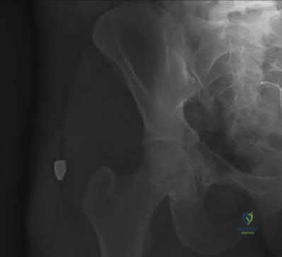
A 28-year-old male presents with a posterior hip dislocation and an associated posterior wall acetabulum fracture. The hip is reduced in the ED. On post-reduction CT scan, there is a 2 mm intra-articular fragment. The posterior wall fracture involves 25% of the articular surface. Examination under anesthesia shows no instability. What is the best management?
Options:
- Skeletal traction for 6 weeks
- Non-operative management with touch-down weight bearing
- Open reduction and internal fixation via a Kocher-Langenbeck approach
- Surgical hip dislocation and primary fragment excision without fixation
- Total hip arthroplasty
Correct Answer: Open reduction and internal fixation via a Kocher-Langenbeck approach
Explanation:
An absolute indication for operative intervention in acetabular fractures is an incarcerated intra-articular fragment, regardless of the size of the posterior wall defect or the dynamic stability of the joint. The Kocher-Langenbeck approach is the standard approach for a posterior wall fracture.
Question 6:
A 40-year-old female is struck by a car and sustains a closed pelvic ring injury. Examination reveals a large, fluctuant, soft tissue swelling over the greater trochanter with ecchymosis and decreased sensation over the area. What is the most appropriate initial management of this specific soft tissue injury?
Options:
- Immediate wide excision of the overlying skin
- Percutaneous aspiration or small incision drainage with a compression dressing
- Observation and warm compresses without compression
- Administration of intralesional corticosteroids
- Emergent fasciotomy of the thigh compartments
Correct Answer: Percutaneous aspiration or small incision drainage with a compression dressing
Explanation:
The patient has a Morel-Lavallée lesion, a closed degloving injury. Early management with percutaneous aspiration or small incision drainage combined with a firm compression dressing is recommended to prevent fluid reaccumulation and subsequent skin necrosis or infection.
Question 7:
A 32-year-old male sustained a Hawkins type II talar neck fracture. Six weeks postoperatively, an AP radiograph of the ankle demonstrates a subchondral radiolucent band in the talar dome. What is the significance of this radiographic finding?
Options:
- It indicates early hardware failure and loss of fixation
- It represents avascular necrosis of the talar body
- It signifies intact vascularity to the talar body
- It is a sign of impending nonunion of the talar neck
- It suggests an underlying joint space infection
Correct Answer: It signifies intact vascularity to the talar body
Explanation:
This finding is known as Hawkins sign. It represents subchondral osteopenia (atrophy) which occurs secondary to hyperemia, confirming that the vascular supply to the talar body is intact. Its presence makes avascular necrosis highly unlikely.
Question 8:
A 45-year-old female falls onto an outstretched hand and sustains a 'terrible triad' injury of the elbow. Which of the following describes the most widely accepted surgical sequence for reconstruction?
Options:
- Coronoid fixation, radial head repair/replacement, lateral collateral ligament (LCL) repair
- LCL repair, coronoid fixation, radial head repair/replacement
- Radial head repair/replacement, coronoid fixation, LCL repair
- Radial head repair/replacement, LCL repair, coronoid fixation
- Coronoid fixation, LCL repair, radial head repair/replacement
Correct Answer: Coronoid fixation, radial head repair/replacement, lateral collateral ligament (LCL) repair
Explanation:
The standard sequence for reconstructing a terrible triad injury works from deep to superficial, and usually medial to lateral (if approached laterally). The sequence is fixation of the coronoid fracture first, followed by radial head repair or replacement, and finally repair of the lateral ulnar collateral ligament (LUCL).
Question 9:
Intramedullary nailing of a proximal third tibial shaft fracture is notoriously associated with which of the following post-operative malalignments?
Options:
- Varus and recurvatum
- Varus and procurvatum
- Valgus and recurvatum
- Valgus and procurvatum
- Neutral alignment with >2 cm shortening
Correct Answer: Valgus and procurvatum
Explanation:
Proximal third tibia fractures have a strong tendency to fall into valgus and procurvatum (apex anterior) during intramedullary nailing. This is due to the wide metaphyseal flare (lack of tight fit of the nail), the pull of the patellar tendon anteriorly, and the anterior starting point of the nail wedge.
Question 10:
A 28-year-old male sustains a vertically oriented, displaced femoral neck fracture (Pauwels type III). Open reduction and internal fixation is planned. What biomechanical construct provides the most stable fixation for this specific fracture pattern to resist shear forces?
Options:
- Three parallel cancellous screws in an inverted triangle configuration
- A sliding hip screw (SHS) with an adjunctive derotational screw
- Two crossed partially threaded cancellous screws
- Proximal femoral locking plate alone
- Multiple fully threaded cortical screws
Correct Answer: A sliding hip screw (SHS) with an adjunctive derotational screw
Explanation:
Pauwels type III fractures are highly vertical and experience massive shear forces. Biomechanical studies have shown that a sliding hip screw (SHS) with a derotational screw provides superior fixation and resists shear forces better than three parallel cancellous screws for these specific injuries in young adults.
Question 11:
When managing a patient with a 'floating knee' (ipsilateral diaphyseal femur and tibia fractures) who is hemodynamically stable, which of the following describes a widely accepted surgical strategy to optimize efficiency and alignment?
Options:
- Antegrade femoral nailing followed by retrograde tibial nailing
- Retrograde femoral nailing and antegrade tibial nailing through a single parapatellar incision
- Plate osteosynthesis of both fractures to avoid entering the knee joint entirely
- External fixation of the femur and intramedullary nailing of the tibia
- Concurrent antegrade nailing of both the femur and the tibia
Correct Answer: Retrograde femoral nailing and antegrade tibial nailing through a single parapatellar incision
Explanation:
For a floating knee in a stable patient, intramedullary nailing of both fractures is preferred. A highly efficient approach is using a single midline knee incision with a medial or lateral parapatellar arthrotomy to perform retrograde femoral nailing and antegrade tibial nailing sequentially.
Question 12:
A 42-year-old male falls from a roof, sustaining a closed, displaced intra-articular calcaneus fracture. He is a heavy smoker. What is the most appropriate management regarding the surgical timing and approach to minimize soft-tissue complications if an extensile lateral approach is chosen?
Options:
- Immediate open reduction and internal fixation within 12 hours
- Delay surgery until the 'wrinkle sign' appears, typically 10-14 days
- Immediate percutaneous pinning without delay
- Perform a primary subtalar arthrodesis within 24 hours
- Delay surgery for 6 weeks until bone healing initiates
Correct Answer: Delay surgery until the 'wrinkle sign' appears, typically 10-14 days
Explanation:
Calcaneus fractures are associated with significant soft-tissue swelling. Operating through swollen, tense skin, particularly via an extensile lateral approach, dramatically increases the risk of wound dehiscence and infection. Surgery should be delayed until soft-tissue swelling subsides, indicated by the 'wrinkle sign', which often takes 10 to 14 days. This is especially critical in smokers.
Question 13:
A 25-year-old male cyclist falls and sustains a midshaft clavicle fracture. Which of the following is considered an absolute indication for operative intervention?
Options:
- 15 mm of shortening
- 100% displacement without skin tenting
- Floating shoulder (ipsilateral scapular neck fracture)
- Open fracture
- Z-type comminution
Correct Answer: Open fracture
Explanation:
Open fractures are an absolute indication for operative intervention (irrigation, debridement, and fixation). Shortening >20 mm, severe displacement, and floating shoulder are relative indications where surgery is often beneficial but not absolutely mandatory.
Question 14:
A 30-year-old male sustains a closed spiral fracture of the distal third of the humeral shaft (Holstein-Lewis fracture). On initial examination, he has an intact radial pulse but demonstrates a wrist drop and inability to extend his fingers. What is the most appropriate initial management?
Options:
- Immediate surgical exploration of the radial nerve and fracture fixation
- Application of a coaptation splint and clinical observation
- Urgent MRI of the humerus to evaluate the nerve continuity
- Electromyography (EMG) and nerve conduction studies on day 1
- Immediate closed reduction and percutaneous pinning
Correct Answer: Application of a coaptation splint and clinical observation
Explanation:
A primary radial nerve palsy associated with a closed humeral shaft fracture (including Holstein-Lewis types) is not an absolute indication for immediate nerve exploration. The majority are neuropraxias that will recover spontaneously. The standard of care is functional bracing or a coaptation splint and observation. If no recovery is noted clinically or on EMG by 3 to 4 months, exploration may be indicated.
Question 15:
A 29-year-old industrial worker suffers a complete traumatic amputation of his dominant index finger. The amputated part is recovered. To maximize the chance of successful replantation, how should the amputated part be transported?
Options:
- Submerged directly in normal saline at room temperature
- Wrapped in saline-moistened gauze, placed in a water-tight plastic bag, and kept on ice water
- Placed directly on ice to rapidly cool the tissue
- Submerged in a 10% betadine solution
- Frozen rapidly in dry ice
Correct Answer: Wrapped in saline-moistened gauze, placed in a water-tight plastic bag, and kept on ice water
Explanation:
The amputated part should be wrapped in saline-moistened gauze, placed inside a sealed plastic bag, and then the bag should be placed in a slurry of ice and water. Direct contact with ice or freezing solutions causes frostbite and tissue necrosis, precluding replantation.
Question 16:
A 22-year-old male presents with a closed tibial shaft fracture after a soccer injury. He complains of severe leg pain. Which of the following is the most sensitive early clinical finding of acute compartment syndrome?
Options:
- Loss of peripheral pulses (dorsalis pedis and posterior tibial)
- Pallor of the foot
- Pain with passive stretch of the toes
- Decreased capillary refill time
- Paralysis of the tibialis anterior muscle
Correct Answer: Pain with passive stretch of the toes
Explanation:
Pain with passive stretch of the muscles in the affected compartment is typically the earliest and most sensitive clinical sign of acute compartment syndrome. Pulselessness, pallor, and paralysis are late signs and indicate irreversible tissue ischemia.
Question 17:
During an ilioinguinal approach for the fixation of an anterior column acetabulum fracture, massive hemorrhage is encountered when dissecting over the superior pubic ramus. Which of the following vascular structures is most likely injured?
Options:
- An anomalous anastomosis between the internal pudendal and superior gluteal arteries
- An anastomosis between the external iliac or inferior epigastric vessels and the obturator vessels
- The main femoral artery
- The internal iliac vein
- The superficial epigastric artery
Correct Answer: An anastomosis between the external iliac or inferior epigastric vessels and the obturator vessels
Explanation:
The structure described is the 'corona mortis' (crown of death). It is a vascular anastomosis between the obturator vessels and the external iliac or inferior epigastric vessels, located on the posterior aspect of the superior pubic ramus. It is at high risk during the ilioinguinal or Stoppa approaches.
Question 18:
A 40-year-old female sustains a pronation-external rotation (PER) type ankle fracture. Intraoperatively, after fixation of the malleoli, the Cotton test is positive, indicating syndesmotic instability. A syndesmotic screw is planned. At what level relative to the ankle joint line should the syndesmotic screw optimally be placed?
Options:
- 1-2 cm below the joint line
- 2-5 cm above the joint line
- 5-8 cm above the joint line
- Exactly at the level of the joint line
- Through the lateral process of the talus
Correct Answer: 2-5 cm above the joint line
Explanation:
A syndesmotic screw should be placed parallel to the tibial plafond, typically 2 to 5 cm above the ankle joint line. It should be directed approximately 20 to 30 degrees anteriorly from the fibula to the tibia to account for the relative position of the bones.
Question 19:
A 25-year-old male sustains a gunshot wound to the abdomen. The bullet transverses the bowel and lodges in the L3 vertebral body. He has a complete neurologic deficit below L3. He undergoes an exploratory laparotomy and bowel repair. What is the most appropriate management of the retained bullet in the spine?
Options:
- Emergent laminectomy and bullet extraction
- Observation with a short course (7-14 days) of broad-spectrum antibiotics
- Anterior corpectomy and spinal fusion
- Long-term (6 months) suppressive intravenous antibiotics
- Epidural steroid injection
Correct Answer: Observation with a short course (7-14 days) of broad-spectrum antibiotics
Explanation:
In the setting of a complete neurologic deficit, removing a bullet from the spinal canal provides no neurologic benefit. Even with transperitoneal bowel perforation, the incidence of spinal infection is low. The standard of care is a short course of broad-spectrum antibiotics (usually 7-14 days) and observation. Extraction is only indicated for incomplete/progressive deficits, copper/lead toxicity in a joint space, or persistent CSF leak.
Question 20:
A 24-year-old male is involved in a high-speed motor vehicle collision. He has a closed unilateral midshaft femur fracture, bilateral pulmonary contusions, and a severe traumatic brain injury (GCS 6, intracranial pressure 25 mmHg). What is the most appropriate orthopaedic management of his femur fracture at this time?
Options:
- Immediate definitive intramedullary nailing
- External fixation as a damage control orthopaedics (DCO) measure
- Skeletal traction for 6 weeks until union
- Plate osteosynthesis to avoid medullary reaming
- Open reduction and internal fixation with dual plates
Correct Answer: External fixation as a damage control orthopaedics (DCO) measure
Explanation:
Patients with severe traumatic brain injury and elevated intracranial pressure are highly susceptible to secondary brain insults (hypotension, hypoxia, embolic showers) that can occur during definitive intramedullary nailing. Damage Control Orthopaedics (DCO) using a temporizing external fixator is indicated to provide rapid stability with minimal physiologic stress.
Question 21:
A 35-year-old male sustains a severe pelvic crush injury. Initial laboratory evaluation in the trauma bay reveals a base deficit of -8.5 mEq/L and a serum lactate of 5.5 mmol/L. He has bilateral comminuted femoral shaft fractures. What is the most appropriate initial management approach for his lower extremity injuries?
Options:
- Early total care with immediate bilateral reamed intramedullary nailing
- Nonoperative management with skeletal traction until discharge
- Damage control orthopedics with bilateral external fixation
- Immediate open reduction and internal fixation with locked plates
- Primary bilateral above-knee amputations
Correct Answer: Damage control orthopedics with bilateral external fixation
Explanation:
This patient presents with signs of profound physiologic derangement (base deficit < -5.5 mEq/L and lactate > 2.5 mmol/L), placing him in the 'unstable' or 'in extremis' category. In such polytrauma patients, early total care (e.g., prolonged reamed IM nailing) risks exacerbating the systemic inflammatory response ('second hit'). Damage control orthopedics (DCO) utilizing rapid external fixation is the standard of care to stabilize fractures while allowing physiological resuscitation.
Question 22:
A 28-year-old pregnant female at 32 weeks gestation sustains an unstable anteroposterior compression (APC III) pelvic ring injury following a motor vehicle collision. She is hemodynamically unstable despite initial aggressive fluid resuscitation. What is the best initial step regarding the management of her pelvic injury?
Options:
- Application of a pelvic binder at the level of the iliac crests
- Immediate emergent Cesarean section prior to any orthopedic intervention
- Application of a pelvic binder at the level of the greater trochanters
- Immediate open reduction and internal fixation of the pubic symphysis
- Bilateral internal iliac artery embolization before any fluid resuscitation
Correct Answer: Application of a pelvic binder at the level of the greater trochanters
Explanation:
In a hemodynamically unstable pregnant patient with an open-book pelvic fracture, mechanical stabilization of the pelvis is a priority and can be safely achieved with a pelvic binder. The binder must be placed accurately at the level of the greater trochanters, which will effectively reduce the pelvic volume without directly compressing the gravid uterus.
Question 23:
A 22-year-old male is admitted with a closed bilateral femoral shaft fracture. On post-injury day 2, he develops confusion, tachypnea, and an axillary rash. According to Gurd and Wilson's criteria for Fat Embolism Syndrome, which of his signs is considered a 'major' criterion?
Options:
- Tachycardia greater than 120 beats per minute
- Fever greater than 39°C
- Petechial rash
- Renal dysfunction
- Jaundice
Correct Answer: Petechial rash
Explanation:
Gurd and Wilson's criteria for Fat Embolism Syndrome (FES) require at least one major and four minor criteria. The major criteria include: 1) Petechial rash (usually in the axillae, conjunctivae, or palate), 2) Respiratory insufficiency, and 3) Cerebral involvement (confusion/coma). Tachycardia, fever, renal changes, and jaundice are considered minor criteria.
Question 24:
A 25-year-old male sustains a low-velocity civilian gunshot wound to his thigh, resulting in a midshaft femur fracture. The entrance and exit wounds are 1 cm and clean without massive tissue devitalization. There is no neurovascular deficit. What is the standard of care for this injury?
Options:
- Intravenous antibiotics for 7 days, immediate tract excision, and intramedullary nailing
- Intravenous antibiotics for 24-48 hours, local wound care, and intramedullary nailing
- Nonoperative management in a spica cast to prevent hardware infection
- External fixation until the bullet tract fully heals, followed by intramedullary nailing
- Bullet removal, formal debridement of all bone fragments, and rigid plating
Correct Answer: Intravenous antibiotics for 24-48 hours, local wound care, and intramedullary nailing
Explanation:
Low-velocity gunshot wounds resulting in femur fractures generally behave like closed fractures regarding infection risk. The standard of care includes local wound care, a short course of intravenous antibiotics (usually a first-generation cephalosporin for 24-48 hours), and standard intramedullary nailing. Formal debridement of the bullet tract and routine bullet removal are not indicated unless the bullet is intra-articular or causing mechanical symptoms.
Question 25:
During the ilioinguinal approach for an anterior column acetabulum fracture, massive arterial bleeding is encountered on the posterior aspect of the superior pubic ramus. Which vascular structure is most likely injured?
Options:
- The anastomosis between the external iliac artery and the obturator artery
- The anastomosis between the internal iliac artery and the superior gluteal artery
- The external iliac vein
- The internal pudendal artery
- The deep circumflex iliac artery
Correct Answer: The anastomosis between the external iliac artery and the obturator artery
Explanation:
Massive bleeding in this location is classic for injury to the corona mortis (crown of death). It is an arterial or venous anastomosis between the external iliac (or inferior epigastric) vessels and the obturator vessels located on the posterior aspect of the superior pubic ramus, typically about 5-6 cm from the pubic symphysis.
Question 26:
Which of the following pediatric fracture patterns is considered to have the highest specificity for non-accidental trauma (child abuse)?
Options:
- Spiral fracture of the tibia in an ambulatory child
- Midshaft clavicle fracture
- Linear skull fracture
- Metaphyseal corner fracture
- Distal radius buckle (torus) fracture
Correct Answer: Metaphyseal corner fracture
Explanation:
Metaphyseal corner fractures, also known as classic metaphyseal lesions (CMLs), are highly specific for child abuse and occur due to sudden twisting or pulling of an infant's extremity. Spiral tibia fractures (Toddler's fractures), clavicle fractures, and linear skull fractures are common accidental injuries in toddlers and young children.
Question 27:
According to the criteria established by Pape et al., which of the following physiological parameters classifies a polytrauma patient into the 'borderline' category, suggesting a need to carefully weigh damage control orthopedics against early total care?
Options:
- Injury Severity Score (ISS) of 15
- Initial serum lactate of 1.5 mmol/L
- Initial serum lactate > 2.5 mmol/L
- Platelet count > 150,000/µL
- Core body temperature of 36.5°C
Correct Answer: Initial serum lactate > 2.5 mmol/L
Explanation:
Pape's criteria for a 'borderline' polytrauma patient include factors such as ISS > 40 (or > 20 with thoracic trauma), expected major blood loss, initial lactate > 2.5 mmol/L, base deficit < -5.5, hypothermia < 35°C, or pulmonary artery pressure > 24 mmHg. A lactate > 2.5 mmol/L is a strong indicator of occult hypoperfusion and places the patient at high risk for complications if subjected to prolonged surgery.
Question 28:
A 30-year-old male is hypotensive (blood pressure 85/50 mmHg) following a severe crush injury to his lower leg. The anterior compartment pressure is measured at 30 mmHg. What is the most appropriate management regarding the diagnosis of acute compartment syndrome?
Options:
- Elevate the leg above the heart and reassess in 4 hours
- Administer intravenous mannitol and observe closely
- Perform an immediate four-compartment fasciotomy
- Apply a compressive dressing to reduce swelling
- Perform a closed reduction and application of a long leg cast
Correct Answer: Perform an immediate four-compartment fasciotomy
Explanation:
Acute compartment syndrome is diagnosed when the Delta P (diastolic blood pressure minus compartment pressure) is less than 30 mmHg. In this hypotensive patient, the diastolic pressure is 50 mmHg and compartment pressure is 30 mmHg, yielding a Delta P of 20 mmHg. This is an absolute indication for emergent four-compartment fasciotomy.
Question 29:
In evaluating a severe lower extremity crush injury for potential amputation, the Mangled Extremity Severity Score (MESS) is calculated. Which of the following components can contribute the highest possible number of points to the MESS score?
Options:
- Patient age over 50 years
- Skeletal and soft tissue injury severity
- Profound shock (blood pressure < 90 mmHg)
- Limb ischemia duration > 6 hours
- Presence of a vascular injury requiring repair
Correct Answer: Limb ischemia duration > 6 hours
Explanation:
The MESS scoring system allocates points for skeletal/soft tissue injury (1-4), shock (0-2), age (0-2), and limb ischemia (1-3). Crucially, the points for ischemia are doubled if the ischemic time exceeds 6 hours. Therefore, severe ischemia > 6 hours can contribute up to 6 points, the highest of any single category, strongly pushing the total score toward the threshold for amputation (>7).
Question 30:
The CRASH-2 trial demonstrated a significant reduction in mortality in bleeding trauma patients treated with tranexamic acid (TXA). To achieve this mortality benefit, TXA must be administered within what maximum timeframe from the time of injury?
Options:
- 1 hour
- 3 hours
- 6 hours
- 12 hours
- 24 hours
Correct Answer: 3 hours
Explanation:
The CRASH-2 trial showed that administration of TXA to bleeding trauma patients within 3 hours of injury significantly reduces the risk of death from hemorrhage. If given after 3 hours, TXA was associated with an increased risk of death due to bleeding, making the 3-hour window a critical guideline in trauma resuscitation.
Question 31:
Based on the findings of the Lower Extremity Assessment Project (LEAP) study, what is the most significant determinant of long-term functional outcome in patients sustaining a mangled lower extremity?
Options:
- The initial decision to amputate versus attempt salvage
- The presence of an intact plantar nerve sensation at presentation
- Patient socioeconomic status, education level, and psychological factors
- The specific type of soft-tissue flap coverage utilized
- The initial Mangled Extremity Severity Score (MESS)
Correct Answer: Patient socioeconomic status, education level, and psychological factors
Explanation:
The LEAP study fundamentally changed the understanding of severe lower extremity trauma by demonstrating that at 2 and 7 years post-injury, functional outcomes were not significantly different between the amputation and limb-salvage groups. Instead, the strongest predictors of poor outcome were lack of high school education, nonwhite race, poverty, poor social support, and poor psychological status (self-efficacy).
Question 32:
A 45-year-old agricultural worker sustains a Gustilo-Anderson Type IIIA open tibia fracture heavily contaminated with soil and manure. According to standard antibiotic prophylaxis guidelines for open fractures, which regimen is most appropriate?
Options:
- First-generation cephalosporin monotherapy
- First-generation cephalosporin and an aminoglycoside
- First-generation cephalosporin, an aminoglycoside, and high-dose penicillin
- Fluoroquinolone and clindamycin
- Vancomycin and ceftriaxone
Correct Answer: First-generation cephalosporin, an aminoglycoside, and high-dose penicillin
Explanation:
For severe open fractures (Type III), coverage is required for both Gram-positive and Gram-negative organisms, typically achieved with a first-generation cephalosporin and an aminoglycoside. However, when there is heavy soil or fecal contamination (e.g., farm injuries), high-dose penicillin should be added to cover Clostridium species to prevent gas gangrene.
Question 33:
In a polytrauma patient with a severe closed head injury (with documented intracranial hemorrhage) and a closed femur fracture, what is the most appropriate initial method for venous thromboembolism (VTE) prophylaxis?
Options:
- Prophylactic placement of an inferior vena cava (IVC) filter
- Aspirin 325 mg daily
- Mechanical prophylaxis with sequential compression devices (SCDs)
- Therapeutic intravenous unfractionated heparin
- Low-molecular-weight heparin (LMWH) starting immediately upon admission
Correct Answer: Mechanical prophylaxis with sequential compression devices (SCDs)
Explanation:
In trauma patients with active or high-risk intracranial hemorrhage, chemical VTE prophylaxis (heparin, LMWH, aspirin) is strictly contraindicated until cleared by neurosurgery. Mechanical prophylaxis with SCDs is the immediate standard of care. Prophylactic IVC filters are no longer recommended as routine initial management for VTE prophylaxis in trauma.
Question 34:
During the ongoing resuscitation of a polytrauma patient in hemorrhagic shock, which of the following markers is considered the most reliable indicator of adequate global tissue perfusion and successful resuscitation?
Options:
- Normalization of blood pressure and heart rate
- Urine output consistently > 0.5 mL/kg/hr
- Clearance of serum lactate and normalization of base deficit
- Central venous pressure reading of 8-12 mmHg
- Hematocrit stabilization at > 30%
Correct Answer: Clearance of serum lactate and normalization of base deficit
Explanation:
While vital signs and urine output are helpful, they can normalize while the patient remains in a state of 'occult hypoperfusion.' The clearance of serum lactate and the normalization of the base deficit are the most reliable and widely accepted biochemical markers of restored global tissue perfusion and the endpoint of shock resuscitation.
Question 35:
When constructing a unilateral external fixator for a tibial shaft fracture, which of the following modifications will most significantly increase the bending stiffness of the construct?
Options:
- Increasing the half-pin diameter from 4 mm to 5 mm
- Increasing the distance between the bone and the connecting longitudinal bar
- Placing all pins in a single bone fragment very close together
- Using pins made of titanium instead of stainless steel
- Increasing the distance from the fracture site to the innermost pins
Correct Answer: Increasing the half-pin diameter from 4 mm to 5 mm
Explanation:
The bending stiffness of an external fixator pin is proportional to the radius of the pin to the fourth power (r^4). Therefore, increasing the pin diameter has an exponential effect and is the single most effective way to increase the stiffness of the construct. Other ways to increase stiffness include placing the bar closer to the bone, spreading pins further apart within a given fragment, and placing inner pins closer to the fracture.
Question 36:
A patient is brought to the emergency department following an industrial explosion. Which of the following injuries is classified as a 'primary' blast injury?
Options:
- Penetrating shrapnel wounds to the lower extremities
- Crush injury from a collapsing structural beam
- Tympanic membrane rupture and pulmonary barotrauma
- Full-thickness thermal burns to exposed skin
- Traumatic brain injury from being thrown against a wall
Correct Answer: Tympanic membrane rupture and pulmonary barotrauma
Explanation:
Blast injuries are categorized into four types. Primary blast injuries are caused directly by the overpressure wave and predominantly affect gas-filled organs (tympanic membrane rupture, blast lung, bowel perforation). Secondary injuries are caused by flying debris (shrapnel). Tertiary injuries occur when the patient is thrown by the blast wind. Quaternary injuries encompass all other explosion-related injuries (burns, crush injuries, toxic gas inhalation).
Question 37:
During the surgical management of a complex lower extremity trauma, a pneumatic tourniquet is applied to minimize blood loss. To prevent irreversible ischemic damage to the muscles and nerves, what is the maximum recommended continuous inflation time before the tourniquet must be released for a 'breathing' period?
Options:
- 60 minutes
- 90 minutes
- 120 minutes
- 150 minutes
- 180 minutes
Correct Answer: 120 minutes
Explanation:
The standard maximum safe tourniquet inflation time for an extremity is 120 minutes (2 hours). If surgery must continue beyond this time, the tourniquet should be deflated for at least 10 to 15 minutes to allow for tissue reperfusion before it can be safely re-inflated.
Question 38:
A trauma patient presents with a heart rate of 130 bpm, blood pressure of 90/60 mmHg, respiratory rate of 35 breaths/min, and confused mental status. Estimated blood loss is approximately 35% of total blood volume. According to the ATLS classification, this patient is in which class of hemorrhagic shock?
Options:
- Class I
- Class II
- Class III
- Class IV
- Class V
Correct Answer: Class III
Explanation:
According to the American College of Surgeons ATLS classification, Class III hemorrhagic shock corresponds to an estimated blood loss of 31-40%. It is clinically characterized by a drop in measurable blood pressure, tachycardia (heart rate > 120), tachypnea (RR 30-40), and altered mental status (confusion). Class IV shock involves >40% blood loss, heart rate >140, and profound lethargy.
Question 39:
A 26-year-old male is involved in a motorcycle collision and sustains a 'floating knee' injury comprising an ipsilateral femur fracture and tibia fracture. Imaging reveals intra-articular extension into the knee joint from both the distal femur and the proximal tibia. According to the Fraser classification, this is categorized as:
Options:
- Type I
- Type IIA
- Type IIB
- Type IIC
- Type III
Correct Answer: Type IIC
Explanation:
The Fraser classification for floating knee injuries is as follows: Type I consists of pure diaphyseal fractures of both the femur and tibia. Type II involves articular fractures: Type IIA involves the femur articular surface, Type IIB involves the tibia articular surface, and Type IIC involves intra-articular extension in both the femur and the tibia.
Question 40:
In the contemporary paradigm of damage control resuscitation for a severely bleeding polytrauma patient, what is the optimal ratio of packed red blood cells (pRBCs) to fresh frozen plasma (FFP) to platelets recommended during massive transfusion?
Options:
- 3:1:1
- 1:1:1
- 4:2:1
- 1:2:2
- 6:1:1
Correct Answer: 1:1:1
Explanation:
Modern damage control resuscitation guidelines strongly recommend a balanced 1:1:1 ratio of packed red blood cells, fresh frozen plasma, and platelets. This strategy, validated by the PROPPR trial, addresses the acute coagulopathy of trauma more effectively than historical crystalloid-heavy or pure RBC resuscitation strategies.
Question 41:
A 28-year-old male is brought to the ED after a motor vehicle collision. He has a closed left femoral shaft fracture, right pulmonary contusion, and grade 2 splenic laceration. On arrival, BP 100/60, HR 110. Base deficit is 7 mmol/L and lactate is 3.5 mmol/L. Which of the following is considered an absolute indication for damage control orthopedics (DCO) with external fixation of the femur rather than early total care (ETC)?
Options:
- Femoral fracture combined with a pulmonary contusion
- Base deficit > 8 mmol/L or worsening lactate
- Injury Severity Score (ISS) > 15
- Glasgow Coma Scale (GCS) < 12
- Age > 25 years
Correct Answer: Base deficit > 8 mmol/L or worsening lactate
Explanation:
The principles of Damage Control Orthopedics (DCO) dictate that patients who are physiologically unstable should undergo temporizing fixation. Absolute indications for DCO include hemodynamic instability, core temperature < 32°C, pH < 7.24, base deficit > 8 mmol/L, and clinical coagulopathy. A femoral fracture with a pulmonary contusion or ISS > 15 represents a 'borderline' patient who may tolerate ETC if adequately resuscitated.
Question 42:
Which of the following is NOT a component of the Mangled Extremity Severity Score (MESS) used in evaluating severe limb trauma?
Options:
- Patient age
- Limb ischemia
- Presence of shock
- Degree of skeletal and soft tissue injury
- Neurological status of the limb
Correct Answer: Neurological status of the limb
Explanation:
The Mangled Extremity Severity Score (MESS) is calculated based on four variables: Skeletal and soft-tissue injury (1-4 points), Limb ischemia (1-3 points), Shock (0-2 points), and Age (0-2 points). Although severe neurological deficit (e.g., absent plantar sensation) is an important clinical factor, it is NOT a formal component of the MESS calculation.
Question 43:
In the management of a polytrauma patient, which of the following markers is considered the most reliable indicator of adequate tissue perfusion and successful systemic resuscitation?
Options:
- Normalization of blood pressure
- Normalization of heart rate
- Urine output > 0.5 mL/kg/hr
- Clearance of serum lactate
- Return of Glasgow Coma Scale to 15
Correct Answer: Clearance of serum lactate
Explanation:
Serum lactate clearance and normalization of base deficit are the most reliable metabolic endpoints of resuscitation. Normalization of vital signs such as heart rate and blood pressure often occur before cellular hypoxia is completely resolved ('occult hypoperfusion').
Question 44:
Patients with severe traumatic brain injury (TBI) often demonstrate accelerated fracture healing and an increased risk of heterotopic ossification due to circulating humoral factors. What is the primary effect of concomitant traumatic brain injury on fracture healing?
Options:
- Delayed union due to hypermetabolism
- Decreased callus volume with increased biomechanical strength
- Increased callus volume and accelerated union times
- High rate of nonunion due to increased corticosteroid release
- Inhibition of endochondral ossification
Correct Answer: Increased callus volume and accelerated union times
Explanation:
Clinical and basic science studies have demonstrated that patients with severe TBI undergo accelerated fracture healing characterized by an increased volume of fracture callus and faster clinical union times. This is mediated by circulating osteogenic factors (such as SDF-1, substance P, and other humoral factors) released following central nervous system injury.
Question 45:
The Reamer-Irrigator-Aspirator (RIA) system is commonly used today for large-volume autogenous bone graft harvest. However, it was originally developed to decrease the incidence of which of the following complications during intramedullary nailing?
Options:
- Thermal necrosis of the diaphysis
- Fat embolism syndrome
- Iatrogenic cortical fracture
- Intramedullary infection
- Hypertrophic nonunion
Correct Answer: Fat embolism syndrome
Explanation:
The RIA system was originally engineered to decrease intramedullary pressure during the reaming of long bones, thereby reducing the intravasation of marrow contents and the subsequent embolic load, which is a key factor in the development of Fat Embolism Syndrome (FES). It was later discovered to yield highly osteogenic graft material.
Question 46:
A 25-year-old male is trapped in a vehicle for 2 hours after a crash in winter. He arrives in the trauma bay intubated, with a pelvic ring injury and bilateral femur fractures. Which of the following accurately describes the 'lethal triad' of trauma that must be aggressively corrected?
Options:
- Hypocalcemia, hyperkalemia, acidosis
- Hypothermia, coagulopathy, acidosis
- Hypoxia, hypotension, coagulopathy
- Acidosis, hypothermia, hypoxia
- Coagulopathy, hyperkalemia, hypothermia
Correct Answer: Hypothermia, coagulopathy, acidosis
Explanation:
The 'lethal triad' in trauma consists of hypothermia, coagulopathy, and metabolic acidosis. These three conditions create a vicious cycle where hypothermia worsens coagulopathy, bleeding leads to reduced perfusion and worsening acidosis, which further depresses myocardial contractility and coagulation enzyme function.
Question 47:
A massively bleeding trauma patient undergoes a rapid thromboelastography (TEG) assay. The results show a significantly prolonged R-time (reaction time) with normal K-time, normal alpha angle, and normal maximum amplitude. Which of the following is the most appropriate targeted therapy based on this specific TEG abnormality?
Options:
- Platelet transfusion
- Cryoprecipitate
- Fresh frozen plasma (FFP)
- Tranexamic acid (TXA)
- Packed red blood cells (pRBCs)
Correct Answer: Fresh frozen plasma (FFP)
Explanation:
In TEG, the R-time represents the initial time for clot formation and is dependent on clotting factors. A prolonged R-time indicates a deficiency in coagulation factors, which is best treated with Fresh Frozen Plasma (FFP). Abnormal K-time/alpha angle reflects fibrinogen deficiency (treated with cryoprecipitate), abnormal MA reflects platelet issues, and abnormal LY30 indicates fibrinolysis (treated with TXA).
Question 48:
A 22-year-old male with a closed femoral shaft fracture develops respiratory distress, confusion, and a petechial rash 48 hours after injury. According to Gurd and Wilson's criteria for Fat Embolism Syndrome, which of the following is considered a MAJOR criterion?
Options:
- Tachycardia > 120 bpm
- Temperature > 39.4°C
- Unexplained drop in hematocrit
- Petechial rash
- Retinal changes
Correct Answer: Petechial rash
Explanation:
Gurd and Wilson's major criteria for Fat Embolism Syndrome (FES) include: 1) Petechial rash (usually across the axilla and thorax), 2) Respiratory symptoms (hypoxia/radiographic changes), and 3) Neurological signs (confusion/coma). Tachycardia, fever, retinal changes, and drops in hematocrit/platelets are considered MINOR criteria.
Question 49:
You are instructing a junior resident on the application of a circumferential pelvic binder for a hemodynamically unstable patient with an open-book pelvic ring injury. To effectively reduce the pelvic volume and stabilize the fracture, over which specific anatomic landmark should the binder be centered?
Options:
- Anterior superior iliac spines (ASIS)
- Iliac crests
- Greater trochanters
- Symphysis pubis
- Umbilicus
Correct Answer: Greater trochanters
Explanation:
A pelvic binder must be applied directly over the greater trochanters to effectively close an open-book pelvic injury. Placing the binder too high (over the iliac crests or ASIS) is a common error that fails to reduce the posterior pelvic ring and can paradoxically open the pelvis further in some fracture patterns.
Question 50:
Based on the CRASH-2 trial regarding the use of Tranexamic Acid (TXA) in bleeding trauma patients, which of the following statements is true regarding its administration?
Options:
- TXA administration should be delayed until the patient reaches the operating room.
- TXA provides the greatest mortality benefit when given within 3 hours of injury.
- Administration of TXA after 3 hours of injury has no effect on mortality but decreases deep vein thrombosis risk.
- TXA increases the risk of fatal pulmonary embolism in polytrauma patients.
- TXA is primarily indicated for patients with isolated traumatic brain injury.
Correct Answer: TXA provides the greatest mortality benefit when given within 3 hours of injury.
Explanation:
The CRASH-2 trial demonstrated that early administration of TXA (within 3 hours of injury) significantly reduces mortality due to bleeding in trauma patients. Importantly, administration after 3 hours was associated with an increased risk of death due to bleeding, making early administration critical.
Question 51:
The Lower Extremity Assessment Project (LEAP) was a large multicenter prospective study comparing amputation to limb salvage for severe lower extremity trauma. Which of the following was a major conclusion of the LEAP study?
Options:
- Limb salvage patients had significantly better functional outcomes at 2 years compared to amputation patients.
- Amputation patients experienced higher rates of major complications and re-hospitalizations.
- There was no significant difference in functional outcomes between limb salvage and amputation at 2 years.
- The Mangled Extremity Severity Score (MESS) accurately predicted the need for amputation with 95% sensitivity.
- Limb salvage was associated with significantly lower overall healthcare costs compared to amputation.
Correct Answer: There was no significant difference in functional outcomes between limb salvage and amputation at 2 years.
Explanation:
A primary finding of the LEAP study was that functional outcomes, as measured by the Sickness Impact Profile (SIP), were not significantly different between patients undergoing amputation and those undergoing limb salvage at 2 years. Furthermore, early scoring systems like the MESS were found to lack predictive validity for limb salvage outcomes.
Question 52:
According to the classic principles described by Godina for the management of complex open extremity injuries requiring free tissue transfer, timing of the flap coverage is most predictive of flap failure and infection. What was the optimal time frame originally recommended by Godina for free flap coverage?
Options:
- Within 24 hours of injury
- Within 72 hours of injury
- Between 5 to 7 days after injury
- Between 7 to 14 days after injury
- After 21 days when granulation tissue is fully formed
Correct Answer: Within 72 hours of injury
Explanation:
Godina's landmark study in 1986 established that early free flap coverage of complex open extremity wounds, specifically within 72 hours (3 days) of injury, resulted in the lowest rates of flap failure (0.75%) and infection (1.5%) compared to delayed coverage.
Question 53:
A 45-year-old male presents with persistent mid-thigh pain 8 months after intramedullary nailing of a femoral shaft fracture. Radiographs reveal an 'elephant foot' appearance with abundant callus that does not bridge the fracture site. What is the primary underlying cause of this specific type of nonunion?
Options:
- Inadequate biological environment
- Poor vascular supply to the fracture fragments
- Inadequate mechanical stability
- Undiagnosed deep infection
- Over-reaming of the medullary canal
Correct Answer: Inadequate mechanical stability
Explanation:
A hypertrophic nonunion (characterized by 'elephant foot' or 'horse hoof' callus) has excellent biology and robust vascular supply but fails to unite primarily due to inadequate mechanical stability. Treatment generally involves increasing construct stiffness, often by exchanging to a larger intramedullary nail or adding a plate.
Question 54:
Bone morphogenetic proteins (BMPs) are utilized in orthopedic trauma to promote fracture healing. Through which of the following mechanisms do BMP-2 and BMP-7 primarily exert their osteoinductive effects?
Options:
- Inhibition of osteoclast activity via the RANKL pathway
- Binding to cell surface receptors to activate intracellular SMAD pathways
- Direct stimulation of angiogenesis via Vascular Endothelial Growth Factor (VEGF) release
- Acting as a structural scaffold for osteoprogenitor cell migration
- Activation of the Wnt/beta-catenin signaling pathway to prevent apoptosis
Correct Answer: Binding to cell surface receptors to activate intracellular SMAD pathways
Explanation:
BMPs belong to the Transforming Growth Factor-beta (TGF-beta) superfamily. They bind to serine/threonine kinase cell surface receptors, which subsequently phosphorylate and activate intracellular SMAD proteins. These activated SMAD complexes translocate to the nucleus to upregulate the transcription of osteogenic genes (e.g., Runx2/Cbfa1).
Question 55:
According to Perren's strain theory of fracture healing, primary (direct) bone healing without callus formation requires the interfragmentary strain to be kept below what specific threshold?
Options:
Correct Answer: 2%
Explanation:
Perren's strain theory states that the type of tissue that forms at a fracture site is determined by the interfragmentary strain. Primary (direct) bone healing via cutting cones requires absolute stability with strain levels below 2%. Strain between 2% and 10% promotes secondary bone healing (callus), while strain >10% leads to fibrous nonunion.
Question 56:
Following severe polytrauma, a patient initially develops Systemic Inflammatory Response Syndrome (SIRS) followed by a Compensatory Anti-inflammatory Response Syndrome (CARS), rendering them highly susceptible to nosocomial infections. Which of the following markers is most characteristic of the CARS phase?
Options:
- Elevated Tumor Necrosis Factor-alpha (TNF-a)
- Decreased HLA-DR expression on monocytes
- Elevated Interleukin-1 (IL-1)
- Increased circulating neutrophil counts
- Increased systemic vascular resistance
Correct Answer: Decreased HLA-DR expression on monocytes
Explanation:
The Compensatory Anti-inflammatory Response Syndrome (CARS) is a state of immune suppression following major trauma. A classic and reliable biomarker for this 'immune paralysis' is the significant down-regulation of HLA-DR expression on the surface of circulating monocytes, which impairs antigen presentation.
Question 57:
A pneumatic tourniquet is routinely used in orthopedic extremity trauma surgery. What is the generally accepted maximum safe continuous tourniquet inflation time before the risk of irreversible muscle damage and nerve injury significantly increases?
Options:
- 60 minutes
- 90 minutes
- 120 minutes
- 180 minutes
- 240 minutes
Correct Answer: 120 minutes
Explanation:
The widely accepted safe limit for continuous tourniquet inflation in a normothermic patient is 120 minutes (2 hours). Extending inflation beyond this time significantly increases the risk of irreversible ischemic muscle damage, reperfusion injury, and compression neurapraxia.
Question 58:
In the pathophysiology of acute compartment syndrome, what is the initial microvascular event that initiates the cascade of tissue ischemia?
Options:
- Arterial occlusion
- Venous outflow obstruction
- Arteriolar spasm
- Capillary endothelial damage
- Lymphatic blockage
Correct Answer: Venous outflow obstruction
Explanation:
The pathophysiology of compartment syndrome begins when tissue pressure rises above the intraluminal pressure of the thin-walled post-capillary venules. This causes venous outflow obstruction, leading to further fluid engorgement, escalating compartment pressure, and eventually exceeding capillary perfusion pressure, causing muscle and nerve ischemia.
Question 59:
The severity of tissue destruction in ballistic trauma is most directly related to the kinetic energy transferred to the tissue. Which of the following parameters of the projectile has the greatest exponential influence on its kinetic energy?
Options:
- Mass of the bullet
- Velocity of the bullet
- Caliber of the weapon
- Density of the target tissue
- Yaw of the bullet upon impact
Correct Answer: Velocity of the bullet
Explanation:
The kinetic energy of a projectile is calculated by the formula KE = 1/2 * mass * velocity^2. Because velocity is squared in this equation, doubling the velocity quadruples the kinetic energy, making velocity the most critical determinant of the wounding potential of a gunshot.
Question 60:
The recent PREVENT CLOT trial compared low-molecular-weight heparin (LMWH) and aspirin for venous thromboembolism (VTE) prophylaxis in patients with extremity fractures. What was the primary finding regarding the use of aspirin compared to LMWH in this patient population?
Options:
- Aspirin was associated with a higher rate of fatal pulmonary embolism.
- Aspirin was non-inferior to LMWH in preventing fatal and non-fatal pulmonary embolisms.
- Aspirin demonstrated a significantly higher rate of deep surgical site infections.
- LMWH provided superior protection against deep vein thrombosis but with a lower bleeding risk.
- Aspirin was inferior to LMWH in preventing overall mortality.
Correct Answer: Aspirin was non-inferior to LMWH in preventing fatal and non-fatal pulmonary embolisms.
Explanation:
The PREVENT CLOT trial (2023) demonstrated that in patients treated operatively for extremity fractures, thromboprophylaxis with aspirin was non-inferior to low-molecular-weight heparin (LMWH) in preventing death and pulmonary embolism, supporting the safety and efficacy of aspirin in this population.
Question 61:
A 25-year-old male sustains a Hawkins type III talar neck fracture following a motor vehicle accident. Which of the following best describes the blood supply to the body of the talus that is disrupted in this injury?
Options:
- Artery of the tarsal canal, artery of the tarsal sinus, and deltoid branches
- Anterior tibial artery, dorsalis pedis, and lateral tarsal artery
- Medial plantar artery, lateral plantar artery, and calcaneal branches
- Peroneal artery, anterior tibial artery, and medial circumflex artery
- Posterior tibial artery, peroneal artery, and dorsalis pedis artery
Correct Answer: Artery of the tarsal canal, artery of the tarsal sinus, and deltoid branches
Explanation:
A Hawkins type III fracture is a fracture of the talar neck with subluxation or dislocation of both the subtalar and tibiotalar joints. This severe injury pattern disrupts all three major sources of blood supply to the talar body: the artery of the tarsal canal (a branch of the posterior tibial artery), the artery of the tarsal sinus (from the perforating peroneal and dorsalis pedis), and the deltoid branches. Consequently, the risk of avascular necrosis (AVN) approaches 100%.
Question 62:
A 40-year-old female presents with a terrible triad injury of the elbow. During surgical reconstruction, after fixing the coronoid process and replacing the comminuted radial head with an arthroplasty, the elbow remains subluxated in extension. What is the most appropriate next step in management?
Options:
- Repair of the medial ulnar collateral ligament (MUCL)
- Application of a hinged external fixator
- Repair of the lateral ulnar collateral ligament (LUCL) to the lateral epicondyle
- Cross-pinning the ulnohumeral joint
- Fasciotomy of the forearm
Correct Answer: Repair of the lateral ulnar collateral ligament (LUCL) to the lateral epicondyle
Explanation:
The standard surgical algorithm for terrible triad injuries involves a deep-to-superficial repair from inside out: 1) Coronoid fixation or reconstruction, 2) Radial head fixation or replacement, and 3) Repair of the lateral ulnar collateral ligament (LUCL) complex to the lateral epicondyle. If the elbow remains unstable after LUCL repair, only then should the MUCL be addressed or a hinged external fixator applied.
Question 63:
A 35-year-old male sustains a high-energy distal femur fracture (OTA 33-C3). A Hoffa fragment is suspected. Which of the following statements regarding Hoffa fractures is most accurate?
Options:
- They are most commonly found on the medial femoral condyle.
- They are best visualized on a standard anteroposterior (AP) radiograph.
- They involve a fracture of the femoral condyle in the coronal plane.
- Nonoperative management is preferred for non-displaced fragments.
- They are typically associated with anterior cruciate ligament (ACL) tears.
Correct Answer: They involve a fracture of the femoral condyle in the coronal plane.
Explanation:
Hoffa fractures are coronal plane fractures of the femoral condyle (OTA 33-B3). They are more common on the lateral condyle. Because they are in the coronal plane, they are frequently missed on standard AP radiographs and are best visualized on lateral radiographs or CT scans. Operative fixation is indicated even if non-displaced due to the high risk of displacement under shear forces and the need to restore intra-articular congruity.
Question 64:
A 28-year-old male sustains a closed, mid-shaft humerus fracture and presents with an isolated radial nerve palsy on initial examination. The fracture is treated with a functional Sarmiento brace. At what time point is surgical exploration of the radial nerve most definitively indicated if there is no clinical or electromyographic (EMG) evidence of recovery?
Options:
- 3 to 4 weeks
- 6 to 8 weeks
- 3 to 4 months
- 6 to 8 months
- 12 months
Correct Answer: 3 to 4 months
Explanation:
For a closed humerus fracture with an associated primary radial nerve palsy treated non-operatively, observation is the initial standard of care because the majority of these are neurapraxias or axonotmesis injuries that spontaneously recover. If there is no clinical recovery or EMG evidence of reinnervation by 3 to 4 months (approximately 12-16 weeks), surgical exploration of the nerve is indicated to evaluate for nerve entrapment or laceration.
Question 65:
Which of the following pelvic ring injury patterns is most highly associated with a massive retroperitoneal hemorrhage requiring angiographic embolization?
Options:
- Lateral compression type I (LC-I)
- Lateral compression type III (LC-III)
- Anteroposterior compression type I (APC-I)
- Anteroposterior compression type III (APC-III)
- Isolated pubic rami fractures
Correct Answer: Anteroposterior compression type III (APC-III)
Explanation:
APC-III (open book) pelvic fractures involve complete disruption of the symphysis pubis and both the anterior and posterior sacroiliac ligaments, leading to massive pelvic volume expansion. This pattern is highly associated with severe retroperitoneal venous and arterial hemorrhage. The internal pudendal and superior gluteal arteries are frequently injured. Angiography is often required if the patient remains hemodynamically unstable despite mechanical stabilization.
Question 66:
A 22-year-old male presents with chronic wrist pain and is diagnosed with a scaphoid nonunion demonstrating a humped back deformity and avascular necrosis of the proximal pole on MRI. Which of the following graft options is most appropriate to restore scaphoid length, correct the deformity, and address the avascular necrosis?
Options:
- Non-vascularized corticocancellous iliac crest graft
- 1,2 intercompartmental supraretinacular artery (1,2 ICSRA) vascularized bone graft
- Free vascularized medial femoral condyle (MFC) bone graft
- Distal radius cancellous autograft
- Pronator quadratus pedicled vascularized bone graft
Correct Answer: Free vascularized medial femoral condyle (MFC) bone graft
Explanation:
A free vascularized medial femoral condyle (MFC) bone graft is highly effective for scaphoid nonunions with proximal pole AVN and structural collapse (humpback deformity). It provides both a robust blood supply to revascularize the necrotic pole and the necessary structural support (cortical bone) to correct the humpback deformity. Pedicled grafts like the 1,2 ICSRA often lack the structural integrity needed to prop open a collapsed scaphoid.
Question 67:
In the setting of a high-energy Schatzker VI tibial plateau fracture, the use of a staged protocol involving initial placement of a spanning external fixator is primarily aimed at minimizing the risk of which of the following complications?
Options:
- Nonunion
- Malunion
- Deep infection and wound breakdown
- Post-traumatic osteoarthritis
- Popliteal artery injury
Correct Answer: Deep infection and wound breakdown
Explanation:
The staged protocol (initial spanning external fixation followed by delayed internal fixation 1-3 weeks later) for high-energy tibial plateau and pilon fractures was developed primarily to allow severe soft tissue swelling to subside. This strategy has significantly reduced the historically high rates of deep infection, wound breakdown, and soft tissue sloughing associated with immediate open reduction and internal fixation.
Question 68:
A 45-year-old construction worker falls from a roof, sustaining a closed, intra-articular calcaneus fracture. Computed tomography reveals a Sanders Type II fracture. Based on large prospective randomized trials, which patient demographic is most likely to benefit from operative over nonoperative management?
Options:
- Patients over 65 years of age receiving worker's compensation
- Women over 50 years of age not receiving worker's compensation
- Young, male patients with a lighter workload who are not receiving worker's compensation
- Young, male patients with a heavy workload receiving worker's compensation
- Patients with severe peripheral neuropathy
Correct Answer: Young, male patients with a lighter workload who are not receiving worker's compensation
Explanation:
In Buckley's landmark randomized controlled trial on calcaneus fractures, while there was no significant difference in functional outcomes overall between operative and nonoperative groups, subgroup analysis demonstrated that young, healthy patients, women, and those not receiving worker's compensation with a lighter workload had significantly better outcomes with operative management. Worker's compensation was a strong predictor of poor outcomes regardless of the treatment modality.
Question 69:
The Kocher-Langenbeck approach for fixation of a posterior wall acetabular fracture places which of the following nervous structures at highest iatrogenic risk during deep dissection and retractor placement?
Options:
- Superior gluteal nerve
- Sciatic nerve
- Lateral femoral cutaneous nerve
- Femoral nerve
- Obturator nerve
Correct Answer: Sciatic nerve
Explanation:
During the Kocher-Langenbeck approach to the posterior column and posterior wall of the acetabulum, the sciatic nerve is at high risk and must be carefully protected. Retractors placed in the greater sciatic notch or excessive traction on the nerve (especially with the hip extended and knee extended) can cause an iatrogenic sciatic nerve palsy. The knee should be flexed and hip extended to relax the nerve during the procedure.
Question 70:
Which of the following clinical or radiographic findings is considered an absolute indication for immediate open reduction and internal fixation of an acute midshaft clavicle fracture?
Options:
- 100% vertical displacement of the fracture fragments
- 2 cm of shortening
- Open fracture
- Presence of a perpendicular Z-fragment
- Female patient with cosmetic concerns
Correct Answer: Open fracture
Explanation:
Absolute indications for ORIF of an acute clavicle fracture include open fractures, skin tenting with impending soft tissue compromise, associated subclavian artery or vein injury, and floating shoulder (sometimes considered relative depending on displacement). Factors such as >2 cm shortening, 100% displacement, and Z-fragments are considered relative indications where surgery may improve functional outcomes but is not absolutely mandated.
Question 71:
According to current guidelines for a Gustilo-Anderson Grade III open tibial shaft fracture with massive soft tissue stripping and heavy farmyard soil contamination, what is the most appropriate initial empiric intravenous antibiotic regimen?
Options:
- First-generation cephalosporin alone
- First-generation cephalosporin and an aminoglycoside
- First-generation cephalosporin, an aminoglycoside, and high-dose penicillin
- Vancomycin and Piperacillin/Tazobactam
- Fluoroquinolone alone
Correct Answer: First-generation cephalosporin, an aminoglycoside, and high-dose penicillin
Explanation:
Grade III open fractures classically require a first-generation cephalosporin (e.g., cefazolin) for Gram-positive coverage and an aminoglycoside (e.g., gentamicin) or a third-generation cephalosporin for Gram-negative coverage. If there is farmyard contamination, massive crush injury, or possible bowel contamination, high-dose penicillin is added specifically to cover Clostridium species to prevent gas gangrene.
Question 72:
According to the Hertel radiographic criteria, which of the following factors is the most accurate predictor of humeral head ischemia following a complex proximal humerus fracture?
Options:
- Medial hinge disruption > 2 mm
- Head-split component
- Calcar length < 8 mm attached to the articular segment
- Varus angulation > 45 degrees
- Displacement of the greater tuberosity > 1 cm
Correct Answer: Calcar length < 8 mm attached to the articular segment
Explanation:
Hertel described several criteria for predicting ischemia of the humeral head in proximal humerus fractures. The most significant individual predictor is a short metaphyseal head extension (calcar length) of less than 8 mm attached to the articular segment. Disruption of the medial hinge > 2 mm and an anatomic neck fracture pattern are also highly predictive, especially when combined.
Question 73:
A 28-year-old male is admitted after a motorcycle accident with a closed, highly comminuted tibial shaft fracture. You suspect compartment syndrome. The patient is hypotensive with a mean arterial pressure (MAP) of 60 mmHg and diastolic blood pressure of 45 mmHg. Intracompartmental pressure testing reveals a pressure of 25 mmHg in the anterior compartment. What is the most appropriate management?
Options:
- Urgent four-compartment fasciotomy
- Elevation of the limb above the level of the heart
- Serial clinical exams and fluid resuscitation
- Application of a long leg cast
- Administration of intravenous mannitol
Correct Answer: Urgent four-compartment fasciotomy
Explanation:
Acute compartment syndrome is diagnosed when the Delta P (diastolic blood pressure minus the intracompartmental pressure) is less than 30 mmHg. In this hypotensive patient, the Delta P is 45 - 25 = 20 mmHg. A Delta P < 30 mmHg indicates a critical lack of tissue perfusion, necessitating urgent four-compartment fasciotomy regardless of the absolute compartment pressure being under 30 mmHg.
Question 74:
A 25-year-old female sustains a vertical, Pauwels type III femoral neck fracture. Which of the following biomechanical characteristics is most associated with this fracture pattern, and what is the preferred fixation strategy to prevent failure?
Options:
- High compressive forces; multiple cancellous screws
- High shear forces; sliding hip screw with a derotation screw
- High tensile forces; cephalomedullary nail
- High shear forces; bipolar hemiarthroplasty
- High compressive forces; dynamic condylar screw
Correct Answer: High shear forces; sliding hip screw with a derotation screw
Explanation:
Pauwels type III fractures are vertically oriented (angle > 50 degrees to the horizontal) and are subjected to high shear forces, making them mechanically unstable and highly prone to varus collapse and nonunion. A fixed-angle device, such as a sliding hip screw (frequently augmented with a derotation screw), provides superior biomechanical stability against these shear forces compared to multiple parallel cancellous screws.
Question 75:
An 80-year-old female presents with a periprosthetic femur fracture around a cemented total hip arthroplasty. Radiographs show a fracture at the tip of the stem. The stem is radiographically loose with subsidence, and the proximal femoral bone stock is extremely poor and fragmented. What is the Vancouver classification and the most appropriate treatment?
Options:
- Vancouver B1; Open reduction and internal fixation with a locking plate
- Vancouver B2; Revision to a long uncemented fully porous-coated stem
- Vancouver B3; Revision to a long fluted, tapered stem or proximal femoral replacement
- Vancouver C; Open reduction and internal fixation with a locking plate
- Vancouver A; Nonoperative management
Correct Answer: Vancouver B3; Revision to a long fluted, tapered stem or proximal femoral replacement
Explanation:
This is a Vancouver type B3 fracture. The fracture is around the stem or just distal to it (Type B), the stem is loose (which distinguishes it from B1), and the bone stock is poor (which distinguishes B3 from B2). The treatment for a B3 fracture is revision arthroplasty, often utilizing a long, fluted, tapered diaphyseal-engaging stem to bypass the fracture, or a proximal femoral replacement (megaprosthesis) if the bone cannot support any stem.
Question 76:
A 32-year-old athlete sustains a hyperplantarflexion injury to his foot. Weight-bearing radiographs show a 3 mm diastasis between the base of the first and second metatarsals. What is the key anatomical stabilizer that is most likely disrupted in this classic Lisfranc injury?
Options:
- The ligament connecting the lateral aspect of the medial cuneiform to the medial base of the second metatarsal
- The ligament connecting the first and second metatarsal bases directly
- The ligament connecting the middle cuneiform to the base of the second metatarsal
- The plantar fascia
- The spring ligament
Correct Answer: The ligament connecting the lateral aspect of the medial cuneiform to the medial base of the second metatarsal
Explanation:
The Lisfranc ligament is a strong interosseous ligament that connects the lateral aspect of the medial cuneiform to the medial base of the second metatarsal. Crucially, there is no direct transverse ligamentous connection between the bases of the first and second metatarsals, making this interval structurally vulnerable to diastasis and dislocation when the Lisfranc ligament is torn.
Question 77:
A 55-year-old female underwent non-operative treatment with a short arm cast for a non-displaced distal radius fracture. Eight weeks later, she suddenly cannot actively extend her thumb interphalangeal joint. What is the most common pathophysiologic mechanism for this complication?
Options:
- Iatrogenic laceration from cast removal
- Ischemic necrosis and mechanical attrition of the extensor pollicis longus (EPL) tendon at Lister's tubercle
- Rupture of the extensor pollicis brevis (EPB) tendon
- Posterior interosseous nerve (PIN) entrapment within the fracture callus
- Flexor pollicis longus (FPL) rupture
Correct Answer: Ischemic necrosis and mechanical attrition of the extensor pollicis longus (EPL) tendon at Lister's tubercle
Explanation:
Extensor pollicis longus (EPL) tendon rupture is a classic complication following non-displaced or minimally displaced distal radius fractures. The pathophysiology involves mechanical attrition of the tendon over the fracture callus or roughened bone near Lister's tubercle, compounded by ischemia within the tight intact third extensor compartment due to fracture hematoma and swelling.
Question 78:
During tension band wiring of a simple transverse olecranon fracture, the figure-of-eight wire is placed to convert which of the following forces into compressive forces at the articular surface during active elbow flexion?
Options:
- Valgus forces
- Varus forces
- Shear forces
- Tensile forces
- Rotational forces
Correct Answer: Tensile forces
Explanation:
The biomechanical principle of tension band wiring relies on placing the implant on the tension side of the bone. In the olecranon, the triceps muscle pulls on the proximal fragment, creating distractive tensile forces across the posterior cortex. The figure-of-eight wire counteracts this and dynamically converts these tensile forces on the posterior cortex into stabilizing compressive forces at the anterior articular surface during elbow flexion.
Question 79:
A patient presents with a high-energy OTA/AO type 43-C3 pilon fracture accompanied by severe soft tissue swelling and hemorrhagic fracture blisters. The patient is placed in a spanning external fixator. Which of the following clinical findings indicates the optimal timing to safely proceed with definitive open reduction and internal fixation?
Options:
- Normalization of the erythrocyte sedimentation rate (ESR)
- Re-epithelialization of hemorrhagic fracture blisters and presence of the skin 'wrinkle sign'
- Resolution of all ecchymosis on the foot
- Visible callus formation on radiographs
- Exactly 7 days post-injury, regardless of swelling
Correct Answer: Re-epithelialization of hemorrhagic fracture blisters and presence of the skin 'wrinkle sign'
Explanation:
Definitive surgical fixation of high-energy pilon fractures must be delayed until the soft tissue envelope has recovered to minimize catastrophic wound complications. The appearance of skin wrinkles (the 'wrinkle sign') on the anterior ankle and the re-epithelialization of fracture blisters are the primary clinical indicators that the interstitial edema has sufficiently subsided to safely allow for surgical incisions.
Question 80:
A 30-year-old male sustains a 'floating knee' injury (ipsilateral femoral and tibial shaft fractures) from a motor vehicle collision. He has been adequately resuscitated and is hemodynamically stable. According to standard orthopedic trauma protocols, which bone should typically be fixed first and what is the primary rationale?
Options:
- Tibia first, to re-establish the mechanical axis of the lower leg.
- Tibia first, because the lower leg is more prone to acute compartment syndrome.
- Femur first, to establish proximal stability, reduce massive blood loss, and facilitate positioning for tibial nailing.
- Femur first, to clear the medullary canal and reduce the risk of fat embolism syndrome.
- Both should be fixed simultaneously using a single antegrade nail spanning the knee joint.
Correct Answer: Femur first, to establish proximal stability, reduce massive blood loss, and facilitate positioning for tibial nailing.
Explanation:
In the management of a floating knee injury in a hemodynamically stable patient, the standard surgical sequence is to stabilize the femur first. Fixation of the femur establishes proximal skeletal control, significantly reduces ongoing hemorrhage from the large thigh compartment, and makes the physical manipulation, traction, and positioning of the leg required for subsequent tibial nailing much easier and safer.
Question 81:
A 45-year-old male is involved in a high-speed motor vehicle collision and sustains an acetabular fracture. Plain radiographs demonstrate disruption of both the iliopectineal and ilioischial lines. An obturator oblique view reveals a classic 'spur sign'. Based on these radiographic findings, what is the correct diagnosis?
Options:
- Transverse acetabular fracture
- T-type acetabular fracture
- Both column acetabular fracture
- Anterior column posterior hemitransverse fracture
- Isolated posterior column fracture
Correct Answer: Both column acetabular fracture
Explanation:
The 'spur sign' is a pathognomonic radiographic feature of a both column acetabular fracture seen on the obturator oblique view. It represents the intact segment of the ilium attached to the axial skeleton, protruding posterosuperiorly to the displaced articular fragment. Disruption of both the iliopectineal and ilioischial lines indicates involvement of both the anterior and posterior columns.
Question 82:
A 28-year-old male is brought to the emergency department after a severe dashboard injury. His right knee is grossly deformed and is immediately reduced. Following reduction, distal pulses are palpable but noticeably diminished compared to the contralateral side. An Ankle-Brachial Index (ABI) is calculated as 0.85. What is the most appropriate next step in management?
Options:
- Immediate vascular surgery consultation for operative exploration
- Computed tomography angiography (CTA) of the lower extremity
- Serial neurovascular examinations every 2 hours for 24 hours
- Duplex ultrasonography of the popliteal vessels
- Discharge with early outpatient orthopedic follow-up
Correct Answer: Computed tomography angiography (CTA) of the lower extremity
Explanation:
In the setting of a knee dislocation, vascular status must be aggressively evaluated. Hard signs of vascular injury (expanding hematoma, pulsatile bleeding, absent pulses, distal ischemia) warrant immediate operative intervention. Soft signs or an ABI between 0.9 and normal require observation, while an ABI < 0.9 without hard signs is a firm indication for a CT angiogram (CTA) to rule out an intimal flap or popliteal artery occlusion.
Question 83:
A 32-year-old female sustains a subtrochanteric femur fracture. Due to the powerful muscle attachments on the proximal femur, the proximal fragment assumes a classic, predictable deformity. Which combination of deforming forces acts on the proximal fragment in this fracture pattern?
Options:
- Flexion, abduction, and internal rotation
- Flexion, abduction, and external rotation
- Extension, adduction, and internal rotation
- Extension, abduction, and external rotation
- Flexion, adduction, and external rotation
Correct Answer: Flexion, abduction, and external rotation
Explanation:
The proximal fragment of a subtrochanteric femur fracture is characteristically held in flexion (by the iliopsoas attaching to the lesser trochanter), abduction (by the gluteus medius and minimus attaching to the greater trochanter), and external rotation (by the short external rotators).
Question 84:
A patient falls from a height and sustains a Hawkins Type III talar neck fracture. Which of the following describes the status of the blood supply to the talar body in this injury?
Options:
- The artery of the tarsal canal is intact, but the sinus tarsi artery is disrupted.
- The deltoid artery branch is the only remaining intact blood supply.
- The arteries of the tarsal canal, sinus tarsi, and the deltoid branch are all likely disrupted.
- The anterior tibial artery branches remain intact providing complete perfusion.
- The posterior tibial artery retrograde flow via the plantar arch preserves viability.
Correct Answer: The arteries of the tarsal canal, sinus tarsi, and the deltoid branch are all likely disrupted.
Explanation:
A Hawkins Type III fracture is characterized by a talar neck fracture with displacement of the talar body from both the subtalar and tibiotalar joints. This extreme displacement typically disrupts all three major sources of blood supply to the talus: the artery of the tarsal canal, the artery of the sinus tarsi, and the deltoid branch, leading to a very high rate of avascular necrosis (AVN), often approaching 100%.
Question 85:
In evaluating a proximal humerus fracture for potential head ischemia, Hertel described several radiographic criteria. Which of the following findings is the most reliable predictor of humeral head ischemia?
Options:
- Calcar length (metaphyseal head extension) less than 8 mm
- Medial hinge disruption less than 2 mm
- A fracture involving the surgical neck alone
- Valgus impaction of the articular segment
- Greater tuberosity displacement of 5 mm
Correct Answer: Calcar length (metaphyseal head extension) less than 8 mm
Explanation:
Hertel identified several key predictors of humeral head ischemia in proximal humerus fractures. The strongest predictors include a calcar length (metaphyseal extension of the articular fragment) of less than 8 mm, disruption of the medial hinge by greater than 2 mm, and an anatomic neck fracture pattern. These findings suggest disruption of the critical arcuate artery network.
Question 86:
A 22-year-old motorcyclist is involved in a high-speed collision. He presents with massive swelling over his left shoulder girdle, and a chest radiograph shows profound lateral displacement of the left scapula relative to the chest wall. The overlying skin is intact. What is the most common neurologic injury associated with this diagnosis?
Options:
- Isolated axillary nerve palsy
- Complete or partial avulsion of the brachial plexus
- Isolated long thoracic nerve palsy
- Musculocutaneous nerve transection
- Spinal accessory nerve palsy
Correct Answer: Complete or partial avulsion of the brachial plexus
Explanation:
The clinical scenario describes a scapulothoracic dissociation, a highly lethal, closed, severe traction injury to the shoulder girdle. It is frequently associated with massive neurovascular compromise, most notably complete or severe partial avulsions of the brachial plexus, as well as subclavian or axillary artery tears.
Question 87:
A 35-year-old male falls onto an outstretched hand, sustaining a severely comminuted radial head fracture, acute wrist pain, and instability of the distal radioulnar joint (DRUJ). The radial head is deemed unsalvageable. What is the most appropriate definitive management of the elbow in this patient?
Options:
- Excision of the radial head and early mobilization
- Excision of the radial head with ulnar shortening osteotomy
- Radial head arthroplasty
- Silicone radial head replacement
- Primary arthrodesis of the radiocapitellar joint
Correct Answer: Radial head arthroplasty
Explanation:
This patient has an Essex-Lopresti injury, characterized by a radial head fracture, tearing of the interosseous membrane, and DRUJ disruption. Excision of the radial head is strictly contraindicated as it will lead to devastating proximal migration of the radius and chronic wrist pain. The correct management is radial head replacement (arthroplasty) with a metallic prosthesis to restore the lateral column length, along with addressing the DRUJ.
Question 88:
A 40-year-old roofer falls 15 feet, landing on his heels. Radiographs demonstrate a severely depressed intra-articular calcaneus fracture with a Bohler's angle of 0 degrees. Which adjacent structure is at greatest risk of impingement secondary to the lateral wall blowout typical of this injury pattern?
Options:
- Sural nerve
- Peroneal tendons
- Flexor hallucis longus tendon
- Posterior tibial tendon
- Medial plantar nerve
Correct Answer: Peroneal tendons
Explanation:
In intra-articular calcaneus fractures, the lateral wall frequently blows out (displaces laterally) due to the impact of the talus. This lateral expansion often impinges on the fibula and traps or irritates the peroneal tendons running posterior and inferior to the lateral malleolus, leading to chronic tenosynovitis or subluxation if not anatomically reduced.
Question 89:
In the context of polytrauma, the decision to proceed with Damage Control Orthopedics (DCO) rather than Early Total Care (ETC) for major long bone fractures is guided by patient physiology. Which of the following admission parameters strongly favors the use of DCO?
Options:
- Arterial pH of 7.35
- Initial serum lactate of 1.2 mmol/L
- Base deficit greater than 6 mmol/L
- Core body temperature of 36.0 degrees Celsius
- Systolic blood pressure consistently above 110 mmHg
Correct Answer: Base deficit greater than 6 mmol/L
Explanation:
Damage Control Orthopedics (DCO) involves provisional stabilization (e.g., external fixation) to avoid the 'second hit' of definitive surgery in an unstable patient. Markers of a 'borderline' or 'in extremis' patient who should receive DCO include: Base deficit > 6, Lactate > 2.5 mmol/L, pH < 7.24, core temperature < 34°C, and prolonged hypotension or coagulopathy.
Question 90:
Nine months after operative fixation of a midshaft femur fracture, a patient presents with persistent thigh pain. Radiographs demonstrate an atrophic nonunion with a 'pencil-point' appearance of the bone ends and no bridging callus. What principle dictates the surgical management of this condition?
Options:
- Improving mechanical stability alone, as the biological environment is highly active
- Surgical debridement, autologous bone grafting to provide biology, and rigid internal fixation
- Exchange nailing with a smaller diameter nail to increase interfragmentary strain
- Pulsed electromagnetic field therapy alone as a non-invasive biological enhancer
- Administration of systemic bisphosphonates to inhibit osteoclast activity at the nonunion site
Correct Answer: Surgical debridement, autologous bone grafting to provide biology, and rigid internal fixation
Explanation:
An atrophic nonunion is fundamentally a biological failure. Unlike hypertrophic nonunions, which have adequate biology ('elephant foot' appearance) but lack stability, atrophic nonunions lack both osteogenic potential and stability. Treatment mandates establishing a healthy biological environment (typically via autologous bone grafting, which provides osteoinduction, osteoconduction, and osteogenesis) combined with stable internal fixation.
Question 91:
A 25-year-old male undergoes a standard two-incision, four-compartment fasciotomy of the lower leg for acute compartment syndrome. During the lateral incision, placed midway between the fibula and the anterior tibial crest, which nerve is at greatest risk of iatrogenic injury as it exits the deep fascia in the distal third of the leg?
Options:
- Deep peroneal nerve
- Superficial peroneal nerve
- Sural nerve
- Saphenous nerve
- Tibial nerve
Correct Answer: Superficial peroneal nerve
Explanation:
The superficial peroneal nerve lies in the lateral compartment and pierces the deep fascia to become subcutaneous at the junction of the middle and distal thirds of the leg. It is highly susceptible to injury during the lateral longitudinal incision of a dual-incision leg fasciotomy if the incision is carried too far anteriorly or performed blindly.
Question 92:
A 35-year-old male sustains a high-energy Pilon fracture. On presentation, the ankle is grossly swollen with multiple tense fracture blisters. What is the standard of care for initial surgical management?
Options:
- Immediate open reduction and internal fixation (ORIF) with dual plating
- Immediate intramedullary nailing of the tibia
- Spanning external fixation across the ankle joint with delayed definitive ORIF
- Primary tibiotalar arthrodesis
- Closed reduction and casting until swelling subsides
Correct Answer: Spanning external fixation across the ankle joint with delayed definitive ORIF
Explanation:
High-energy Pilon (distal tibia) fractures are notoriously associated with severe soft tissue envelopes that cannot tolerate acute open incisions (high risk of necrosis and deep infection). The gold standard approach is staged management: acute application of a joint-spanning external fixator to restore length and alignment, followed by definitive ORIF once the soft tissues have stabilized (typically 10-21 days later, indicated by the 'wrinkle sign').
Question 93:
A 50-year-old female falls onto her outstretched hand and presents with elbow pain. A lateral radiograph reveals a 'double arc sign'. What specific fracture pattern does this classic radiographic sign indicate?
Options:
- Radial head fracture with an associated coronoid tip fracture
- Coronal shear fracture involving the capitellum and the lateral trochlear ridge
- Anterior dislocation of the radial head with an intact ulna
- Olecranon fracture with comminution
- Supracondylar humerus fracture in an adult
Correct Answer: Coronal shear fracture involving the capitellum and the lateral trochlear ridge
Explanation:
The 'double arc sign' on a true lateral radiograph of the elbow is pathognomonic for a Type IV (McKee modification of Bryan and Morrey) coronal shear fracture of the distal humerus. One arc represents the capitellum, and the second parallel arc represents the lateral ridge of the trochlea, which has been sheared off in continuity with the capitellum.
Question 94:
A 42-year-old female sustains a rare ankle injury known as a Bosworth fracture-dislocation. Closed reduction in the emergency department is unsuccessful. Anatomically, what is blocking the reduction of this specific injury?
Options:
- The proximal fibular fragment is trapped behind the anterior tubercle of the tibia (Chaput fragment)
- The proximal fibular fragment is trapped behind the posterior lateral ridge (posterior tubercle) of the distal tibia
- The deltoid ligament is avulsed and interposed in the medial gutter
- The anterior tibial tendon is interposed in the syndesmosis
- The medial malleolus is incarcerated within the talar dome
Correct Answer: The proximal fibular fragment is trapped behind the posterior lateral ridge (posterior tubercle) of the distal tibia
Explanation:
A Bosworth fracture is a rare fracture-dislocation of the ankle where the proximal fragment of the fractured fibula dislocates posteriorly and becomes locked or incarcerated behind the posterolateral ridge (posterior tubercle) of the distal tibia. This mechanical block makes closed reduction virtually impossible, requiring emergent open reduction.
Question 95:
An 80-year-old female with a prior cemented total hip arthroplasty falls and sustains a periprosthetic femur fracture. Radiographs show a fracture around the tip of the stem. The stem appears radiographically loose, but there is adequate surrounding bone stock. According to the Vancouver classification, how is this classified and primarily treated?
Options:
- Vancouver A; nonoperative management
- Vancouver B1; ORIF with locking plates and cables
- Vancouver B2; revision to a long uncemented diaphyseal-engaging stem
- Vancouver B3; proximal femoral replacement (megaprosthesis)
- Vancouver C; ORIF leaving the stem in place
Correct Answer: Vancouver B2; revision to a long uncemented diaphyseal-engaging stem
Explanation:
This is a Vancouver B2 periprosthetic fracture. Type B fractures occur around the stem or just below it. B1 indicates a well-fixed stem (treated with ORIF). B2 indicates a loose stem with adequate bone stock (treated with revision to a long bypass stem). B3 indicates a loose stem with poor bone stock (often requiring proximal femoral replacement).
Question 96:
The Mangled Extremity Severity Score (MESS) is historically used as a tool to help determine whether to amputate or salvage a severely injured limb. Which of the following combinations of variables comprises the MESS criteria?
Options:
- Skeletal/soft tissue injury, Limb ischemia time, Presence of shock, and Patient age
- Mechanism of injury, Contamination level, Nerve transection, and Lactate level
- Fracture comminution, Vascular injury level, Gender, and Time to surgery
- Soft tissue loss size, Associated head injury, Patient age, and Venous outflow
- Skin defect size, Base deficit, Muscle necrosis, and Bone loss
Correct Answer: Skeletal/soft tissue injury, Limb ischemia time, Presence of shock, and Patient age
Explanation:
The Mangled Extremity Severity Score (MESS) is calculated using four variables: M (Skeletal and soft tissue injury extent), E (Limb Ischemia duration and severity), S (Shock presence and duration), and A (Age of the patient). While no score perfectly dictates treatment, historically a score of 7 or higher suggests a low probability of successful salvage.
Question 97:
A 26-year-old male trauma patient presents with an isolated closed ipsilateral femoral shaft fracture and tibial shaft fracture (a 'floating knee'). He is hemodynamically stable. According to best practice guidelines for minimizing complications, what is the most appropriate sequence of operative intervention?
Options:
- Simultaneous bilateral external fixation only
- Intramedullary nailing of the tibia followed by intramedullary nailing of the femur
- Intramedullary nailing of the femur followed by intramedullary nailing of the tibia
- Plate fixation of the femur followed by external fixation of the tibia
- Nonoperative casting of the tibia and intramedullary nailing of the femur
Correct Answer: Intramedullary nailing of the femur followed by intramedullary nailing of the tibia
Explanation:
In a stable patient with a floating knee, it is recommended to fix the femur first. Intramedullary nailing of the femur stabilizes the largest long bone, rapidly decreases the systemic inflammatory response and risk of fat emboli, and crucially provides a stable base against which the knee can be flexed to facilitate subsequent intramedullary nailing of the tibia.
Question 98:
A 14-year-old boy presents after a skateboard injury with a Salter-Harris type III fracture of the anterolateral aspect of his distal tibia (Tillaux fracture). Which ligament is responsible for the avulsion of this specific bony fragment?
Options:
- Posterior inferior tibiofibular ligament (PITFL)
- Anterior talofibular ligament (ATFL)
- Anterior inferior tibiofibular ligament (AITFL)
- Calcaneofibular ligament (CFL)
- Deltoid ligament
Correct Answer: Anterior inferior tibiofibular ligament (AITFL)
Explanation:
A Tillaux fracture is a juvenile Salter-Harris III avulsion fracture of the anterolateral distal tibial epiphysis. It occurs because the central and medial aspects of the distal tibial physis close before the lateral aspect. External rotation forces cause the anterior inferior tibiofibular ligament (AITFL) to pull off this anterolateral quadrant.
Question 99:
A 32-year-old male arrives in the trauma bay hemodynamically unstable following a crush injury. Radiographs show a widened pubic symphysis consistent with an APC-II (open book) pelvic ring injury. A decision is made to apply a pelvic binder. To maximize mechanical advantage and effectively reduce the pelvic volume, over which specific anatomic landmark should the binder be centered?
Options:
- Anterior superior iliac spines (ASIS)
- Iliac crests
- Greater trochanters
- Symphysis pubis
- Umbilicus
Correct Answer: Greater trochanters
Explanation:
To effectively reduce pelvic volume and stabilize an 'open book' pelvic fracture, a pelvic binder (or sheet) must be centered directly over the greater trochanters of the femurs. Placing it higher, over the iliac crests or ASIS, can ironically act as a fulcrum to splay the pelvis further open and fails to provide adequate compression to the posterior pelvic ring.
Question 100:
A 45-year-old farmer sustains a severe open midshaft tibia fracture (Gustilo-Anderson Type III) after his leg is caught in a tractor power take-off mechanism in a muddy field. Which of the following prophylactic antibiotic regimens is most appropriate for this specific injury setting?
Options:
- First-generation cephalosporin alone
- First-generation cephalosporin and an aminoglycoside
- First-generation cephalosporin, an aminoglycoside, and high-dose penicillin
- Vancomycin and Piperacillin-tazobactam only
- Clindamycin and Metronidazole
Correct Answer: First-generation cephalosporin, an aminoglycoside, and high-dose penicillin
Explanation:
For a Gustilo-Anderson Type III open fracture, standard antibiotic prophylaxis includes Gram-positive coverage (first-generation cephalosporin) and Gram-negative coverage (an aminoglycoside or a third-generation cephalosporin like ceftriaxone). However, 'barnyard' or highly contaminated soil injuries carry a high risk for anaerobic infection, specifically Clostridium perfringens. Therefore, high-dose Penicillin is added to the regimen.