Full Question & Answer Text (for Search Engines)
Question 1:
A polytrauma patient presents with a severe head injury, multiple rib fractures, a pulmonary contusion, and bilateral femur fractures. What is the most widely accepted physiological parameter indicating the need for damage control orthopedics (DCO) over early total care (ETC)?
Options:
- Arterial pH of 7.35
- Lactate level > 4.0 mmol/L
- Base deficit of -2 mEq/L
- Core body temperature of 36°C
- Platelet count of 100,000/mcL
Correct Answer: Arterial pH of 7.35
Explanation:
Damage control orthopedics is indicated in borderline or unstable patients to avoid the 'second hit' phenomenon, which can exacerbate systemic inflammatory response syndrome (SIRS). Parameters suggesting instability include serum lactate > 2.5-4.0 mmol/L, base deficit > -4 to -6 mEq/L, pH < 7.25, temperature < 35°C, or significant coagulopathy. A lactate > 4.0 mmol/L is a strong indicator of inadequate tissue perfusion and warrants DCO.
Question 2:
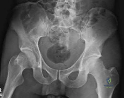
A 35-year-old male is brought in after a motorcycle accident with an unstable pelvis and hemodynamic shock. EMS placed a pelvic binder. Upon evaluation in the ED, the binder is noted to be centered at the level of the anterior superior iliac spines (ASIS). What is the appropriate next step regarding the binder?
Options:
- Leave it in place and proceed with fluid resuscitation
- Reposition the binder to be centered over the greater trochanters
- Remove the binder immediately to assess the skin
- Tighten the binder further to reduce the volume of the false pelvis
- Replace the binder with a C-clamp immediately before any imaging
Correct Answer: Leave it in place and proceed with fluid resuscitation
Explanation:
A pelvic binder must be centered over the greater trochanters, not the iliac crests or ASIS, to effectively close an open book pelvic ring injury (APC) and reduce the volume of the true pelvis. Placement over the iliac wings can potentially act as a fulcrum, exacerbating the deformity in certain injury patterns, and is less biomechanically effective at closing the symphysis.
Question 3:
A 25-year-old farmer sustains a severe open tibial fracture (Gustilo-Anderson IIIB) severely contaminated with soil and manure. According to standard guidelines regarding antibiotic prophylaxis for this specific mechanism, which of the following regimens is historically the most appropriate initial choice?
Options:
- First-generation cephalosporin alone
- First-generation cephalosporin and an aminoglycoside
- Third-generation cephalosporin alone
- First-generation cephalosporin, an aminoglycoside, and high-dose penicillin
- Vancomycin and metronidazole
Correct Answer: First-generation cephalosporin alone
Explanation:
For severe open fractures with heavy soil or barnyard contamination, there is a high risk for Clostridium perfringens (gas gangrene). Classic teaching and guidelines recommend adding high-dose penicillin for anaerobic coverage, in addition to a first-generation cephalosporin (for Gram-positives) and an aminoglycoside (for Gram-negatives). While modern protocols may use broad-spectrum agents like piperacillin/tazobactam, the combination involving penicillin remains a standard answer for farm injuries.
Question 4:
A 65-year-old female sustains a proximal humerus fracture. Radiographs and CT show a 4-part fracture with complete lateral hinge disruption and a short calcar segment (<8 mm). What is the most appropriate definitive surgical management?
Options:
- Non-operative management in a sling
- Open reduction and internal fixation with a locking plate
- Hemiarthroplasty
- Reverse total shoulder arthroplasty
- Percutaneous pinning
Correct Answer: Non-operative management in a sling
Explanation:
In elderly patients with a 4-part proximal humerus fracture, especially with strong predictors of humeral head ischemia (disrupted medial hinge, short calcar <8mm, disrupted anatomic neck), reverse total shoulder arthroplasty (RTSA) provides more predictable outcomes and better forward elevation than ORIF or hemiarthroplasty. RTSA relies on the deltoid and does not depend on tuberosity healing for basic overhead function, though tuberosity repair improves rotation.
Question 5:
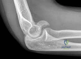
A 32-year-old male sustains a closed, spiral fracture of the distal third of his humeral shaft while arm wrestling. He is unable to actively extend his wrist or fingers in the emergency department. Which of the following is the most appropriate initial management?
Options:
- Immediate surgical exploration of the radial nerve and plate fixation
- Application of a coaptation splint and clinical observation of the nerve palsy
- Immediate intramedullary nailing of the humerus
- EMG/NCS in the emergency department prior to reduction
- External fixation
Correct Answer: Immediate surgical exploration of the radial nerve and plate fixation
Explanation:
A Holstein-Lewis fracture is a spiral fracture of the distal third of the humeral shaft. Radial nerve palsy occurs in up to 18% of humeral shaft fractures. Primary radial nerve palsy in a closed fracture is not an absolute indication for immediate surgical exploration, as over 85% will recover spontaneously. The standard of care is functional bracing/splinting and observation. Exploration is indicated if there is an open fracture, vascular injury, or if the palsy occurs *after* closed reduction.
Question 6:
A 22-year-old male presents with radial-sided wrist pain after a fall onto an outstretched hand. X-rays reveal a displaced fracture of the proximal pole of the scaphoid. What is the predominant blood supply to the scaphoid that makes this specific fracture pattern highly prone to nonunion and avascular necrosis?
Options:
- Volar carpal branch of the radial artery entering distally
- Dorsal carpal branch of the radial artery entering distally
- Volar carpal branch of the ulnar artery entering proximally
- Dorsal carpal branch of the ulnar artery entering proximally
- Intraosseous branches from the capitate
Correct Answer: Volar carpal branch of the radial artery entering distally
Explanation:
The scaphoid receives its primary blood supply (up to 80%) from the dorsal carpal branch of the radial artery, which enters the bone at the dorsal ridge (distal to the waist) and flows in a retrograde fashion to the proximal pole. Fractures at the waist or proximal pole disrupt this retrograde supply, leading to a high rate of avascular necrosis and nonunion for proximal pole fragments.
Question 7:
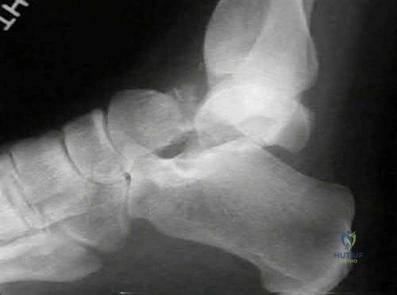
A 28-year-old male is involved in a high-speed MVA and sustains a closed fracture of the talar neck. Imaging shows a displaced fracture of the talar neck with subluxation of the subtalar joint, while the tibiotalar and talonavicular joints remain congruent. What is the Hawkins classification for this injury?
Options:
- Type I
- Type II
- Type III
- Type IV
- Type V
Correct Answer: Type I
Explanation:
The Hawkins classification is used for talar neck fractures. Type I: Non-displaced. Type II: Displaced with subtalar subluxation or dislocation. Type III: Displaced with subtalar and tibiotalar dislocation. Type IV: Displaced with subtalar, tibiotalar, and talonavicular dislocation. The scenario describes a Type II fracture, which carries an avascular necrosis risk of approximately 20-50%.
Question 8:
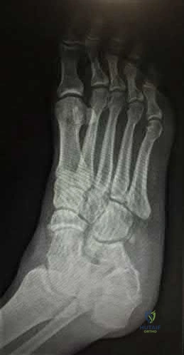
A 40-year-old male misses a step and presents with midfoot pain and swelling. X-rays show a small bony avulsion in the first intermetatarsal space ('Fleck sign'). Weight-bearing films demonstrate widening of the space between the 1st and 2nd metatarsal bases. What ligament is primarily injured?
Options:
- Plantar ligament connecting the 1st cuneiform to the 1st metatarsal
- Dorsal ligament connecting the 2nd cuneiform to the 2nd metatarsal
- Interosseous ligament connecting the medial cuneiform to the base of the 2nd metatarsal
- Interosseous ligament connecting the lateral cuneiform to the base of the 3rd metatarsal
- Plantar ligament connecting the navicular to the medial cuneiform
Correct Answer: Plantar ligament connecting the 1st cuneiform to the 1st metatarsal
Explanation:
The Lisfranc ligament is a strong interosseous ligament that runs obliquely from the lateral aspect of the medial cuneiform to the medial aspect of the base of the second metatarsal. It is the critical stabilizer of the midfoot arch. A 'fleck sign' represents a bony avulsion of this ligament and is pathognomonic for a Lisfranc injury.
Question 9:
A 30-year-old male sustains a displaced, highly vertical (Pauwels III) femoral neck fracture following a fall from a height. What is the most appropriate surgical management for this patient to minimize the risk of nonunion and optimize biomechanical stability?
Options:
- Immediate hemiarthroplasty
- Total hip arthroplasty
- Closed reduction and percutaneous pinning with 3 parallel cannulated screws
- Open reduction and internal fixation with a sliding hip screw (DHS) or cephalomedullary nail
- Non-operative management with skeletal traction
Correct Answer: Immediate hemiarthroplasty
Explanation:
In young adults, femoral neck fractures are considered a surgical emergency to preserve the native hip joint. A Pauwels III fracture is characterized by a vertical fracture line (high shear angle), making it highly unstable. Fixation with a fixed-angle device like a sliding hip screw (often with a derotational screw) or a cephalomedullary nail offers vastly superior biomechanical stability against vertical shear forces compared to parallel cannulated screws, reducing the high risk of nonunion.
Question 10:
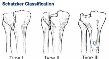
A 45-year-old female sustains a tibial plateau fracture. A CT scan reveals a depression of the lateral articular surface with an associated split fracture of the lateral condyle, as well as an avulsion fracture of the fibular head. Based on the Schatzker classification, what type of fracture is this, and what nerve is most at risk?
Options:
- Schatzker I, Common peroneal nerve
- Schatzker II, Common peroneal nerve
- Schatzker II, Tibial nerve
- Schatzker III, Saphenous nerve
- Schatzker IV, Common peroneal nerve
Correct Answer: Schatzker I, Common peroneal nerve
Explanation:
A split-depression fracture of the lateral tibial plateau is classified as a Schatzker II fracture. The concomitant avulsion of the fibular head suggests a lateral collateral ligament (LCL) injury or biceps femoris avulsion. The common peroneal nerve courses directly around the fibular neck and is at high risk of stretch or direct injury in this specific lateral-sided injury pattern.
Question 11:
Six weeks after an initially uncomplicated volar plating of a distal radius fracture, a 55-year-old female presents with a sudden inability to actively extend her thumb interphalangeal joint. What is the most likely cause of this complication?
Options:
- Attritional rupture of the Extensor Pollicis Longus (EPL) tendon
- Iatrogenic laceration of the Extensor Pollicis Brevis (EPB) tendon
- Posterior interosseous nerve (PIN) entrapment
- Flexor Pollicis Longus (FPL) tendon rupture
- De Quervain's tenosynovitis
Correct Answer: Attritional rupture of the Extensor Pollicis Longus (EPL) tendon
Explanation:
Attritional rupture of the Extensor Pollicis Longus (EPL) tendon is a well-known complication following distal radius fractures, both operatively and non-operatively treated. In the setting of volar plating, prominent dorsal screws that penetrate the dorsal cortex can act as a frictional saw, irritating and eventually rupturing the EPL tendon as it courses around Lister's tubercle in the third dorsal compartment.
Question 12:
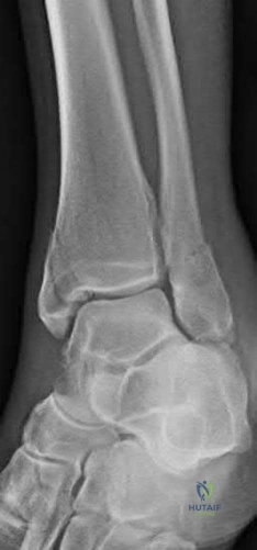
A patient twists their ankle during a soccer match. Radiographs reveal a short oblique fracture of the fibula extending posterosuperiorly from the joint line, and a transverse avulsion fracture of the medial malleolus. What is the most likely Lauge-Hansen classification for this injury mechanism?
Options:
- Supination-Adduction (SAD)
- Supination-External Rotation (SER)
- Pronation-Abduction (PAB)
- Pronation-External Rotation (PER)
- Pronation-Adduction (PAD)
Correct Answer: Supination-Adduction (SAD)
Explanation:
The Supination-External Rotation (SER) mechanism is the most common cause of ankle fractures. Stage I: Anterior inferior tibiofibular ligament (AITFL) sprain. Stage II: Short oblique or spiral fracture of the distal fibula. Stage III: Posterior inferior tibiofibular ligament (PITFL) tear or posterior malleolus fracture. Stage IV: Transverse fracture of the medial malleolus or deltoid ligament rupture. The described pattern is a classic SER IV injury.
Question 13:
A 25-year-old male motorcyclist is struck by a car and sustains a 'floating knee' injury. Radiographs reveal a diaphyseal fracture of the femur and a proximal intra-articular fracture of the tibia extending into the plateau. How is this injury classified according to the Blake and McBryde (Fraser) classification?
Options:
- Type I
- Type IIA
- Type IIB
- Type IIC
- Type III
Correct Answer: Type I
Explanation:
The Fraser classification describes floating knee injuries. Type I: True diaphyseal fractures of both the femur and tibia. Type IIA: Diaphyseal tibia with an intra-articular fracture of the femur. Type IIB: Diaphyseal femur with an intra-articular fracture of the tibia. Type IIC: Intra-articular fractures involving both the femur and tibia. The scenario describes a Type IIB floating knee.
Question 14:
A 70-year-old female sustains a subtrochanteric femur fracture. Following the injury, the proximal fragment typically assumes a position of flexion, abduction, and external rotation. Which muscle is primarily responsible for the abduction deformity of the proximal fragment?
Options:
- Iliopsoas
- Gluteus medius
- Short external rotators
- Adductor longus
- Rectus femoris
Correct Answer: Iliopsoas
Explanation:
In a subtrochanteric femur fracture, the proximal fragment is highly influenced by strong muscle attachments. It is flexed by the iliopsoas (attaching to the lesser trochanter), abducted by the gluteus medius and minimus (attaching to the greater trochanter), and externally rotated by the short external rotators. The distal fragment is pulled proximally by the hamstrings and adducted by the adductor complex.
Question 15:
A 6-year-old boy falls off monkey bars and sustains a widely displaced extension-type supracondylar humerus fracture (Gartland Type III). During neurological examination, he is unable to actively flex his thumb interphalangeal joint and the distal interphalangeal joint of his index finger. Which nerve is most likely injured?
Options:
- Radial nerve
- Ulnar nerve
- Anterior Interosseous Nerve (AIN)
- Posterior Interosseous Nerve (PIN)
- Musculocutaneous nerve
Correct Answer: Radial nerve
Explanation:
The Anterior Interosseous Nerve (AIN), a purely motor branch of the median nerve, is the most commonly injured nerve in extension-type supracondylar humerus fractures (often tented over the proximal fragment). Clinically, it presents with an inability to make the 'A-OK' sign due to weakness of the flexor pollicis longus (thumb IP flexion) and the flexor digitorum profundus to the index finger (index DIP flexion).
Question 16:
A hemodynamically unstable trauma patient presents with an Anteroposterior Compression (APC) Type III pelvic ring injury. Following the placement of a pelvic binder, the patient's blood pressure remains 70/40 mmHg despite receiving 2 units of uncrossmatched packed RBCs. A FAST scan is negative. What is the most appropriate next step in management?
Options:
- Retrograde urethrogram
- Pre-peritoneal pelvic packing and/or angioembolization
- Exploratory laparotomy
- Open reduction and internal fixation of the symphysis pubis
- Immediate application of a C-clamp
Correct Answer: Retrograde urethrogram
Explanation:
In a hemodynamically unstable patient with an unstable pelvic ring injury and a negative FAST (ruling out massive intra-abdominal hemorrhage), the source of life-threatening hemorrhage is assumed to be the pelvis (most commonly the presacral venous plexus, but also arterial branches). If mechanical stabilization (binder) and initial resuscitation fail, emergent intervention with pre-peritoneal pelvic packing and/or pelvic angiography with embolization is required.
Question 17:
A 40-year-old roofer falls from a ladder and sustains a severely displaced intra-articular calcaneus fracture (Sanders Type III). He is scheduled to undergo open reduction and internal fixation via an extensile lateral approach. Which complication is most frequently and classically associated with this specific surgical approach?
Options:
- Sural nerve neuroma
- Wound edge necrosis and dehiscence
- Tibial nerve entrapment
- Flexor hallucis longus (FHL) tendon laceration
- Avascular necrosis of the tuberosity
Correct Answer: Sural nerve neuroma
Explanation:
The extensile lateral approach to the calcaneus is notorious for severe soft tissue complications, specifically wound edge necrosis, dehiscence, and deep infection, occurring in 10-25% of cases. The flap relies heavily on the lateral calcaneal artery. Proper timing (waiting for the 'wrinkle sign'), careful subperiosteal elevation creating a full-thickness 'no-touch' flap, and minimal retraction are critical to mitigate this risk.
Question 18:
A 35-year-old male sustains a severe axial load injury to his flexed knee during a motor vehicle collision. A CT scan reveals an isolated coronal plane fracture of the lateral femoral condyle. What is the correct eponym for this fracture, and what is the generally accepted optimal trajectory for lag screw fixation?
Options:
- Segond fracture; Lateral to medial
- Barton fracture; Anterior to posterior
- Hoffa fracture; Anterior to posterior
- Hoffa fracture; Medial to lateral
- Tillaux fracture; Inferior to superior
Correct Answer: Segond fracture; Lateral to medial
Explanation:
A coronal plane fracture of the femoral condyle (most commonly lateral) is termed a Hoffa fracture. It is a highly unstable intra-articular shear injury. Biomechanical studies and surgical techniques recommend lag screw placement perpendicular to the fracture plane. Anterior-to-posterior (AP) screws (often countersunk beneath the articular cartilage) or posterior-to-anterior (PA) screws provide stable fixation. Medial-to-lateral screws would be parallel to the fracture and provide no compression.
Question 19:
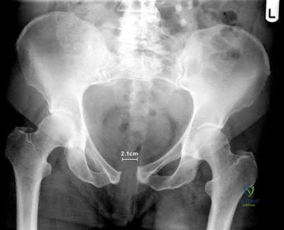
A 29-year-old male sustains an acetabular fracture in an MVC. Plain radiographs reveal a disruption of the iliopectineal line, a teardrop that is displaced medially, an intact ilioischial line, and an intact posterior wall. Based on the Judet-Letournel classification, which type of acetabular fracture is this?
Options:
- Posterior column
- Anterior column
- Transverse
- Both column
- T-shaped
Correct Answer: Posterior column
Explanation:
In radiographic evaluation of the acetabulum, the iliopectineal line represents the anterior column, and the ilioischial line represents the posterior column. Disruption of the iliopectineal line with an intact ilioischial line firmly indicates an isolated anterior column fracture. The radiographic teardrop is often displaced or disrupted in anterior column or anterior wall injuries.
Question 20:
A 24-year-old cyclist falls directly onto his shoulder and sustains a completely displaced midshaft clavicle fracture. Which of the following is considered an ABSOLUTE indication for acute open reduction and internal fixation?
Options:
- 1.5 cm of shortening
- 100% vertical displacement of the fragments
- Z-deformity with a comminuted butterfly fragment
- Skin tenting that threatens skin integrity
- Patient preference for a faster return to competitive sports
Correct Answer: 1.5 cm of shortening
Explanation:
Absolute indications for acute operative fixation of a clavicle fracture include: open fracture, associated neurovascular injury, skin tenting threatening to progress to an open fracture, and 'floating shoulder' (ipsilateral displaced scapular neck fracture). Factors like 100% displacement, shortening > 2 cm, and severe comminution are considered RELATIVE indications, often discussed with the patient regarding the risk of symptomatic nonunion versus surgical risks.
Question 21:
A 35-year-old polytrauma patient undergoes damage control orthopedics (DCO) with external fixation of bilateral femur fractures. Which parameter is the MOST reliable indicator that the patient has been adequately resuscitated and is physiologically optimized for definitive intramedullary nailing?
Options:
- Normalization of heart rate and mean arterial pressure
- Urine output consistently > 0.5 mL/kg/hr for 12 hours
- Serum lactate < 2.0 mmol/L and base deficit between -2 and +2
- Systemic vascular resistance > 800 dynes/sec/cm^-5
- Central venous pressure > 8 mmHg
Correct Answer: Normalization of heart rate and mean arterial pressure
Explanation:
Clearance of serum lactate (target < 2.0 mmol/L) and normalization of base deficit (between -2 and +2) are the most reliable indicators of adequate tissue perfusion and successful resuscitation in polytrauma patients. While heart rate, blood pressure, and urine output are useful clinical markers, they can normalize before cellular hypoxia has resolved (occult hypoperfusion).
Question 22:
In a patient presenting with severe traumatic brain injury (GCS 6) and an ipsilateral closed comminuted femur fracture, what is the major physiological rationale for performing damage control external fixation rather than early intramedullary nailing within the first 24 hours?
Options:
- To prevent the acceleration of severe heterotopic ossification in the thigh
- To avoid the 'second hit' of intraoperative hypoxia and hypotension which exacerbates secondary brain injury
- To definitively prevent systemic fat embolization syndrome
- To wait for clearance of the cervical spine prior to definitive positioning
- To prevent arrest of fracture callus formation due to the systemic inflammatory response
Correct Answer: To prevent the acceleration of severe heterotopic ossification in the thigh
Explanation:
In severe TBI, early total care (e.g., reamed intramedullary nailing) can act as a 'second hit' due to intraoperative hypotension, hypoxia, and fat embolization. These physiological insults significantly exacerbate secondary brain injury and increase intracranial pressure. Therefore, Damage Control Orthopedics (DCO) with rapid external fixation is often preferred to stabilize the fracture while minimizing surgical physiological burden.
Question 23:
A hemodynamically unstable 42-year-old male is brought in after a motorcycle crash. Pelvic radiographs demonstrate an anteroposterior compression (APC-III) pelvic ring injury. Where is the anatomically correct position to place a pelvic circumferential compression device (binder) for optimal reduction and hemorrhage control?
Options:
- Level of the anterior superior iliac spines (ASIS)
- Level of the iliac crests
- Level of the greater trochanters
- Midway between the umbilicus and the pubic symphysis
- Proximal third of the femurs
Correct Answer: Level of the anterior superior iliac spines (ASIS)
Explanation:
A pelvic binder must be placed at the level of the greater trochanters to effectively close the pelvic ring and reduce pelvic volume. Placement higher (e.g., at the iliac crests or ASIS) is a common error and fails to adequately close the symphysis pubis, and can sometimes paradoxically open the pelvic floor or impede abdominal access.
Question 24:
A 28-year-old sustains an ultra-low velocity knee dislocation during a sporting event. Following prompt closed reduction, the dorsalis pedis and posterior tibial pulses are palpable and symmetrical. The Ankle-Brachial Index (ABI) is calculated to be 0.85. What is the most appropriate next step in management?
Options:
- Serial clinical vascular examinations every 2 hours for 24 hours
- Immediate discharge with close outpatient orthopedic follow-up
- Immediate operative exploration of the popliteal artery
- CT angiography of the lower extremity
- Duplex ultrasound of the superficial femoral artery
Correct Answer: Serial clinical vascular examinations every 2 hours for 24 hours
Explanation:
In the setting of a knee dislocation, an ABI < 0.9 is highly indicative of a vascular injury, even if pulses are palpable. The appropriate next step is advanced imaging with CT angiography (CTA) to rule out an intimal flap or popliteal artery injury. If the ABI is > 0.9, serial examinations are appropriate.
Question 25:
A 40-year-old male sustains a Gustilo-Anderson Type IIIB open fracture of the distal third of the tibia. After serial debridements resulting in a clean wound bed with exposed bone devoid of periosteum, soft tissue coverage is required. Which of the following is the most appropriate soft tissue flap for this specific zone?
Options:
- Medial gastrocnemius rotational flap
- Soleus rotational flap
- Latissimus dorsi or Anterolateral thigh (ALT) free flap
- Sural artery cross-leg flap
- Fasciocutaneous advancement flap
Correct Answer: Medial gastrocnemius rotational flap
Explanation:
Coverage for open tibia fractures is classically divided by thirds: the proximal third is covered by the gastrocnemius flap, the middle third by the soleus flap, and the distal third typically requires a free tissue transfer (such as an ALT or latissimus dorsi flap) due to the lack of adequate local muscle bulk.
Question 26:
A 25-year-old male sustains a low-velocity civilian gunshot wound to the right thigh, resulting in a comminuted midshaft femur fracture. The entrance and exit wounds are 1 cm each, with no expanding hematoma or signs of ischemia. What is the most appropriate definitive management strategy?
Options:
- Immediate formal open debridement of the fracture site and skeletal traction
- Local wound care, intravenous antibiotics, and intramedullary nailing without formal track debridement
- Formal track debridement, external fixation, and delayed primary closure
- Local wound care, tetanus prophylaxis, and nonoperative management in a spica cast
- Exploratory laparotomy and external fixation
Correct Answer: Immediate formal open debridement of the fracture site and skeletal traction
Explanation:
Low-velocity gunshot wounds resulting in femur fractures without intra-articular extension or significant vascular injury are treated similarly to closed fractures. Management involves local wound care (superficial debridement), antibiotics, and definitive fixation, typically with an intramedullary nail. Formal open debridement of the bullet track is not routinely indicated.
Question 27:
According to the findings of the Lower Extremity Assessment Project (LEAP) study, which of the following factors was found to be the most reliable predictor of poor functional outcome at 2 years following a severe, limb-threatening lower extremity injury?
Options:
- The initial decision to pursue limb salvage rather than amputation
- An initial Mangled Extremity Severity Score (MESS) greater than 7
- The presence of an insensate plantar surface at initial presentation
- Lack of adequate soft-tissue coverage within 7 days
- Patient characteristics such as lower educational level, lack of private insurance, and poor social support
Correct Answer: The initial decision to pursue limb salvage rather than amputation
Explanation:
The LEAP study demonstrated that functional outcomes between limb salvage and amputation were statistically similar at 2 years. The strongest predictors of poor functional outcomes were not injury characteristics (like an insensate foot or MESS score), but rather patient demographic and socioeconomic factors, including lack of private insurance, lower educational level, poor social support, and smoking.
Question 28:
A 32-year-old male undergoes reamed intramedullary nailing for a closed tibial shaft fracture. In the recovery room, he complains of worsening, intractable leg pain. His diastolic blood pressure is 65 mmHg. The anterior compartment pressure is measured directly at 40 mmHg. What is the calculated "delta P" and the most appropriate next step?
Options:
- Delta P is 25 mmHg; perform urgent four-compartment fasciotomies.
- Delta P is 25 mmHg; elevate the leg above the level of the heart and observe.
- Delta P is 40 mmHg; administer intravenous mannitol and recheck in 1 hour.
- Delta P is 105 mmHg; remove the intramedullary nail and place an external fixator.
- Delta P is 25 mmHg; apply ice and prescribe patient-controlled analgesia.
Correct Answer: Delta P is 25 mmHg; perform urgent four-compartment fasciotomies.
Explanation:
Delta P is calculated as the Diastolic Blood Pressure minus the Compartment Pressure. Here, 65 mmHg - 40 mmHg = 25 mmHg. A Delta P of 30 mmHg or less (some sources say < 30 mmHg) is a widely accepted threshold indicating acute compartment syndrome requiring emergent fasciotomy. Elevation above the heart is contraindicated as it decreases arterial perfusion pressure.
Question 29:
In the staged management of severe pilon fractures (OTA/AO 43-C3), spanning external fixation is initially applied. What is the primary clinical indicator used to determine when it is safe to proceed with definitive open reduction and internal fixation (ORIF)?
Options:
- Normalization of serum inflammatory markers (CRP and ESR)
- Appearance of the "wrinkle sign" on the skin overlying the fracture
- Radiographic evidence of soft fracture callus
- Achievement of exactly 14 days post-injury
- Negative wound cultures from the external fixator pin sites
Correct Answer: Normalization of serum inflammatory markers (CRP and ESR)
Explanation:
Staged management of high-energy pilon fractures involves early spanning external fixation to allow for soft tissue recovery. The primary clinical indicator that soft tissue swelling has subsided enough to safely permit definitive ORIF (minimizing wound dehiscence risk) is the appearance of the "wrinkle sign" in the skin.
Question 30:
A 45-year-old male presents with a deep laceration over the 3rd metacarpophalangeal joint sustained during a fistfight (human bite/clenched fist injury). He presents 24 hours later with erythema and purulent drainage. Which organism is most specifically associated with this injury type, and what is the appropriate empiric antibiotic therapy?
Options:
- Pasteurella multocida; Amoxicillin-clavulanate
- Eikenella corrodens; Amoxicillin-clavulanate
- Capnocytophaga canimorsus; Penicillin G
- Bartonella henselae; Azithromycin
- Staphylococcus aureus; Cephalexin
Correct Answer: Pasteurella multocida; Amoxicillin-clavulanate
Explanation:
Human bite wounds (clenched fist injuries) are characteristically associated with Eikenella corrodens, along with staph and strep species. The empiric oral antibiotic of choice is Amoxicillin-clavulanate. Pasteurella multocida is associated with dog and cat bites.
Question 31:
A 35-year-old male sustains a severe blunt force trauma to the lateral hip. Two weeks later, examination reveals a fluctuant, ballotable mass over the greater trochanter with overlying skin hypesthesia. Aspiration yields serosanguinous fluid. What is the specific pathophysiology of this lesion?
Options:
- Herniation of the tensor fasciae latae muscle through a fascial defect
- Subperiosteal hematoma secondary to an occult intertrochanteric fracture
- Traumatic shearing separation of the subcutaneous tissue from the underlying deep fascia
- Laceration of the superior gluteal artery leading to a pseudoaneurysm
- Infection of the trochanteric bursa
Correct Answer: Herniation of the tensor fasciae latae muscle through a fascial defect
Explanation:
The patient has a Morel-Lavallée lesion, which is a closed degloving injury. The pathophysiology involves a severe shearing force that separates the subcutaneous fat from the underlying deep fascia, creating a potential space that fills with blood, lymph, and necrotic fat.
Question 32:
A 50-year-old smoker presents 8 months after nonoperative management of a midshaft humerus fracture. Radiographs demonstrate an atrophic nonunion with no bridging callus and pencil-point sclerosis of the fracture ends. Which of the following is the most critical component of surgical management for this specific type of nonunion?
Options:
- Rigid internal fixation to enhance mechanical stability alone
- Provision of biological stimulation (e.g., autologous bone grafting) coupled with rigid internal fixation
- Dynamization of the fracture site to stimulate micromotion and callus formation
- Noninvasive pulsed electromagnetic field (PEMF) bone stimulation
- Extracorporeal shock wave therapy (ESWT)
Correct Answer: Rigid internal fixation to enhance mechanical stability alone
Explanation:
An atrophic nonunion lacks the biological capacity to heal. Therefore, management requires BOTH biological stimulation (e.g., autologous iliac crest bone graft or RIA) and rigid mechanical stability. In contrast, a hypertrophic nonunion (elephant shoe appearance) has adequate biology but lacks mechanical stability, and can be treated with rigid fixation alone.
Question 33:
A 24-year-old motorcyclist presents after a high-speed collision. Clinical exam reveals massive swelling over the shoulder girdle, a pulseless ipsilateral upper extremity, and complete flaccid paralysis of the arm. Radiographs show significant lateral displacement of the scapula. What is the most immediate life-threatening complication associated with this injury pattern?
Options:
- Tension pneumothorax
- Exsanguinating hemorrhage from subclavian or axillary vessel disruption
- Irreversible brachial plexus avulsion leading to a flail limb
- Systemic fat embolism syndrome
- Cervical spine fracture-dislocation
Correct Answer: Tension pneumothorax
Explanation:
The diagnosis is scapulothoracic dissociation, characterized by lateral displacement of the scapula, complete brachial plexus avulsion, and severe vascular injury. The most immediate life-threatening complication is massive hemorrhage from avulsion/disruption of the subclavian or axillary vessels. Immediate operative control of hemorrhage or endovascular intervention is critical.
Question 34:
A 22-year-old male is recovering in the ICU 36 hours after sustaining bilateral closed femur fractures. He develops acute hypoxemia, confusion, and a non-blanching rash over his axilla and chest. According to Gurd and Wilson's criteria for Fat Embolism Syndrome (FES), which of his symptoms is considered a MAJOR criterion?
Options:
- Tachycardia > 120 bpm
- Pyrexia > 39.5°C
- Petechial rash
- Sudden drop in hemoglobin
- Oliguria
Correct Answer: Tachycardia > 120 bpm
Explanation:
Gurd and Wilson's criteria for Fat Embolism Syndrome classify symptoms into major and minor criteria. The major criteria are: 1) Respiratory insufficiency (hypoxemia), 2) Cerebral involvement (confusion/coma), and 3) Petechial rash (typically over the thorax, axilla, and conjunctiva). Tachycardia, fever, and drop in hemoglobin are minor criteria.
Question 35:
During a traumatic below-knee amputation, the surgeon is isolating the superficial peroneal nerve. To prevent the formation of a symptomatic stump neuroma, what is the widely recommended surgical technique for managing the nerve?
Options:
- Ligate the nerve with non-absorbable suture and leave it directly at the fascial incision line
- Cauterize the nerve ending and wrap it in a local skin flap
- Apply gentle distal traction, transect the nerve sharply, and allow it to retract deep into healthy soft tissues
- Inject the nerve stump with a heavy concentration of phenol
- Anastomose the distal end of the nerve to a nearby small vein
Correct Answer: Ligate the nerve with non-absorbable suture and leave it directly at the fascial incision line
Explanation:
To prevent a symptomatic stump neuroma in amputations, the standard technique is traction neurectomy. This involves applying gentle distal traction to the nerve, transecting it sharply, and allowing the proximal stump to retract deep into a healthy, well-cushioned muscle belly away from the scar, incision line, and weight-bearing areas.
Question 36:
In a hemodynamically borderline polytrauma patient, Damage Control Orthopedics (DCO) principles are being applied. Which of the following inflammatory markers has been most extensively validated as a strong predictor of the systemic inflammatory response magnitude and multiorgan failure, often used to guide the timing of definitive fracture fixation?
Options:
- Interleukin-1 (IL-1)
- Interleukin-6 (IL-6)
- Tumor Necrosis Factor-alpha (TNF-a)
- C-Reactive Protein (CRP)
- Erythrocyte Sedimentation Rate (ESR)
Correct Answer: Interleukin-1 (IL-1)
Explanation:
Interleukin-6 (IL-6) is the most heavily studied cytokine in trauma and orthopedics for measuring the systemic inflammatory response syndrome (SIRS). Its levels peak early and correlate tightly with the risk of multiorgan failure, making it a valuable marker when deciding if a "borderline" patient is ready to transition from DCO to early total care.
Question 37:
Deflation of a pneumatic tourniquet after 2 hours of inflation during a complex lower extremity reconstruction is most likely to cause which of the following acute physiological changes?
Options:
- Increase in core body temperature
- Increase in systemic vascular resistance
- Decrease in end-tidal carbon dioxide (ETCO2)
- Transient decrease in systemic blood pressure
- Decrease in serum potassium levels
Correct Answer: Increase in core body temperature
Explanation:
Upon tourniquet deflation, the sudden release of ischemic, acidotic blood containing vasoactive metabolites back into systemic circulation causes a transient decrease in systemic vascular resistance and blood pressure. It also causes a surge in end-tidal CO2 (due to metabolic acidosis) and a decrease in core body temperature.
Question 38:
A 30-year-old farmer sustains an open midshaft femur fracture (Gustilo-Anderson Type IIIA) from a tractor accident. The wound is heavily contaminated with soil and organic matter. In addition to a first-generation cephalosporin and an aminoglycoside, what specific antibiotic coverage must be added?
Options:
- High-dose Penicillin
- Clindamycin
- Vancomycin
- Metronidazole
- Doxycycline
Correct Answer: High-dose Penicillin
Explanation:
In the setting of a farm injury or heavy soil contamination, there is a high risk of Clostridium perfringens infection, which can lead to gas gangrene. The standard of care mandates the addition of high-dose Penicillin to the regimen of a cephalosporin and aminoglycoside.
Question 39:
A 28-year-old male suffers a severe crush injury to the foot. He presents with excruciating midfoot pain out of proportion to examination, tense swelling, and pain with passive toe extension. If a fasciotomy is deemed necessary for impending compartment syndrome, how many discrete fascial compartments are anatomically recognized in the foot?
Options:
Correct Answer: 4
Explanation:
The foot contains 9 recognized anatomical compartments: Medial, Lateral, Superficial Central, Deep Central (Calcaneal), and 4 Interosseous compartments. Standard releases are often performed via a dual dorsal incision approach or a combination of medial and dorsal incisions.
Question 40:
Secondary bone healing (healing by callus formation) is characterized by a sequential progression of tissue types following a fracture. What is the correct order of the predominant tissue types bridging the fracture gap during this physiological process?
Options:
- Hematoma -> Woven bone -> Cartilage -> Lamellar bone
- Hematoma -> Granulation tissue -> Cartilage -> Woven bone -> Lamellar bone
- Granulation tissue -> Hematoma -> Woven bone -> Cartilage -> Lamellar bone
- Hematoma -> Cartilage -> Granulation tissue -> Lamellar bone -> Woven bone
- Woven bone -> Hematoma -> Granulation tissue -> Cartilage -> Lamellar bone
Correct Answer: Hematoma -> Woven bone -> Cartilage -> Lamellar bone
Explanation:
Secondary fracture healing progresses sequentially through distinct phases: Inflammation (Hematoma followed by Granulation tissue), Soft Callus formation (predominantly Cartilage), Hard Callus formation (calcification into Woven bone), and finally Remodeling (replacement of woven bone with mature Lamellar bone along lines of stress).
Question 41:
A 25-year-old male is brought in after a high-speed motor vehicle collision. He is intubated, with a blood pressure of 85/50 mmHg, a serum lactate of 5.0 mmol/L, and a core temperature of 34°C. Secondary survey reveals a closed midshaft femur fracture. According to the principles of Damage Control Orthopedics (DCO), what is the most appropriate initial management for his femur fracture?
Options:
- Early total care with intramedullary nailing
- Immediate plate osteosynthesis
- Temporary external fixation
- Skeletal traction pin and bed rest only
- Primary amputation
Correct Answer: Early total care with intramedullary nailing
Explanation:
This patient is in extremis and exhibiting signs of the 'lethal triad' of trauma (hypothermia, acidosis, and coagulopathy, although coagulopathy is unstated, it is presumed). In an unstable polytrauma patient, Early Total Care (ETC) such as IM nailing provides a 'second hit' that can be fatal. Damage Control Orthopedics (DCO) principles dictate rapid, temporary stabilization with external fixation to control hemorrhage and stabilize soft tissues, allowing for physiologic resuscitation in the ICU before definitive fracture care.
Question 42:
A motorcyclist presents after a severe accident with massive swelling around the shoulder girdle, a pulseless ipsilateral upper extremity, and a profoundly widened scapulothoracic articulation on a chest radiograph. What associated injury in this specific injury pattern carries the highest immediate mortality rate?
Options:
- Complete brachial plexus avulsion
- Subclavian or axillary artery disruption
- Massive tension pneumothorax
- Ipsilateral flail chest
- Traumatic brain injury
Correct Answer: Complete brachial plexus avulsion
Explanation:
The clinical presentation describes a scapulothoracic dissociation, which is characterized by complete disruption of the scapulothoracic articulation. The most life-threatening complication associated with this severe traction injury is a massive hemorrhage resulting from the disruption of the subclavian or axillary artery, leading to rapid exsanguination if not identified and treated emergently.
Question 43:
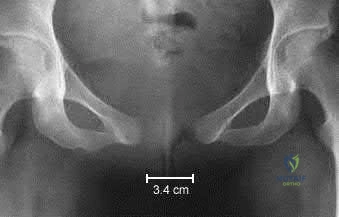
During an anterior intrapelvic (modified Stoppa) approach for the fixation of an anterior column acetabular fracture, massive hemorrhage occurs while dissecting over the superior pubic ramus. Which anatomical vascular anastomosis has likely been disrupted?
Options:
- Anastomosis between the obturator vessels and the external iliac or inferior epigastric vessels
- Anastomosis between the internal pudendal and the inferior gluteal vessels
- Anastomosis between the deep circumflex iliac and the ascending branch of the lateral femoral circumflex artery
- Anastomosis between the internal iliac and the median sacral vessels
- Anastomosis between the superior gluteal artery and the medial femoral circumflex artery
Correct Answer: Anastomosis between the obturator vessels and the external iliac or inferior epigastric vessels
Explanation:
The "corona mortis" (crown of death) is a significant vascular anastomosis between the obturator vessels (from the internal iliac system) and the external iliac or inferior epigastric vessels. It crosses the superior pubic ramus at an average distance of 5-6 cm from the symphysis pubis and is at high risk of iatrogenic injury during anterior intrapelvic exposures.
Question 44:
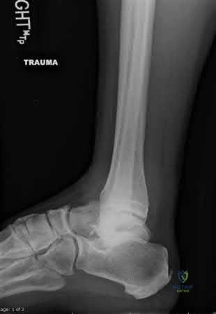
A 40-year-old construction worker falls from a height, sustaining a severe, closed OTA/AO 43-C3 pilon fracture with massive soft tissue swelling, fracture blisters, and extreme tension over the anterior ankle. What is the most appropriate initial, evidence-based management strategy?
Options:
- Immediate definitive open reduction and internal fixation (ORIF) with dual plating
- Joint-spanning external fixation across the ankle with leg elevation
- Placement of a calcaneal traction pin and delayed primary closure
- Primary below-knee amputation
- Fasciotomies of the lower leg followed by immediate ORIF
Correct Answer: Immediate definitive open reduction and internal fixation (ORIF) with dual plating
Explanation:
For high-energy, complex pilon fractures with severe soft tissue compromise, standard of care dictates a staged protocol. The first stage involves temporary joint-spanning external fixation (often with concurrent fibular fixation if anatomically feasible) to restore length and alignment while allowing soft tissue swelling to subside over 10-14 days. Definitive ORIF is performed once the soft tissue envelope recovers (evidenced by the 'wrinkle test').
Question 45:
A 30-year-old male sustains a high-energy trauma to the knee. A CT scan reveals a supracondylar distal femur fracture with an associated coronal plane shear fracture of the lateral femoral condyle (Hoffa fragment). Biomechanically, what is the most stable screw trajectory for independent lag screw fixation of this specific coronal fragment?
Options:
- Anterior-to-posterior placement
- Posterior-to-anterior placement
- Medial-to-lateral placement
- Lateral-to-medial placement
- Inferior-to-superior placement
Correct Answer: Anterior-to-posterior placement
Explanation:
A Hoffa fracture is a coronal shear fracture of the femoral condyle. When placing lag screws to fix this fragment, an anterior-to-posterior (AP) trajectory is biomechanically superior to a posterior-to-anterior (PA) trajectory. This is because AP screws engage the denser cortical bone of the posterior condyle, yielding significantly higher pull-out strength and compression across the fracture site.
Question 46:
A patient presents with a knee dislocation and an associated posteromedial tibial plateau fracture. Following closed reduction, the foot remains pulseless, and a CT angiogram confirms a complete occlusion of the popliteal artery. Irreversible skeletal muscle necrosis in the lower extremity typically begins after what duration of warm ischemia time?
Options:
- 2 hours
- 4 hours
- 6 hours
- 10 hours
- 12 hours
Correct Answer: 2 hours
Explanation:
In the setting of vascular occlusion (e.g., popliteal artery injury with knee dislocation), warm ischemia time is critical. Irreversible muscle necrosis begins at approximately 6 hours. Immediate vascular intervention is necessary to reestablish perfusion and limb salvage.
Question 47:
A Gustilo-Anderson Type IIIB open tibia fracture results in a 6x6 cm soft tissue defect overlying the middle third of the tibia, leaving cortical bone exposed. Following aggressive debridement, which of the following is the most reliable local rotational muscle flap for coverage of this specific defect?
Options:
- Medial gastrocnemius rotational flap
- Soleus rotational flap
- Reverse superficial sural artery flap
- Sural nerve fasciocutaneous flap
- Anterolateral thigh free flap
Correct Answer: Medial gastrocnemius rotational flap
Explanation:
For soft tissue coverage of the lower extremity, local options are categorized by thirds of the tibia. Proximal third defects are typically covered by the medial (or lateral) gastrocnemius flap. Middle third defects are optimally covered by a soleus muscle rotational flap. Distal third defects usually require a free tissue transfer or specialized reverse-flow local flaps.
Question 48:
A 'terrible triad' injury of the elbow involves an elbow dislocation, a radial head fracture, and a coronoid process fracture. When undertaking surgical repair, which of the following represents the standard, sequential order of fixation to progressively restore elbow stability?
Options:
- Coronoid fixation, followed by Radial head fixation/replacement, followed by Lateral collateral ligament (LCL) repair
- Radial head fixation/replacement, followed by Coronoid fixation, followed by LCL repair
- LCL repair, followed by Coronoid fixation, followed by Radial head fixation
- Coronoid fixation, followed by Medial collateral ligament (MCL) repair, followed by Radial head fixation
- LCL repair, followed by Radial head fixation, followed by Medial collateral ligament repair
Correct Answer: Coronoid fixation, followed by Radial head fixation/replacement, followed by Lateral collateral ligament (LCL) repair
Explanation:
The standard surgical protocol for a terrible triad injury involves a systematic approach working deep to superficial and typically from medial (via deep exposure through lateral side) to lateral. The order is: 1) Coronoid fixation or capsular repair, 2) Radial head fixation or arthroplasty, and 3) Lateral collateral ligament (LUCL) complex repair to the lateral epicondyle.
Question 49:
A 25-year-old male falls onto an outstretched hand. A lateral radiograph of the wrist demonstrates a dorsally dislocated capitate, while the lunate is displaced volarly into the carpal tunnel, resembling a 'spilled teacup'. Which specific nerve deficit is most commonly associated with this acute injury pattern?
Options:
- Inability to forcefully extend the thumb IP joint
- Numbness over the dorsal first web space
- Weakness in finger abduction and adduction
- Numbness over the volar aspect of the thumb, index, and middle fingers
- Inability to flex the distal phalanx of the thumb
Correct Answer: Inability to forcefully extend the thumb IP joint
Explanation:
The injury described is a lunate dislocation (volar displacement of the lunate). The displaced lunate protrudes into the carpal tunnel, frequently compressing the median nerve. This results in acute carpal tunnel syndrome, characterized by numbness and tingling in the median nerve distribution (volar aspect of thumb, index, middle, and radial half of the ring finger).
Question 50:
A polytrauma patient develops a massive, tense thigh following a blunt crush injury. Thigh compartment pressures are measured at 45 mmHg with a diastolic blood pressure of 60 mmHg. If surgical fasciotomy of the thigh is indicated, which compartment is most commonly the primary source of the compartment syndrome?
Options:
- Posterior compartment
- Medial compartment
- Anterior compartment
- Lateral compartment
- Adductor compartment
Correct Answer: Posterior compartment
Explanation:
While compartment syndrome of the thigh is relatively rare compared to the leg, when it occurs, the anterior compartment (containing the quadriceps) is the most frequently affected. A standard lateral incision is typically utilized to decompress the anterior compartment.
Question 51:
A 45-year-old patient presents with abdominal colic, unexplained anemia, peripheral neuropathy, and a history of a gunshot wound to the hip 10 years ago. Radiographs reveal dissolution of a retained intra-articular bullet in the hip joint. What is the pathophysiologic explanation for this presentation?
Options:
- Copper toxicity causing localized nerve necrosis
- Systemic lead absorption facilitated by synovial fluid acting as a solvent
- Late-onset subacute pyogenic arthritis secondary to bullet retention
- Metallosis-induced heterotopic ossification
- Intra-articular mechanical wear debris leading to systemic inflammatory response
Correct Answer: Copper toxicity causing localized nerve necrosis
Explanation:
Bullets retained within synovial joints or bursae are continuously bathed in synovial fluid. The fluid acts as a mild solvent, dissolving the lead over time, which is then systemically absorbed. This can lead to classic symptoms of plumbism (systemic lead toxicity), including abdominal colic, encephalopathy, anemia, and neuropathies. For this reason, intra-articular bullets are an absolute indication for removal.
Question 52:
A large-for-gestational-age infant is delivered via vaginal birth complicated by severe shoulder dystocia. On physical examination, the neonate has an asymmetric Moro reflex and palpable crepitus over the right clavicle. No other neurological deficits are noted. What is the standard orthopedic management?
Options:
- Surgical open reduction and internal fixation
- Rigid figure-of-eight bracing for 4 weeks
- Reassurance and non-operative management with the sleeve pinned to the shirt
- Application of a unilateral shoulder spica cast
- Closed reduction under sedation and casting
Correct Answer: Surgical open reduction and internal fixation
Explanation:
Neonatal clavicle fractures secondary to birth trauma possess immense remodeling potential. They heal rapidly without functional deficits. The standard of care is entirely non-operative, consisting of parental reassurance, gentle handling, and minimizing arm movement by pinning the infant's sleeve to their shirt for a week or two.
Question 53:
A 78-year-old woman with a cemented total hip arthroplasty placed 10 years ago sustains a mechanical fall. Radiographs demonstrate a displaced spiral fracture of the femur occurring entirely distal to the tip of her well-fixed femoral stem. According to the Vancouver classification, how is this fracture categorized?
Options:
- Vancouver Type A
- Vancouver Type B1
- Vancouver Type B2
- Vancouver Type C
- Vancouver Type B3
Correct Answer: Vancouver Type A
Explanation:
The Vancouver classification for periprosthetic hip fractures is based on location and stem stability. Type A is in the trochanteric region. Type B fractures are around or just below the stem (B1 = well fixed, B2 = loose, B3 = loose with poor bone stock). Type C fractures occur well below the tip of the femoral stem. Since this fracture is entirely distal to the stem, it is a Vancouver Type C.
Question 54:
Following a high-energy dashboard injury, a patient is diagnosed with a posterior wall acetabular fracture and posterior hip dislocation. Post-reduction, the patient exhibits a foot drop and is unable to dorsiflex the toes. Which neural structure is most characteristically injured in this trauma mechanism?
Options:
- Tibial division of the sciatic nerve
- Peroneal (fibular) division of the sciatic nerve
- Femoral nerve
- Sural nerve
- Superior gluteal nerve
Correct Answer: Tibial division of the sciatic nerve
Explanation:
The sciatic nerve is at high risk during posterior hip dislocations and posterior wall acetabular fractures. The peroneal (fibular) division is disproportionately affected compared to the tibial division due to its lateral and more fixed anatomical position, tethering it as the femoral head displaces posteriorly.
Question 55:
A patient is evaluated 8 weeks after surgical fixation of a Hawkins Type II talar neck fracture. An AP radiograph of the ankle demonstrates a subchondral radiolucent band extending across the dome of the talus. What is the clinical significance of this radiographic finding?
Options:
- It represents early-stage avascular necrosis (AVN) of the talar body
- It confirms the development of a talar neck nonunion
- It is an indicator of preserved or re-established blood supply to the talar body
- It is a pathognomonic sign of intra-articular osteomyelitis
- It suggests rapid chondrolysis of the ankle joint
Correct Answer: It represents early-stage avascular necrosis (AVN) of the talar body
Explanation:
The presence of a subchondral radiolucent band in the talar dome at 6-8 weeks post-injury is known as the Hawkins sign. This radiolucency represents subchondral bone atrophy secondary to hyperemic bone resorption. It is an excellent prognostic sign indicating that the talar body is vascularized, thereby ruling out avascular necrosis (AVN).
Question 56:
A 35-year-old roofer falls and lands directly on his heels, sustaining a severely displaced intra-articular calcaneus fracture. On the lateral foot radiograph, Bohler's angle is measured at 5 degrees. What is the normal anatomic range for Bohler's angle?
Options:
- 0 to 10 degrees
- 10 to 20 degrees
- 20 to 40 degrees
- 40 to 60 degrees
- -10 to 0 degrees
Correct Answer: 0 to 10 degrees
Explanation:
Bohler's angle is formed by the intersection of a line drawn from the highest point of the anterior process to the highest point of the posterior facet, and a line drawn from the highest point of the posterior facet to the superior edge of the calcaneal tuberosity. The normal range is 20 to 40 degrees. A decrease in this angle indicates collapse of the posterior facet.
Question 57:
A collegiate football player sustains an axial load and twisting injury to a plantarflexed foot. Weight-bearing radiographs reveal a 3 mm diastasis between the base of the first and second metatarsals. What is the primary ligamentous stabilizer of this joint complex?
Options:
- Dorsal Lisfranc ligament
- Interosseous Lisfranc ligament
- Plantar Lisfranc ligament
- Spring ligament
- Bifurcate ligament
Correct Answer: Dorsal Lisfranc ligament
Explanation:
The Lisfranc ligament complex stabilizes the midfoot. The interosseous Lisfranc ligament, connecting the medial cuneiform to the base of the second metatarsal, is the largest, thickest, and primary stabilizer of the Lisfranc joint. The plantar ligament is the second strongest, while the dorsal ligament is the weakest.
Question 58:
A 24-year-old athlete sustains a fracture at the metaphyseal-diaphyseal junction of the 5th metatarsal (Jones fracture). This specific fracture pattern is notorious for a high risk of delayed union or nonunion. What anatomical factor is the primary reason for this complication?
Options:
- Constant avulsion forces from the inserting peroneus brevis tendon
- The fracture occurs in a vascular watershed area
- Overpull from the lateral band of the plantar fascia
- Lack of surrounding soft tissue envelope to provide secondary healing cells
- Excessive localized weight-bearing forces during the heel-strike phase
Correct Answer: Constant avulsion forces from the inserting peroneus brevis tendon
Explanation:
A true Jones fracture occurs at the metaphyseal-diaphyseal junction of the 5th metatarsal. The primary reason for the high nonunion rate is that this region is a vascular watershed area. The proximal portion is supplied by a retrograde metaphyseal artery, while the distal diaphysis is supplied by antegrade nutrient arteries, leaving the junction relatively avascular.
Question 59:
A hypotensive polytrauma patient presents in the emergency department following a crush injury. Pelvic radiographs show a significant anteroposterior compression (APC) injury with massive diastasis of the pubic symphysis. To optimally reduce the pelvic volume and help control venous hemorrhage, where should the pelvic binder be centered anatomically?
Options:
- Over the anterior superior iliac spines (ASIS)
- Over the iliac crests
- Over the greater trochanters
- Around the mid-thigh level
- Directly over the umbilicus
Correct Answer: Over the anterior superior iliac spines (ASIS)
Explanation:
To be biomechanically effective in closing the pelvic ring and reducing internal volume (thereby assisting in tamponade of venous bleeding), a pelvic binder must be applied directly centered over the greater trochanters. Application over the iliac crests can paradoxicially open the pelvic ring further or fail to provide adequate closure.
Question 60:
A 35-year-old agricultural worker sustains a severely contaminated open tibial shaft fracture (Gustilo-Anderson IIIA) after his leg is caught in a tractor. The wound contains significant amounts of soil and organic matter. Current prophylactic antibiotic guidelines recommend a first-generation cephalosporin, an aminoglycoside, and which additional antibiotic to cover specific soil-borne anaerobic pathogens?
Options:
- Oral Ciprofloxacin
- Intravenous Vancomycin
- High-dose Penicillin
- Intravenous Daptomycin
- Oral Azithromycin
Correct Answer: Oral Ciprofloxacin
Explanation:
In the setting of farm-related injuries or massive soil contamination, there is a significant risk of Clostridium perfringens infection (gas gangrene). Standard protocols recommend adding high-dose intravenous Penicillin (or Metronidazole for penicillin-allergic patients) for anaerobic coverage, in addition to the standard Gram-positive (cephalosporin) and Gram-negative (aminoglycoside) coverage.
Question 61:
A 25-year-old male presents after an MVC with closed bilateral femur fractures, pulmonary contusions, and a GCS of 9. His admission lactate is 4.5 mmol/L, pH is 7.20, and base excess is -8. What is the most appropriate management for his femur fractures?
Options:
- Bilateral reamed antegrade intramedullary nailing
- Bilateral unreamed antegrade intramedullary nailing
- Bilateral external fixation (Damage Control Orthopedics)
- Skeletal traction and observation in ICU
- Unilateral nailing of the more displaced side and external fixation of the other
Correct Answer: Bilateral reamed antegrade intramedullary nailing
Explanation:
In a borderline or unstable polytrauma patient (lactate >4.0, pH <7.24, base excess worse than -6), Damage Control Orthopedics (DCO) with external fixation is indicated to minimize the second hit of systemic inflammatory response (SIRS) associated with intramedullary nailing.
Question 62:
A 35-year-old male sustains an ankle fracture. Radiographs show a vertical medial malleolus fracture and a transverse lateral malleolus fracture below the level of the syndesmosis. According to the Lauge-Hansen classification, which of the following is the mechanism of injury?
Options:
- Supination-External Rotation
- Pronation-External Rotation
- Supination-Adduction
- Pronation-Abduction
- Pronation-Adduction
Correct Answer: Supination-External Rotation
Explanation:
The Supination-Adduction (SAD) injury pattern classically presents with a transverse fracture of the fibula below the level of the syndesmosis (Stage 1) and a vertical shear fracture of the medial malleolus (Stage 2).
Question 63:
A 78-year-old female presents with a periprosthetic femur fracture around a cemented polished taper slip hip stem. Radiographs show a fracture around the tip of the stem. The stem is loose, but the bone stock is adequate. What is the Vancouver classification and appropriate treatment?
Options:
- Vancouver B1; ORIF with locking plate and cables
- Vancouver B2; Revision to a long uncemented diaphyseal-fitting stem
- Vancouver B3; Revision with proximal femoral replacement
- Vancouver C; ORIF with locking plate
- Vancouver A; Nonoperative management
Correct Answer: Vancouver B1; ORIF with locking plate and cables
Explanation:
Vancouver B2 fractures occur around the stem with a loose implant but adequate bone stock. The standard treatment is revision arthroplasty using a long uncemented diaphyseal-engaging stem (often modular), bypassing the fracture site by at least 2 cortical diameters.
Question 64:
A 29-year-old male falls from a height and sustains a talar neck fracture with subluxation of the subtalar joint but a congruous tibiotalar and talonavicular joint. According to the Hawkins classification, what is the risk of avascular necrosis (AVN)?
Options:
- 0-10%
- 20-50%
- 70-100%
- 100%
- AVN does not occur in this pattern
Correct Answer: 0-10%
Explanation:
This is a Hawkins Type II fracture (talar neck fracture with subtalar dislocation). The risk of AVN for Type I is 0-10%, Type II is 20-50%, Type III (subtalar and tibiotalar dislocation) is 50-100%, and Type IV (Type III plus talonavicular dislocation) is near 100%.
Question 65:
A 22-year-old male is admitted with a closed tibia fracture. He complains of pain out of proportion to his injury. His diastolic blood pressure is 70 mmHg. Intracompartmental pressure testing of the anterior compartment yields a pressure of 45 mmHg. What is his Delta pressure, and what is the next best step?
Options:
- Delta pressure 25 mmHg; Urgent four-compartment fasciotomy
- Delta pressure 45 mmHg; Urgent four-compartment fasciotomy
- Delta pressure 25 mmHg; Elevation and observation
- Delta pressure 45 mmHg; Elevation and observation
- Delta pressure 70 mmHg; Urgent four-compartment fasciotomy
Correct Answer: Delta pressure 25 mmHg; Urgent four-compartment fasciotomy
Explanation:
Delta pressure = Diastolic BP - Intracompartmental pressure. Here, 70 - 45 = 25 mmHg. A Delta pressure of less than 30 mmHg is an objective indication for urgent fasciotomy in the setting of suspected acute compartment syndrome.
Question 66:
A 45-year-old male sustains a high-energy closed pilon fracture with severe soft tissue swelling and fracture blisters. What is the most appropriate initial management?
Options:
- Immediate open reduction and internal fixation of both tibia and fibula
- Immediate fibula fixation and spanning external fixation of the tibia
- Spanning external fixation and delayed ORIF of the tibia at 10-14 days once soft tissues permit
- Closed reduction and casting for 6 weeks
- Primary arthrodesis of the ankle
Correct Answer: Immediate open reduction and internal fixation of both tibia and fibula
Explanation:
For high-energy pilon fractures with significant soft tissue compromise, the standard of care is a staged approach: immediate joint-spanning external fixation followed by delayed definitive ORIF of the tibia once the 'wrinkle sign' appears, typically at 10-14 days, to minimize soft tissue complications.
Question 67:
A 30-year-old athlete complains of midfoot pain after a twisting injury. Non-weight-bearing radiographs appear normal. A 'fleck sign' is noted on careful inspection. What is the most appropriate next imaging step?
Options:
- MRI of the midfoot
- CT scan of the foot
- Weight-bearing radiographs of the foot
- Ultrasound of the Lisfranc ligament
- Bone scan
Correct Answer: MRI of the midfoot
Explanation:
In suspected Lisfranc injuries with normal non-weight-bearing radiographs, weight-bearing radiographs (anteroposterior and lateral) are the critical next step to evaluate for dynamic instability. A 'fleck sign' represents an avulsion of the Lisfranc ligament from the base of the second metatarsal.
Question 68:
A 50-year-old male falls off a ladder and sustains a bicondylar tibial plateau fracture with diaphyseal dissociation. What is the Schatzker classification, and what surgical approach is often required for the posteromedial fragment?
Options:
- Schatzker V; Anterolateral approach alone
- Schatzker VI; Dual approaches (Anterolateral and Posteromedial)
- Schatzker IV; Medial approach alone
- Schatzker VI; Single midline anterior approach
- Schatzker V; Dual approaches (Anterolateral and Posteromedial)
Correct Answer: Schatzker V; Anterolateral approach alone
Explanation:
A bicondylar tibial plateau fracture with complete dissociation of the metaphysis from the diaphysis is a Schatzker VI fracture. Because of the common posteromedial shear fragment, dual incisions (anterolateral and posteromedial) are frequently required to achieve stable fixation and avoid extensive soft tissue stripping.
Question 69:
A 40-year-old roof worker falls and sustains an intra-articular calcaneus fracture. The coronal CT scan shows a single primary fracture line through the posterior facet, dividing it into two pieces (one lateral, one medial). According to the Sanders classification, what type is this?
Options:
- Type I
- Type II
- Type III
- Type IV
- Type V
Correct Answer: Type I
Explanation:
The Sanders classification is based on coronal CT cuts of the posterior facet. Type I is non-displaced. Type II has one fracture line (two articular fragments). Type III has two fracture lines (three fragments). Type IV is highly comminuted (four or more fragments).
Question 70:
A 28-year-old restrained driver presents after an MVC with a posterior hip dislocation. A post-reduction CT scan reveals a posterior wall acetabular fracture involving 45% of the articular surface. The hip is stable on dynamic stress fluoroscopy. What is the most appropriate management?
Options:
- Skeletal traction for 6 weeks
- Open reduction and internal fixation (ORIF) via a Kocher-Langenbeck approach
- ORIF via an ilioinguinal approach
- Touch-down weight-bearing for 6 weeks
- Total hip arthroplasty
Correct Answer: Skeletal traction for 6 weeks
Explanation:
Posterior wall fractures involving >40-50% of the posterior articular surface are typically unstable and require surgical fixation. The Kocher-Langenbeck approach is standard. Even if the hip appears stable on initial examination, the large defect size is an indication for ORIF to prevent late subluxation.
Question 71:
A 35-year-old male sustains an open tibia fracture with a 12 cm laceration. After thorough surgical debridement, the bone can be adequately covered by local soft tissue without the need for a rotational or free flap. There is no arterial injury. What is the Gustilo-Anderson classification?
Options:
- Type II
- Type IIIA
- Type IIIB
- Type IIIC
- Type I
Correct Answer: Type II
Explanation:
A Gustilo-Anderson Type IIIA fracture is a high-energy open fracture with extensive soft tissue laceration or stripping, but adequate soft tissue coverage of the fractured bone. Type IIIB requires a flap for coverage, and Type IIIC involves an arterial injury requiring repair.
Question 72:
An 82-year-old female with a history of atrial fibrillation presents with a displaced femoral neck fracture. She is taking Apixaban (a direct factor Xa inhibitor). What is the optimal timing for surgery to minimize mortality and bleeding risk?
Options:
- Delay surgery for 5 days to allow complete washout of Apixaban
- Proceed with surgery within 48 hours; consider holding Apixaban for 24-48 hours depending on renal function
- Administer Vitamin K and Fresh Frozen Plasma, then proceed to surgery immediately
- Administer Protamine sulfate and proceed to surgery immediately
- Perform non-operative management only
Correct Answer: Delay surgery for 5 days to allow complete washout of Apixaban
Explanation:
Current guidelines advocate surgery within 48 hours for geriatric hip fractures. For patients on direct oral anticoagulants like Apixaban, surgery can typically be safely performed after holding the medication for 24-48 hours. Vitamin K and FFP do not reverse DOACs.
Question 73:
A 34-year-old male sustains a distal femur fracture. CT imaging reveals a coronal plane fracture of the lateral femoral condyle. What is the eponym for this fracture, and what is the optimal fixation strategy?
Options:
- Barton fracture; Anteroposterior lag screws
- Hoffa fracture; Posteroanterior lag screws
- Chopart fracture; Medial-to-lateral lag screws
- Segond fracture; Suture anchors
- Tillaux fracture; K-wires
Correct Answer: Barton fracture; Anteroposterior lag screws
Explanation:
A coronal plane fracture of the femoral condyle is a Hoffa fracture. It must be fixed with screws placed perpendicular to the fracture line. Posterior-to-anterior (PA) screws have been shown biomechanically to provide stronger fixation than AP screws.
Question 74:
A 26-year-old football player sustains a multi-ligamentous knee injury that spontaneously reduced. Distal pulses are palpable. What is the most appropriate next step to evaluate for vascular injury?
Options:
- Immediate surgical exploration of the popliteal artery
- Ankle-Brachial Index (ABI); if >0.9, observe
- Venous duplex ultrasound
- Angiography in all cases regardless of exam
- Ankle-Brachial Index (ABI); if <0.9, CT angiography
Correct Answer: Immediate surgical exploration of the popliteal artery
Explanation:
In knee dislocations, initial assessment should include an ABI. If the ABI is >0.9, serial examinations are generally sufficient. If the ABI is <0.9 or if there are asymmetric pulses, CT angiography (CTA) is mandated.
Question 75:
A 45-year-old smoker presents 9 months after an intramedullary nailing of a tibial shaft fracture with persistent pain. Radiographs show no bridging callus, rounding of the fracture ends, and a persistent fracture gap. What is the primary cause of this nonunion, and what is the required surgical strategy?
Options:
- Inadequate stability; requires exchange nailing only
- Inadequate biology; requires debridement, bone grafting, and potentially exchange nailing
- Infection; requires 6 weeks of IV antibiotics only
- Hypertrophic nonunion; requires dynamization
- Excessive strain; requires application of a circular frame without grafting
Correct Answer: Inadequate stability; requires exchange nailing only
Explanation:
The radiographic appearance (no callus, rounded fracture ends) is classic for an atrophic nonunion, representing a failure of biology. Treatment requires improving the biological environment through debridement, adding bone graft, and ensuring stable fixation.
Question 76:
A 60-year-old female is struck by a vehicle from the side. Pelvic radiographs demonstrate a transverse fracture of the pubic rami and an impaction fracture of the anterior sacrum on the same side. What type of injury is this, and what is the typical initial management if she is hemodynamically stable?
Options:
- APC-II; Application of a pelvic binder
- LC-I; Mobilization with weight-bearing as tolerated
- VS; Immediate application of an external fixator
- LC-III; Skeletal traction
- APC-III; Angioembolization
Correct Answer: APC-II; Application of a pelvic binder
Explanation:
This is a Lateral Compression Type I (LC-I) injury, characterized by transverse pubic rami fractures and a sacral impaction fracture on the side of impact. In a stable patient, these are generally rotationally and vertically stable and treated with mobilization and weight-bearing as tolerated.
Question 77:
A patient with multiple severe midfoot crush injuries presents with a tense, swollen foot and severe pain with passive toe dorsiflexion. If foot fasciotomies are indicated, what surgical approaches are commonly utilized to release all 9 compartments?
Options:
- A single dorsal midline incision
- Two dorsal incisions (over the 2nd and 4th metatarsals) and a medial utility incision
- A single plantar transverse incision
- Medial and lateral sub-malleolar incisions
- Four intermetatarsal plantar incisions
Correct Answer: A single dorsal midline incision
Explanation:
The foot has 9 compartments. The classic surgical approach includes two longitudinal dorsal incisions to release the interosseous compartments, and a medial utility incision to release the deep and superficial plantar compartments.
Question 78:
A 30-year-old female sustains a transverse patella fracture with 4 mm of displacement. She undergoes ORIF with tension band wiring. What biomechanical principle allows this construct to promote fracture healing?
Options:
- It converts tensile forces on the anterior surface into compressive forces at the articular surface during knee flexion
- It acts as a static neutralization plate
- It applies rigid compression via lag screw technique only
- It distracts the fracture to allow secondary bone healing
- It acts as a buttress against shear forces
Correct Answer: It converts tensile forces on the anterior surface into compressive forces at the articular surface during knee flexion
Explanation:
The principle of the tension band is to convert tensile forces into compressive forces. Placing the tension band on the anterior (tension) surface ensures that when the knee flexes, it resists distraction and converts the force into compression at the posterior articular surface.
Question 79:
A 22-year-old basketball player presents with lateral foot pain after a pivot injury. Radiographs reveal a transverse fracture at the metaphyseal-diaphyseal junction of the 5th metatarsal, extending into the 4th-5th intermetatarsal articulation. What is the diagnosis, and why is surgical fixation often recommended in elite athletes?
Options:
- Avulsion fracture; high risk of nonunion
- Jones fracture; high risk of delayed union/nonunion due to the watershed blood supply
- Pseudo-Jones fracture; early return to play
- Stress fracture of the diaphysis; high risk of infection
- Lisfranc injury; high risk of arch collapse
Correct Answer: Avulsion fracture; high risk of nonunion
Explanation:
A fracture at the metaphyseal-diaphyseal junction of the 5th metatarsal is a Jones fracture. This area represents a vascular watershed zone, making these fractures prone to nonunion. In elite athletes, early intramedullary screw fixation is recommended to decrease time to union.
Question 80:
A 24-year-old cyclist falls onto his shoulder. Radiographs show a completely displaced, shortened (>2 cm) midshaft clavicle fracture with skin tenting that does not blanch. What is the most definitive indication for immediate surgical intervention in this scenario?
Options:
- Midshaft location
- Patient age
- >2 cm shortening
- Impending open fracture (skin tenting with ischemia)
- Sports participation
Correct Answer: Midshaft location
Explanation:
While >2 cm of shortening is a relative indication for surgery to improve outcomes, an 'impending open fracture' characterized by severe skin tenting with blanching/ischemia is an absolute indication for urgent ORIF to prevent skin necrosis and conversion to an open fracture.
Question 81:
In the context of Damage Control Orthopedics (DCO) for a polytrauma patient, the 'two-hit hypothesis' is a widely accepted pathophysiological model. Which of the following best describes the 'first hit' and 'second hit' respectively?
Options:
- First hit: Initial traumatic insult; Second hit: Extensive definitive surgery (e.g., intramedullary nailing)
- First hit: Hemorrhage; Second hit: Hypoxia
- First hit: Initial surgical intervention; Second hit: Post-operative sepsis
- First hit: Systemic Inflammatory Response Syndrome (SIRS); Second hit: Nosocomial infection
- First hit: Fat embolism; Second hit: Acute Respiratory Distress Syndrome (ARDS)
Correct Answer: First hit: Initial traumatic insult; Second hit: Extensive definitive surgery (e.g., intramedullary nailing)
Explanation:
The 'two-hit hypothesis' in polytrauma proposes that the initial trauma ('first hit') primes the patient's immune system, leading to a systemic inflammatory response. A subsequent major physiological stressor, such as lengthy, definitive orthopedic surgery like reamed intramedullary nailing ('second hit'), can exacerbate this inflammatory cascade, precipitating Acute Respiratory Distress Syndrome (ARDS), Multiple Organ Dysfunction Syndrome (MODS), and potentially death. This is the core rationale for Damage Control Orthopedics (DCO) over Early Total Care (ETC) in borderline or unstable patients.
Question 82:
A hemodynamically unstable 45-year-old male arrives in the trauma bay following a high-speed motor vehicle collision. He has a mechanically unstable pelvic ring injury (APC Type III). Despite placement of a pelvic binder and aggressive fluid/blood resuscitation, he remains hypotensive. FAST exam is negative. What is the most common anatomical source of retroperitoneal hemorrhage in this specific injury pattern?
Options:
- Superior gluteal artery
- Presacral venous plexus and bleeding from cancellous bone surfaces
- Internal pudendal artery
- Corona mortis
- External iliac vein
Correct Answer: Presacral venous plexus and bleeding from cancellous bone surfaces
Explanation:
The vast majority (80-90%) of bleeding in severe pelvic ring disruptions is of venous or osseous origin, most commonly from the presacral venous plexus and the raw cancellous bone ends of the fractured pelvis. While arterial bleeding (such as from the superior gluteal artery or internal pudendal artery) can occur and is often the target for angioembolization, it accounts for only 10-20% of cases. Venous and osseous bleeding is initially managed by reducing pelvic volume (pelvic binder) and may require preperitoneal pelvic packing if hemodynamic instability persists.
Question 83:
A 30-year-old construction worker sustains an isolated Gustilo-Anderson Type IIIA open tibial shaft fracture. Assuming the patient receives appropriate early intravenous antibiotics in the emergency department, what does current orthopedic trauma literature dictate regarding the optimal timing of the initial surgical debridement?
Options:
- Mandatory debridement within 6 hours to significantly reduce the risk of osteomyelitis
- Debridement within 24 hours has comparable infection rates to debridement within 6 hours
- Immediate soft tissue coverage (flap) is required within 12 hours regardless of wound evolution
- Debridement can be safely delayed past 48 hours without any increase in infection risk
- Timing of debridement is irrelevant as long as local antibiotic beads are placed
Correct Answer: Debridement within 24 hours has comparable infection rates to debridement within 6 hours
Explanation:
Historically, the '6-hour rule' was strictly taught for open fractures. However, modern evidence, including multiple systematic reviews and large prospective cohort studies, demonstrates that if appropriate early intravenous antibiotics are administered, surgical debridement within 24 hours does not increase the risk of deep infection or nonunion compared to debridement within 6 hours. This allows for safe optimization of the patient and performance of the procedure during daylight hours by experienced personnel.
Question 84:
An intubated, obtunded polytrauma patient is admitted to the ICU with a severely comminuted closed tibial shaft fracture. The nursing staff notes a tense calf. Which of the following criteria is the most widely accepted and accurate threshold for diagnosing acute compartment syndrome in this obtunded patient and indicating a four-compartment fasciotomy?
Options:
- Absolute compartment pressure > 20 mmHg
- Absolute compartment pressure > 30 mmHg
- Delta pressure (Diastolic Blood Pressure - Compartment Pressure) < 30 mmHg
- Delta pressure (Mean Arterial Pressure - Compartment Pressure) < 30 mmHg
- Delta pressure (Systolic Blood Pressure - Compartment Pressure) < 40 mmHg
Correct Answer: Delta pressure (Diastolic Blood Pressure - Compartment Pressure) < 30 mmHg
Explanation:
In obtunded or unexaminable patients, objective compartment pressure monitoring is required. The most accurate predictor of acute compartment syndrome is the delta pressure (∆P), defined as the Diastolic Blood Pressure minus the Intra-compartmental Pressure. A delta pressure of less than 30 mmHg indicates inadequate tissue perfusion and is the standard threshold to proceed with fasciotomy. Relying on absolute pressure alone can lead to unnecessary fasciotomies, as normal perfusion gradients vary with the patient's systemic blood pressure.
Question 85:
During a massive transfusion protocol (MTP) for a patient with bilateral femoral shaft fractures and a crushed pelvis, the anesthesiologist notes worsening hypotension and coagulopathy. Which of the following electrolyte abnormalities is an expected complication of massive transfusion that directly exacerbates coagulopathy and requires active replacement?
Options:
- Hyperkalemia
- Hypocalcemia
- Hypernatremia
- Hypomagnesemia
- Hypercalcemia
Correct Answer: Hypocalcemia
Explanation:
Hypocalcemia is a frequent and dangerous complication of massive blood transfusion. Banked blood products contain citrate, which is used as an anticoagulant. Citrate chelates serum calcium in the patient's bloodstream, leading to profound hypocalcemia. Calcium is a vital cofactor (Factor IV) in the coagulation cascade. Its depletion exacerbates coagulopathy and impairs myocardial contractility (worsening shock), making proactive calcium administration critical during MTP.
Question 86:
The Mangled Extremity Severity Score (MESS) was developed to help surgeons objectively differentiate between limbs that are salvageable and those that require primary amputation. Which of the following variables is NOT a component of the MESS evaluation?
Options:
- Skeletal and soft-tissue injury extent
- Limb ischemia duration and severity
- Patient age
- Presence of a severe head injury (GCS < 8)
- Systemic hypotension (Shock)
Correct Answer: Presence of a severe head injury (GCS < 8)
Explanation:
The Mangled Extremity Severity Score (MESS) comprises four distinct categories: 1) Skeletal / Soft-tissue injury (energy level), 2) Limb ischemia, 3) Shock (systemic hemodynamics), and 4) Patient age. A score of 7 or higher historically suggested amputation, though modern limb salvage techniques have altered its predictive value. The presence of an isolated severe head injury or Glasgow Coma Scale (GCS) is not a component of the MESS.
Question 87:
A 22-year-old male sustains an isolated, closed midshaft femur fracture. Due to operating room unavailability, he is placed in skeletal traction overnight. 36 hours later, he develops acute confusion, tachypnea, and a petechial rash over his axilla and conjunctiva. Which of the following interventions has definitively been shown to reduce the incidence of this specific syndrome?
Options:
- Administration of high-dose systemic corticosteroids on admission
- Early operative stabilization of the long-bone fracture (within 24 hours)
- Placement of a prophylactic inferior vena cava (IVC) filter
- Aggressive early administration of fresh frozen plasma
- Empiric therapeutic anticoagulation with intravenous heparin
Correct Answer: Early operative stabilization of the long-bone fracture (within 24 hours)
Explanation:
The patient is presenting with the classic triad of Fat Embolism Syndrome (FES): hypoxemia, neurological abnormalities, and a petechial rash. The most effective, evidence-based strategy to prevent FES in patients with long-bone fractures is early operative stabilization (ideally within 24 hours). While corticosteroids have been studied and may reduce the incidence of hypoxemia in some trials, they are not universally recommended as standard prophylaxis over early fracture fixation.
Question 88:
An 82-year-old female sustains an open distal femur fracture. Her medical history includes atrial fibrillation, for which she takes rivaroxaban (a direct Factor Xa inhibitor). She requires urgent surgical debridement and stabilization. If she experiences severe, life-threatening hemorrhage during surgery, what is the most specific and appropriate reversal agent?
Options:
- Idarucizumab
- Andexanet alfa
- Protamine sulfate
- Vitamin K and Fresh Frozen Plasma
- Desmopressin (DDAVP)
Correct Answer: Andexanet alfa
Explanation:
Rivaroxaban (Xarelto) and apixaban (Eliquis) are direct Factor Xa inhibitors. The specific reversal agent for life-threatening bleeding in patients taking these medications is Andexanet alfa, a recombinant modified human Factor Xa decoy protein. Idarucizumab is the specific reversal agent for dabigatran (a direct thrombin inhibitor). Protamine sulfate reverses heparin, and Vitamin K/FFP (or Prothrombin Complex Concentrate) is used for warfarin reversal.
Question 89:
An obtunded polytrauma patient (GCS 6) has a high-quality, multi-detector CT scan of the cervical spine with multiplanar reconstructions that is completely normal. According to current Eastern Association for the Surgery of Trauma (EAST) guidelines, what is the most appropriate next step regarding the patient's cervical spine precautions?
Options:
- Maintain the cervical collar until an MRI can be obtained to rule out ligamentous injury
- Maintain the cervical collar until the patient can be extubated and is alert for a clinical exam
- Remove the cervical collar based solely on the normal high-quality CT scan
- Perform dynamic flexion-extension fluoroscopy in the intensive care unit
- Transition from a rigid collar to a soft collar for 6 weeks
Correct Answer: Remove the cervical collar based solely on the normal high-quality CT scan
Explanation:
Current EAST guidelines state that in an obtunded trauma patient, a normal, high-quality multi-detector CT scan is sufficient to clear the cervical spine. The incidence of clinically significant ligamentous injuries missed by a modern high-quality CT scan is exceedingly low. Prolonged use of cervical collars in obtunded patients leads to significant morbidity, including pressure ulcers, increased intracranial pressure, and impaired venous return. Therefore, collar removal without an MRI is recommended.
Question 90:
Based on the landmark CRASH-2 trial, what is the optimal dosing regimen and timing for the administration of Tranexamic Acid (TXA) to confer a survival benefit in bleeding trauma patients?
Options:
- 1g loading dose over 10 minutes followed by 1g infusion over 8 hours, administered within 3 hours of injury
- 1g loading dose followed by 1g infusion over 8 hours, administered within 6 hours of injury
- 2g IV push upon arrival, regardless of time from injury
- 1g loading dose followed by 2g infusion over 24 hours, administered within 3 hours of injury
- 1g given only if Thromboelastography (TEG) demonstrates evidence of hyperfibrinolysis
Correct Answer: 1g loading dose over 10 minutes followed by 1g infusion over 8 hours, administered within 3 hours of injury
Explanation:
The CRASH-2 (Clinical Randomisation of an Antifibrinolytic in Significant Haemorrhage) trial demonstrated that TXA significantly reduces overall mortality and death due to bleeding in trauma patients if administered within 3 hours of the injury. The protocol-defined dosing regimen is a 1g loading dose infused over 10 minutes, followed by an intravenous infusion of 1g over 8 hours. Administration after 3 hours was associated with an increased risk of death due to bleeding.