Patient Presentation & History
A 48-year-old male presented with a two-year history of a progressively enlarging, occasionally painful, soft tissue mass located in the posteromedial aspect of his distal right thigh, just superior to the popliteal fossa. He initially noted a small, pea-sized lump which has gradually increased in size, becoming more noticeable and causing intermittent, dull aching pain that occasionally radiated distally into the calf. The pain was exacerbated by prolonged standing or activities requiring knee flexion, such as squatting or climbing stairs. He denied any acute trauma to the limb.
Over the last six months, he reported paresthesia in the sural nerve distribution, specifically along the lateral aspect of his calf and foot, which he described as numbness and tingling. He had no motor weakness or gait disturbance. The patient's past medical history included well-controlled hypertension and dyslipidemia. He was an active individual with no history of previous orthopedic surgeries or chronic neurological conditions. Family history was negative for neurofibromatosis or other syndromic conditions. He was a non-smoker and consumed alcohol occasionally.
Clinical Examination
Upon inspection of the right lower extremity, a discrete, ovoid soft tissue mass was observed in the posteromedial thigh, approximately 10 cm proximal to the medial femoral condyle. The overlying skin appeared normal, without erythema, discoloration, or trophic changes. No skin lesions, such as café-au-lait macules or neurofibromas, were noted elsewhere.
Palpation revealed a firm, well-circumscribed, non-tender, mobile mass measuring approximately 4 x 3 cm. It exhibited some mobility perpendicular to the long axis of the limb but was notably less mobile along the longitudinal axis, consistent with a nerve sheath tumor (positive Tinel's sign was elicited with percussion over the mass, radiating distally). There was no warmth or fluctuance. Regional lymphadenopathy was absent.
Range of motion (ROM) of the right knee and hip was full and pain-free. There was no joint effusion.
Neurological assessment revealed:
*
Motor:
Intact motor strength (5/5 MRC scale) in all muscle groups of the right lower extremity. No focal weakness or muscle atrophy was appreciated.
*
Sensory:
Diminished light touch and pinprick sensation in the distribution of the sural nerve (lateral aspect of the calf and dorsum of the foot). Sensation was otherwise intact throughout the limb.
*
Reflexes:
Patellar and Achilles reflexes were bilaterally symmetrical and normoreflexic (2+).
*
Vascular:
Peripheral pulses (femoral, popliteal, dorsalis pedis, posterior tibial) were all palpable, strong, and symmetrical bilaterally. Capillary refill was brisk.
Imaging & Diagnostics
Initial plain radiographs of the right distal femur and knee were obtained to rule out any osseous involvement. These demonstrated no cortical erosion, periosteal reaction, soft tissue calcifications, or other bony abnormalities. The images were essentially unremarkable.
Given the clinical suspicion of a soft tissue mass, particularly with neurological symptoms and a positive Tinel's sign, magnetic resonance imaging (MRI) was performed with and without intravenous contrast.
MRI findings were characteristic:
*
T1-weighted images:
Demonstrated a well-circumscribed, encapsulated, ovoid mass isointense to muscle, located adjacent to the tibial nerve within the adductor canal region. The mass appeared homogenous.
*
T2-weighted images:
Showed a hyperintense mass with an internal heterogeneous signal. A distinct 'target sign' was identified, characterized by a central low signal surrounded by a high signal periphery. The mass also displayed a 'split fat sign,' where a rim of fat was visible surrounding the lesion, indicating its origin from a nerve sheath and its extra-neural location.
*
STIR sequences:
Confirmed the hyperintense nature of the lesion, suggesting a high water content.
*
Post-contrast T1-weighted images:
Revealed intense and heterogeneous enhancement of the mass. Areas of cystic degeneration and hemorrhage were suggested by non-enhancing foci. The main trunk of the tibial nerve was clearly visualized entering and exiting the tumor capsule, though the nerve fibers appeared to be eccentrically displaced and splayed around the mass rather than directly incorporated into its substance, a hallmark differentiating feature from neurofibromas.
This comprehensive imaging profile strongly suggested a benign peripheral nerve sheath tumor, most consistent with a schwannoma. The lack of aggressive features such as ill-defined margins, bone invasion, or extensive perilesional edema further supported a benign etiology.
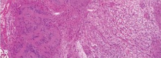
Figure 1: Axial T2-weighted MRI demonstrating a hyperintense, well-circumscribed mass with characteristic 'target sign' and 'split fat sign', indicative of a schwannoma.
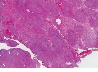
Figure 2: Coronal T1-weighted MRI post-contrast showing intense and heterogeneous enhancement of the nerve sheath tumor, confirming its vascularity.
Electromyography (EMG) and nerve conduction studies (NCS) were considered but ultimately deferred given the clear imaging findings and the decision to proceed with excisional biopsy for definitive diagnosis and symptomatic relief. However, in cases with more ambiguous imaging or pronounced neurological deficits, these studies would be crucial to assess nerve function and differentiate between demyelinating versus axonal injury.
Differential Diagnosis
The clinical presentation and initial imaging findings necessitated a thorough differential diagnosis for a solitary soft tissue mass in the thigh with suspected neural involvement. The primary differentials considered were:
| Feature | Schwannoma (Neurilemmoma) | Neurofibroma | Malignant Peripheral Nerve Sheath Tumor (MPNST) | Lipoma | Ganglion Cyst |
|---|---|---|---|---|---|
| Origin | Schwann cells, usually eccentric to the nerve | Schwann cells, fibroblasts, perineurial cells; involves nerve fascicles directly | Schwann cells, fibroblasts, perineurial cells; high-grade malignancy | Adipose tissue | Synovial fluid from joint/tendon sheath |
| Encapsulation | Yes, well-encapsulated | No, unencapsulated (nerve fibers directly incorporated) | Poorly encapsulated or infiltrative | Yes, thin capsule (pseudocapsule) | Yes, pseudocapsule |
| Clinical Presentation | Solitary, slow-growing, often painful mass; Tinel's sign; possible neurological deficits | Solitary or multiple (NF1), can be painful; 'plexiform' type in NF1; often asymptomatic | Rapidly growing, painful mass; often with neurological deficit; may arise from NF1 | Soft, mobile, often asymptomatic; 'doughy' feel; common in extremities/trunk | Firm, mobile, often non-tender; typically near joints/tendons; can fluctuate in size |
| MRI Characteristics | T1: Isointense to muscle. T2: Hyperintense, heterogeneous, 'target sign', 'split fat sign'. Intense, heterogeneous contrast enhancement. Nerve splayed around tumor. | T1: Isointense to muscle. T2: Hyperintense, 'target sign' (less common/distinct). Diffuse contrast enhancement. Nerve runs through center. | T1: Variable. T2: Heterogeneous, ill-defined margins, perilesional edema, necrosis, hemorrhage. Diffuse, often irregular enhancement. Infiltration. | T1: Hyperintense (fat signal). T2: Intermediate to fat. Suppresses with fat-sat. | T1: Hypointense. T2: Hyperintense, homogenous. No enhancement (unless inflamed). |
| Histology | Antoni A (dense cellularity, Verocay bodies), Antoni B (loose hypocellularity), thick capsule | Mixed cellularity, diffuse growth, no capsule, often myxoid stroma | High mitotic activity, pleomorphism, necrosis, infiltrative growth, atypical cells | Mature adipocytes | Fibrous capsule with mucinous content, no epithelial lining |
| Management | Surgical excision (enucleation) with nerve preservation | Excision if symptomatic, painful, or cosmesis; challenging due to nerve involvement | Wide en bloc resection with clear margins; adjuvant radiotherapy/chemotherapy | Surgical excision if symptomatic, large, or for cosmesis | Aspiration, observation, or surgical excision if symptomatic or recurrent |
| Malignant Potential | Very low (<1%), rare malignant transformation (malignant schwannoma) | Low (5-15% lifetime risk in NF1 for MPNST) | High (aggressive tumor with high recurrence and metastatic potential) | None (benign) | None (benign) |
The distinctive MRI features, particularly the 'target sign,' 'split fat sign,' and the clear visualization of the nerve splayed around the lesion, were highly suggestive of a schwannoma and helped differentiate it from a neurofibroma where the nerve fibers are intimately interspersed within the tumor. The absence of rapid growth, ill-defined margins, or significant perilesional edema also lowered the suspicion for MPNST.
Surgical Decision Making & Classification
Given the patient's progressive symptoms, including pain and paresthesia, and the characteristic imaging findings consistent with a symptomatic benign peripheral nerve sheath tumor (most likely a schwannoma), operative intervention was indicated. The primary goals were:
1.
Symptomatic relief:
Decompression of the affected nerve and removal of the pain-generating mass.
2.
Definitive histological diagnosis:
While imaging was highly suggestive, excisional biopsy provides definitive confirmation and rules out malignancy.
3.
Prevention of further neurological deficit:
Continued compression could lead to irreversible nerve damage.
Non-operative management, such as observation and pain management, was not appropriate in this case due to the progressive nature of the symptoms and the size of the mass causing nerve compression.
Classification:
Peripheral nerve sheath tumors are broadly classified into benign and malignant. Benign tumors include schwannomas and neurofibromas.
*
Schwannomas
are typically solitary and sporadic, though multiple schwannomas can occur in association with Neurofibromatosis Type 2 (NF2) or schwannomatosis. This patient's presentation was consistent with a solitary, sporadic schwannoma.
*
Neurofibromas
are more commonly associated with Neurofibromatosis Type 1 (NF1) and can be solitary or plexiform.
The decision was to proceed with a planned excisional biopsy with the intent of complete tumor enucleation, aiming for nerve preservation.
Surgical Technique / Intervention
Pre-operative Planning:
Detailed MRI images were reviewed to precisely localize the mass, its relationship to the tibial nerve, and its proximity to major vascular structures (femoral artery and vein, popliteal artery and vein). The dimensions of the tumor and the expected course of the nerve were mapped.
Patient Positioning:
The patient was positioned supine on the operating table. The entire right lower extremity was prepped and draped free to allow for full range of motion of the knee and hip, and access to the posteromedial thigh. A tourniquet was applied to the proximal thigh but not inflated unless required for hemostasis.
Surgical Approach:
A longitudinal skin incision, approximately 8 cm in length, was made in the posteromedial aspect of the distal thigh, centered over the palpable mass. This directly approached the adductor canal region. The incision was designed to be extensile if needed.
Subcutaneous tissues were incised, and meticulous blunt and sharp dissection was performed to identify the fascia overlying the muscle compartments. The deep fascia was incised. The mass was identified deep to the sartorius muscle, within the adductor canal.
Dissection and Nerve Identification:
Careful dissection proceeded, identifying the anatomical landmarks of the adductor canal, including the sartorius muscle anteriorly, adductor longus and magnus medially, and the vastus medialis laterally. The saphenous nerve and artery were identified and protected.
The encapsulated mass was located, clearly arising from the tibial nerve, which was identified both proximally and distally to the tumor. The nerve fibers were splayed out and flattened over the surface of the tumor capsule. This eccentric position of the nerve fibers is characteristic of a schwannoma.
Microsurgical principles were employed throughout. A nerve stimulator was used intraoperatively to confirm the neural origin of the tumor and to identify functional nerve fascicles. The stimulator confirmed the tibial nerve origin and allowed for precise identification of the nerve trunk and its fascicles.
Tumor Enucleation:
The well-defined capsule of the schwannoma was meticulously incised longitudinally. The tumor was then carefully enucleated from its bed. The nerve fascicles were seen to run over the surface of the tumor and were gently dissected off the capsule without transection. This allowed for complete removal of the tumor while preserving the integrity of the main nerve trunk.
No neural tissue was sacrificed. Intraoperative findings confirmed a benign-appearing, yellowish-white, lobulated tumor with a distinct capsule, consistent with a schwannoma. The excised specimen measured approximately 4.5 x 3.5 x 3 cm.
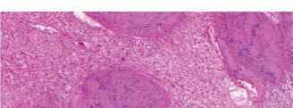
Figure 3: Intraoperative view after meticulous dissection, showing the well-encapsulated schwannoma being carefully enucleated from the splayed nerve fibers, emphasizing nerve preservation.
Hemostasis and Closure:
Thorough hemostasis was achieved using bipolar electrocautery. The wound was irrigated with copious amounts of saline. No drain was deemed necessary. The deep fascia was closed with absorbable sutures. The subcutaneous tissue was approximated, and the skin was closed with non-absorbable sutures in a running subcuticular fashion. A sterile dressing was applied.
Post-Operative Protocol & Rehabilitation
Immediate Post-operative Period (Days 0-7):
*
Pain Management:
Multimodal analgesia including NSAIDs, paracetamol, and short-term opioids as needed.
*
Wound Care:
Daily wound inspection for signs of infection, hematoma, or seroma. Dressing changes as per protocol. Skin sutures typically removed at 10-14 days.
*
Neurological Monitoring:
Frequent assessment of motor and sensory function in the tibial and sural nerve distributions. Patients are counselled about potential transient neurological deficits (e.g., temporary increase in paresthesia or mild sensory loss) due to nerve manipulation.
*
Mobility:
Early ambulation encouraged with partial weight-bearing as tolerated, progressing to full weight-bearing once comfort allows. No specific restrictions on knee ROM initially, but strenuous activities are avoided.
*
Histopathology:
Specimen sent for urgent histopathological examination. Results confirmed benign schwannoma with Antoni A and Antoni B areas, and Verocay bodies. No evidence of malignancy was found.
Early Rehabilitation (Weeks 2-6):
*
Physical Therapy:
*
Gentle Nerve Gliding Exercises:
Initiated to prevent adhesions and promote nerve mobility. Examples include seated sciatic nerve glides and sural nerve glides.
*
Range of Motion:
Full knee and hip range of motion maintained and encouraged.
*
Strengthening:
Gradual, progressive strengthening exercises for the lower extremity, avoiding excessive strain on the surgical site.
*
Proprioception and Balance:
Exercises to regain balance and proprioception.
*
Activity Restriction:
Avoid heavy lifting, strenuous exercise, and high-impact activities for the first 4-6 weeks to allow for soft tissue healing.
Late Rehabilitation and Return to Activity (Weeks 6-12+):
*
Progression of Strengthening:
Continue with advanced strengthening and conditioning exercises.
*
Functional Training:
Tailored exercises to meet specific occupational or recreational demands.
*
Return to Sport/Work:
Gradual return to full activities and sports, guided by pain levels, functional recovery, and physician clearance. Most patients can expect a full recovery and return to previous activity levels within 3-4 months.
*
Follow-up:
Clinical follow-up at 3 months, 6 months, and 1 year to assess functional outcome, monitor for recurrence, and address any persistent neurological symptoms. Further imaging is usually not required unless new symptoms or signs develop.
Pearls & Pitfalls (Crucial for FRCS/Board Exams)
Pearls:
- Clinical Suspicion: Any progressively enlarging, painful, or neurologically symptomatic soft tissue mass, particularly with a positive Tinel's sign, should raise suspicion for a peripheral nerve sheath tumor.
- MRI is Gold Standard: MRI with contrast is indispensable for diagnosis, anatomical localization, and surgical planning. Look for the 'target sign' and 'split fat sign' which are highly suggestive of schwannoma.
- Differentiation: Understand the key differences between schwannoma and neurofibroma on imaging and intraoperatively (e.g., schwannomas typically "push" nerve fibers eccentrically, while neurofibromas "blend" with them centrally). This dictates nerve preservation strategy.
- Microsurgical Technique: Schwannoma excision demands meticulous microsurgical technique. The goal is complete enucleation from the nerve sheath, preserving functional nerve fascicles.
- Intraoperative Nerve Stimulation: This tool is invaluable for confirming nerve identity, assessing fascicular integrity, and guiding safe tumor removal.
- Histopathology is Definitive: While imaging provides a strong presumptive diagnosis, definitive diagnosis and exclusion of malignancy require histopathological examination.
- Syndromic Associations: Always screen for signs of Neurofibromatosis Type 1 (NF1), Neurofibromatosis Type 2 (NF2), or schwannomatosis, especially if multiple lesions are present or family history is suggestive.
Pitfalls:
- Misdiagnosis as other Soft Tissue Tumors: Failing to recognize the neural origin can lead to inappropriate biopsy or surgical approaches that compromise the nerve.
- Incomplete Excision vs. Nerve Injury: Aggressive attempts at excision without careful identification and preservation of nerve fascicles can lead to iatrogenic nerve injury. Conversely, incomplete excision (especially of a neurofibroma) may lead to recurrence.
-
Biopsy Considerations:
- Incisional Biopsy for Suspected Schwannoma: Generally avoided as excisional biopsy is often diagnostic and therapeutic. If biopsy is needed for large, deep, or highly suspicious lesions, a core needle biopsy is preferred over incisional biopsy due to less tissue disruption.
- Biopsy of MPNST: If MPNST is suspected (rapid growth, large size, deep location, ill-defined margins, necrosis on MRI), a well-planned incisional biopsy (or core biopsy) is crucial prior to definitive wide resection.
- Malignant Transformation: While rare for solitary schwannomas (<1%), the potential for malignant transformation (to MPNST) exists, especially in the context of NF1 for neurofibromas. Any rapid growth or recurrence of a "benign" tumor warrants re-evaluation and potentially re-biopsy.
- Post-operative Neuroma Formation: While uncommon with careful enucleation, nerve handling can sometimes lead to localized neuroma formation or persistent dysesthesia.
- Recurrence: Incomplete excision, although rare for schwannomas due to their encapsulation, can lead to recurrence. Careful intraoperative inspection of the nerve and tumor bed is essential.
