Optimizing the Management of Difficult Metastatic Lesions
Key Takeaway
Looking for accurate information on Optimizing the Management of Difficult Metastatic Lesions? Management of difficult metastatic lesions, like those affecting the hip, often necessitates hip arthroplasty, especially with significant femoral head/neck disease and fractures, to address mechanical symptoms. For sacral lesions, proactively screening for bladder/bowel dysfunction is crucial due to potential nerve root encroachment. Prophylactic stabilization or curettage are typically inappropriate treatments for these complex metastatic cases.
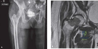
A 62-year-old patient presents with a painful proximal femur lesion. Radiographs demonstrate a large lytic lesion. The patient has a history of treated renal cell carcinoma. How do you assess the stability of this lesion, and what are the clinical indications for surgical intervention?
Candidate: I would use the Mirels scoring system to assess fracture risk. If the score is 7 or greater, I would consider prophylactic surgery. Indications for surgery include pain, impending fracture, or an established pathological fracture. In this case, because it is renal cell carcinoma, I would also consider pre-operative embolization to reduce bleeding risk.
The candidate focuses only on the Mirels score and fails to discuss the "Big Picture": Patient prognosis (Tokuhashi/Tomita), performance status (ECOG), and systemic disease burden. They often forget to mention the multidisciplinary team (MDT) or that the Mirels score is only a guide, not a strict protocol.
Structured approach: 1. Stability: Assess using the Mirels score (site, size, nature, pain). 2. Prognosis: Evaluate life expectancy (Tokuhashi/Tomita) and performance status; surgical goal must match expected survival. 3. Biology: Specifically acknowledge the high vascularity of renal cell metastases and mandate preoperative embolization. 4. Decision: Surgery is indicated for failure of conservative care, impending/actual fracture, or neurological compromise. 5. MDT: Highlight the role of the oncology MDT in timing surgery relative to systemic therapy/radiation.
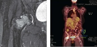
You have decided to proceed with surgical stabilization of a large, lytic, metastatic femoral lesion. You notice significant cortical destruction on the CT scan. What are the biomechanical principles you must apply during your fixation?
Candidate: The goal is load-bearing rather than load-sharing, as the bone is compromised. I would perform internal fixation—usually an intramedullary nail—and augment the defect with PMMA cement to provide immediate stability, improve screw purchase, and potentially induce local tumor necrosis.
Ignoring the "bypass" principle. Candidates often forget to mention that the construct must extend well into healthy bone (2 cortical diameters) proximal and distal to the lesion, or fail to discuss why cement is used (thermal necrosis/filler).
Biomechanical Principles: 1. Load-Bearing: Assume the bone will not heal; the implant must bear the physiological load alone. 2. Span: Bypass the lesion by at least two cortical diameters into healthy diaphyseal bone. 3. Augmentation: Use PMMA cement to fill the lytic cavity, increasing torsional and compressive stiffness. 4. Fixation: Use intramedullary devices for diaphyseal lesions and consider modular tumor endoprostheses if there is periarticular bone loss exceeding 50% of the joint surface.
