Lower Limb Trauma: Decipher Anteroposterior (AP) Figure for Exams
Key Takeaway
In this comprehensive guide, we discuss everything you need to know about Lower Limb Trauma: Decipher Anteroposterior (AP) Figure for Exams. A reverse-obliquity inter-trochanteric fracture with subtrochanteric extension is an unstable femoral fracture. As seen in the figure anteroposterior ap radiograph, it presents with proximal displacement of the lesser trochanter and loss of the medial buttress. Initial management includes medical optimization and operative repair, often within 36 hours, using a cephalomedullary device for better outcomes compared to fixed-angle plating.
A 78-year-old woman presents after a fall. She is independently mobile. Examine this radiograph. What is the injury, and how would you manage this patient?
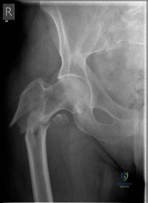
Candidate: This is a reverse-obliquity intertrochanteric fracture with subtrochanteric extension. There is proximal displacement of the lesser trochanter, indicating a loss of the medial buttress, making this an inherently unstable pattern. I would optimize the patient medically and aim for surgery within 36 hours. I would recommend a cephalomedullary nail rather than a fixed-angle plate due to the fracture configuration.
Candidates often miss the "reverse obliquity" classification and simply call it an "intertrochanteric fracture." Failing to recognize the instability caused by the loss of the medial buttress is a critical error, as it dictates the choice of implant.
The perfect answer categorizes the injury (Reverse obliquity), recognizes the stability status (Unstable/Loss of medial buttress), mandates specific timing (NICE guidelines < 36h), and provides a biomechanical justification for the implant (Cephalomedullary nail vs. lateral plate).
The patient was treated with a fixed-angle locking plate. She presents 4 months later with this appearance. What went wrong?
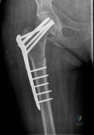
Candidate: This is a mechanical failure due to a combination of biological and biomechanical factors. The lateral locking plate acts as a tension band, but the fracture gap and lack of medial support mean the construct is subjected to excessive cyclical load. The plate acts as a cantilever; because it is lateral, it increases the moment arm, leading to fatigue failure at the stress riser—the junction of the proximal locking holes and the plate shaft.
Candidates often focus only on the implant breaking. A high-scoring answer must specifically mention the biomechanical disadvantage of lateral plating in reverse-obliquity fractures and the stress-shielding/fatigue mechanisms.
Structure the answer into "Biology" (lack of medial support, fracture gap) and "Biomechanics" (lever arm increase, lateral tension side vs. medial compression side). Explicitly identify the "fatigue failure" occurring at the "stress riser" of the plate holes.
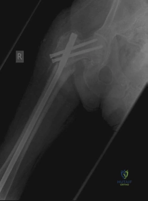
You have revised the fracture with a cephalomedullary nail. Looking at this follow-up radiograph, what is your assessment?
Candidate: The nail is in slight varus, and there is persistent translation at the fracture site. The proximal screws are positioned somewhat superiorly in the femoral head. I would correlate this with lateral imaging to confirm screw placement. Ideally, bone grafting should have been performed to address the non-union biology.
Failure to critically appraise one's own surgical alignment. Always acknowledge the "varus" position if present, as varus collapse is a known mode of failure in hip fracture fixation.
Acknowledge the radiological parameters: Alignment (varus/translation), Implant positioning (screw depth/location), and the need for biological augmentation (bone graft) given this is a revision scenario for non-union.
