Mastering Lower Limb Trauma: AP & Oblique X-ray Diagnosis
Key Takeaway
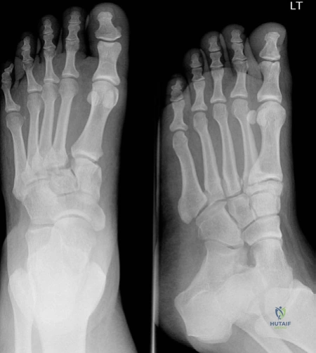
For anyone wondering about Mastering Lower Limb Trauma: AP & Oblique X-ray Diagnosis, AP and oblique radiographs are crucial for diagnosing Lisfranc tarsometatarsal fracture dislocation. They reveal diastasis (gap) between the first and second metatarsals, often alongside a 'fleck sign'—an avulsion fracture from the Lisfranc ligament insertion. These findings are highly suggestive of a Lisfranc injury. Further imaging like CT or MRI may confirm the diagnosis.
A 49-year-old lady presents to A&E following a fall on the stairs. She has a painful, swollen, and bruised foot and is unable to bear weight. The CT1 is unsure of the diagnosis. Examine the images provided and discuss your findings.
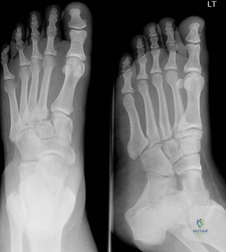
Candidate: I see AP and oblique radiographs of the left foot. There is a diastasis greater than 2mm between the bases of the first and second metatarsals, which is diagnostic of a Lisfranc injury. I also note a 'fleck sign'—an avulsion fragment in the intermetatarsal space—suggestive of an avulsion of the Lisfranc ligament from the base of the second metatarsal. I would request a lateral view to assess for dorsal subluxation of the metatarsal bases.
Failing to mention the "fleck sign" or ignoring the 2mm threshold for diastasis. Borderline candidates often jump straight to "it's a fracture" without systematically describing the tarsometatarsal alignment or the specific radiographic landmarks.
Systematically describe the view, identify the loss of congruity between the medial cuneiform and base of the second metatarsal, acknowledge the pathognomonic 'fleck sign', and emphasize that the lateral view is essential to rule out occult dorsal subluxation.
The radiographs are highly suspicious of a Lisfranc injury, but standard views are inconclusive regarding the extent of the instability. How would you proceed with your diagnostic workup?
Candidate: If clinical suspicion remains high despite plain films, I would proceed with weight-bearing or gravity-stress radiographs to demonstrate dynamic instability. If still inconclusive, I would order a CT scan to evaluate for occult fractures or minor subluxations. MRI is reserved for cases where we suspect a ligamentous-only injury without bony displacement.
Suggesting an MRI immediately. While sensitive for soft tissues, it is not the first-line investigation for mechanical instability. Missing the concept of "weight-bearing" or "stress" views to demonstrate the injury is a significant oversight.
A structured, stepwise approach: Stress views (demonstrating dynamic instability) → CT (defining bony anatomy/comminution) → MRI (for purely ligamentous injuries). High-scoring candidates mention that weight-bearing is the "gold standard" for functional assessment.
How do you approach the management of a confirmed, displaced Lisfranc fracture-dislocation, and what are the specific surgical goals?
Candidate: Management is surgical for displaced injuries. The goals are an anatomical reduction of the TMT joints and stable fixation. This is typically achieved via Open Reduction and Internal Fixation (ORIF) using plates and screws. For severely comminuted fractures or late-stage injury, primary arthrodesis of the medial and central columns is the gold standard to prevent post-traumatic arthritis.
Overlooking the role of primary arthrodesis in severely comminuted injuries. Candidates often forget to mention the importance of addressing the soft tissue envelope, which is often compromised in high-energy injuries.
Distinguish between ORIF (for reducible, acute fractures) and primary arthrodesis (for comminution/unstable variants). Discussing the "column" concept of the foot (medial, central, lateral) and the need for anatomical reduction of the "keystone" (the base of the 2nd metatarsal) demonstrates consultant-level thinking.
What is your strategy if the patient presents with signs of foot compartment syndrome?
Candidate: Compartment syndrome of the foot is a clinical diagnosis. Upon suspicion, I would immediately remove all splints/dressings, elevate the foot to the level of the heart, and prepare for urgent surgical decompression. The foot has 9 compartments. I use a 3-incision approach—two dorsal incisions over the 2nd and 3rd metatarsals, and one medial incision—to fully release the compartments. The patient will then require a return to theatre for delayed primary closure or skin grafting.
Using compartment pressure monitoring as a substitute for clinical judgment or delaying for advanced imaging. Failing to mention that the foot has 9 compartments (not 4 as in the leg) is a common failure point.
Emphasize that this is a clinical emergency. Describe the anatomy confidently (9 compartments, 3 incisions). Highlight the secondary nature of the intervention (delayed closure) and the requirement for multi-disciplinary communication (theatre team/anaesthetics).
